国际口腔医学杂志 ›› 2026, Vol. 53 ›› Issue (2): 274-280.doi: 10.7518/gjkq.2026115
• 综述 • 上一篇
余铭轩1( ),何茹逸1,鄢荣曾1,2()
Mingxuan Yu1(),Ruyi He1,Rongzeng Yan1,2()
摘要:
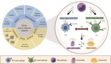
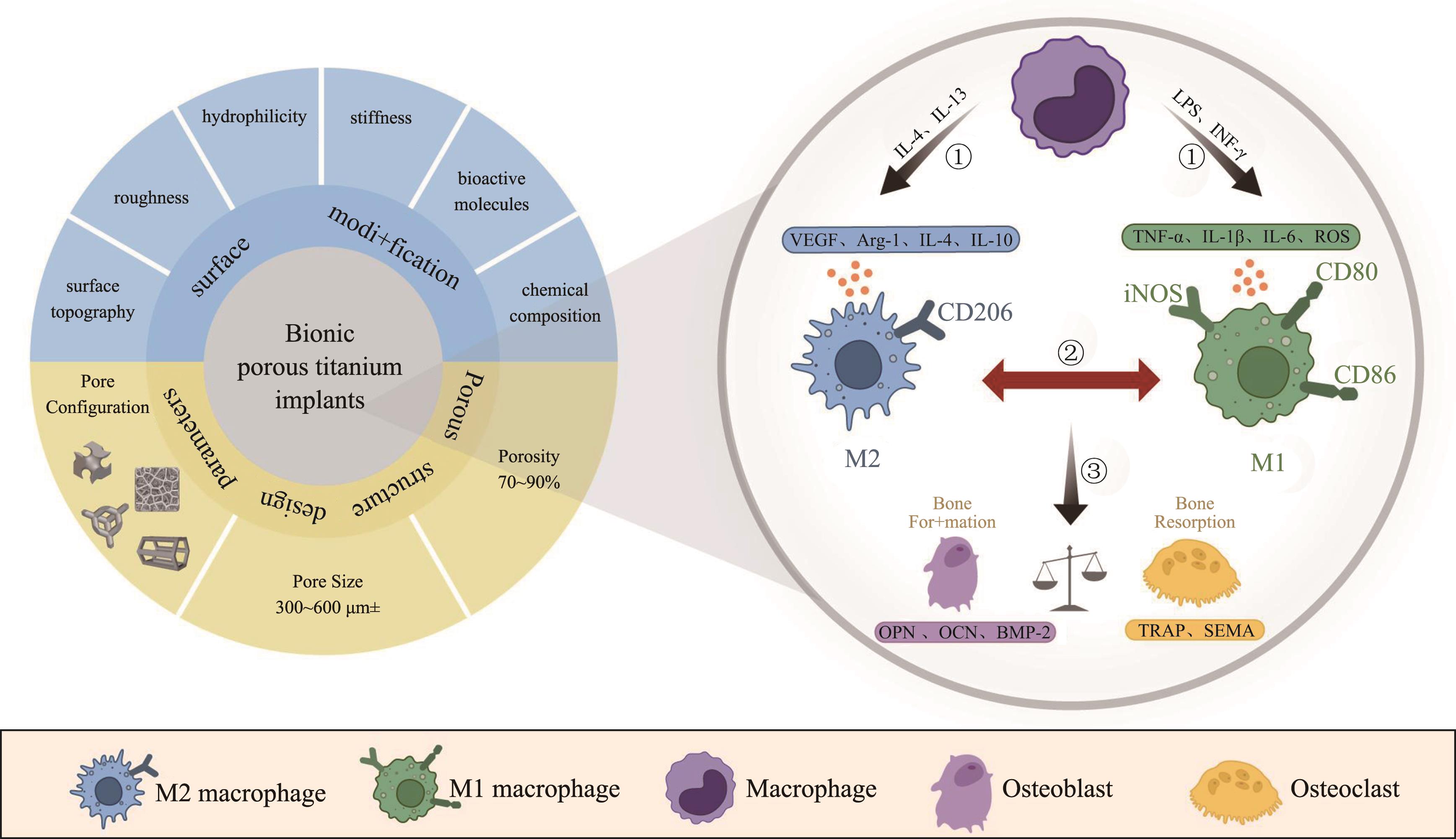
钛种植体植入机体后会引起宿主免疫炎症反应,其进程影响骨组织的修复与再生。以巨噬细胞为核心的骨免疫调控近年来备受关注。仿生多孔钛种植体表面形貌可调控其周围免疫微环境中巨噬细胞的生物学行为,促进血管生成和加速骨改建。本文阐述了巨噬细胞在仿生多孔钛种植体植入后免疫反应中的作用及其表面处理后骨免疫调控能力,重点介绍表面仿生多孔结构设计对巨噬细胞生物学行为的影响。深入理解钛种植体与机体界面相互作用,为临床仿生设计、制备具有免疫调节功能的多孔钛种植体提供新思路。
中图分类号:
| [1] | Zhang LC, Chen LY. A review on biomedical tita-nium alloys: recent progress and prospect[J]. Adv Eng Mater, 2019, 21(4): 1801215. |
| [2] | Xiao F, Ye JH, Huang CX, et al. Gradient gyroid Ti6Al4V scaffolds with TiO2 surface modification: promising approach for large bone defect repair[J]. Biomater Adv, 2024, 161: 213899. |
| [3] | Wang ZH, Zhang M, Liu ZW, et al. Biomimetic design strategy of complex porous structure based on 3D printing Ti-6Al-4V scaffolds for enhanced osseointegration[J]. Mater Des, 2022, 218: 110721. |
| [4] | Lee J, Byun H, Perikamana SKM, et al. Current advances in immunomodulatory biomaterials for bone regeneration[J]. Adv Healthc Mater, 2019, 8(4): e1801106. |
| [5] | Davenport Huyer L, Pascual-Gil S, Wang YF, et al. Advanced strategies for modulation of the mate-rial-macrophage interface[J]. Adv Funct Materials, 2020, 30(44): 1909331. |
| [6] | Zhu YZ, Liang H, Liu XM, et al. Regulation of macrophage polarization through surface topography design to facilitate implant-to-bone osteointegration[J]. Sci Adv, 2021, 7(14): eabf6654. |
| [7] | Liu Y, Rui ZY, Cheng W, et al. Characterization and evaluation of a femtosecond laser-induced osseointegration and an anti-inflammatory structure genera-ted on a titanium alloy[J]. Regen Biomater, 2021, 8(2): rbab006. |
| [8] | Xia Y, He XT, Xu XY, et al. Exosomes derived from M0, M1 and M2 macrophages exert distinct in-fluences on the proliferation and differentiation of mesenchymal stem cells[J]. PeerJ, 2020, 8: e8970. |
| [9] | Amengual-Peñafiel L, Córdova LA, Jara-Sepúlveda MC, et al. Osteoimmunology drives dental implant osseointegration: a new paradigm for implant dentistry[J]. Jpn Dent Sci Rev, 2021, 57: 12-19. |
| [10] |
Ji ZB, Wan Y, Wang HW, et al. Effects of surface morphology and composition of titanium implants on osteogenesis and inflammatory responses: a review[J]. Biomed Mater, 2023, 18(4). doi: 10.1088/1748-605X/acd976 .
doi: 10.1088/1748-605X/acd976 |
| [11] | Yang Y, Zhang T, Jiang MY, et al. Effect of the immune responses induced by implants in a integrated three-dimensional micro-nano topography on osseointegration[J]. J Biomed Mater Res A, 2021, 109(8): 1429-1440. |
| [12] | Razzi F, Fratila-Apachitei LE, Fahy N, et al. Im-munomodulation of surface biofunctionalized 3D printed porous titanium implants[J]. Biomed Mater, 2020, 15(3): 035017. |
| [13] | Liu ZG, Liu Y, Liu S, et al. The effects of TiO2 nanotubes on the biocompatibility of 3D printed Cu-bea-ring TC4 alloy[J]. Mater Des, 2021, 207: 109831. |
| [14] | Zhao XY, Wang BB, Lai WJ, et al. Improved tribological properties, cyto-biocompatibility and anti-inflammatory ability of additive manufactured Ti-6Al-4V alloy through surface texturing and nitriding[J]. Surf Coat Technol, 2021, 425: 127686. |
| [15] | Li G, Liu WT, Liang LX, et al. Preparing Sr-contai-ning nano-structures on micro-structured titanium alloy surface fabricated by additively manufactu-ring to enhance the anti-inflammation and osteoge-nesis[J]. Colloids Surf B Biointerfaces, 2022, 218: 112762. |
| [16] | Wang W, Xiong YZ, Zhao RL, et al. A novel hierarchical biofunctionalized 3D-printed porous Ti6Al4V scaffold with enhanced osteoporotic osseointegration through osteoimmunomodulation[J]. J Nanobiotechnology, 2022, 20(1): 68. |
| [17] | Wang YP, Feng ZJ, Liu X, et al. Titanium alloy composited with dual-cytokine releasing polysaccharide hydrogel to enhance osseointegration via osteogenic and macrophage polarization signaling pathways[J]. Regen Biomater, 2022, 9: rbac003. |
| [18] | Wu YP, Shi XW, Wang JJ, et al. A surface metal ion-modified 3D-printed Ti-6Al-4V implant with direct and immunoregulatory antibacterial and osteogenic activity[J]. Front Bioeng Biotechnol, 2023, 11: 1142264. |
| [19] | Wu H, Dong H, Tang Z, et al. Electrical stimulation of piezoelectric BaTiO3 coated Ti6Al4V scaffolds pro-motes anti-inflammatory polarization of macropha-ges and bone repair via MAPK/JNK inhibition and OXPHOS activation[J]. Biomaterials, 2023, 293: 121990. |
| [20] | Li JX, Zhong HZ, Cao BJ, et al. Comparative study of 3D-printed porous titanium alloy with rod designs of three different geometric structures for orthopaedic implantation[J]. Acta Metall Sin Engl Lett, 2024, 37(1): 54-66. |
| [21] | Pei X, Wang LN, Zhou CC, et al. Ti6Al4V orthopedic implant with biomimetic heterogeneous structure via 3D printing for improving osteogenesis[J]. Mater Des, 2022, 221: 110964. |
| [22] | Liang HX, Yang YW, Xie DQ, et al. Trabecular-like Ti-6Al-4V scaffolds for orthopedic: fabrication by selective laser melting and in vitro biocompatibility[J]. J Mater Sci Technol, 2019, 35(7): 1284-1297. |
| [23] |
Pei X, Wu LN, Zhou CC, et al. 3D printed titanium scaffolds with homogeneous diamond-like structures mimicking that of the osteocyte microenvironment and its bone regeneration study[J]. Biofabrication, 2020, 13(1). doi: 10.1088/1758-5090/abc060 .
doi: 10.1088/1758-5090/abc060 |
| [24] | Zhang YN, Sun N, Zhu MR, et al. The contribution of pore size and porosity of 3D printed porous tita-nium scaffolds to osteogenesis[J]. Biomater Adv, 2022, 133: 112651. |
| [25] | Liu QY, Wei F, Coathup M, et al. Effect of porosity and pore shape on the mechanical and biological properties of additively manufactured bone scaffolds[J]. Adv Healthc Mater, 2023, 12(30): e2301111. |
| [26] | Liu Y, Cao LY, Zhang S, et al. Effect of hierarchical porous scaffold on osteoimmunomodulation and bone formation[J]. Appl Mater Today, 2020, 20: 100779. |
| [27] | Wang C, Wu J, Liu LY, et al. Improving osteoinduction and osteogenesis of Ti6Al4V alloy porous scaffold by regulating the pore structure[J]. Front Chem, 2023, 11: 1190630. |
| [28] | Wu YQ, Liu Z, Xu ZC, et al. Macrophage responses to selective laser-melted Ti-6Al-4V scaffolds of different pore geometries and the corresponding osteoimmunomodulatory effects toward osteogenesis[J]. J Biomed Mater Res A, 2022, 110(4): 873-883. |
| [29] | Deng FY, Liu LL, Li Z, et al. 3D printed Ti6Al4V bone scaffolds with different pore structure effects on bone ingrowth[J]. J Biol Eng, 2021, 15(1): 4. |
| [30] | Zhao HY, Han YF, Pan C, et al. Design and mecha-nical properties verification of gradient voronoi scaffold for bone tissue engineering[J]. Micromachines (Basel), 2021, 12(6): 664. |
| [31] | Feng JW, Fu JZ, Yao XH, et al. Triply periodic minimal surface (TPMS) porous structures: from multi-scale design, precise additive manufacturing to multidisciplinary applications[J]. Int J Extreme Manuf, 2022, 4(2): 022001. |
| [32] | He YD, Li Z, Ding X, et al. Nanoporous titanium implant surface promotes osteogenesis by suppres-sing osteoclastogenesis via integrin β1/FAKpY397/MAPK pathway[J]. Bioact Mater, 2021, 8: 109-123. |
| [33] | Dai XH, Bai YY, Heng BC, et al. Biomimetic hierarchical implant surfaces promote early osseointegration in osteoporotic rats by suppressing macrophage activation and osteoclastogenesis[J]. J Mater Chem B, 2022, 10(11): 1875-1885. |
| [34] | Zhou Y, Tang CZ, Deng JL, et al. Micro/nano topography of selective laser melting titanium inhibits osteoclastogenesis via mediation of macrophage pola-rization[J]. Biochem Biophys Res Commun, 2021, 581: 53-59. |
| [1] | 刘翼,邱江珊,申道南,关鑫,丁一. 不同脉冲能量Er: YAG激光对钛盘表面形貌及生物学性能的影响[J]. 国际口腔医学杂志, 2024, 51(6): 713-721. |
| [2] | 李佳敏,李毓晨,葛张洁,廖凌子,郭鑫,郭晓龙,周平. 抗菌肽在口腔钛种植体涂层中的研究进展[J]. 国际口腔医学杂志, 2024, 51(5): 572-584. |
| [3] | 张政,杨锋,李家锋,曹焜. 钛种植体抗菌化修饰的研究进展[J]. 国际口腔医学杂志, 2024, 51(5): 585-595. |
| [4] | 朱俊瑾,王剑. 钛种植体表面银纳米颗粒负载方法的进展[J]. 国际口腔医学杂志, 2021, 48(3): 334-340. |
| [5] | 赵玉洁,管晓燕,李小兰,陈琦君,王倩,刘建国. 巨噬细胞极化参与正畸牙移动的研究进展[J]. 国际口腔医学杂志, 2020, 47(4): 478-483. |
| [6] | 刘晔,洪润丹,王志国,刘涵云,孟琛达,王茹,徐全臣. 人单核细胞和外周血单个核细胞衍生的巨噬细胞极化特性的比较[J]. 国际口腔医学杂志, 2020, 47(3): 286-292. |
| [7] | 刘育豪,袁泉,张士文. 基于共价接枝的钛种植体载药抗菌涂层的研究进展[J]. 国际口腔医学杂志, 2019, 46(2): 228-233. |
| [8] | 祁星颖,郑国莹,隋磊. 钛种植体表面形貌对成骨的影响[J]. 国际口腔医学杂志, 2018, 45(5): 527-533. |
| [9] | 樊牮,邹耿森,陈江. 钛种植体表面纳米改性及其与机体免疫应答[J]. 国际口腔医学杂志, 2014, 41(6): 691-693. |
| [10] | 庄秀妹 邓飞龙. 钛表面及其涂层纳米化对骨结合的影响和机制[J]. 国际口腔医学杂志, 2014, 41(4): 427-430. |
| [11] | 刘媛媛1 李果1 任家银1 赵书平1 聂晶2 王虎1. 纳米钛膜种植体-骨界面的骨整合研究[J]. 国际口腔医学杂志, 2012, 39(3): 312-316. |
| [12] | 张静超,莫安春,. 钛种植体表面微结构对成骨细胞影响的研究进展[J]. 国际口腔医学杂志, 2007, 34(03): 207-209. |
| [13] | 张悦,夏海斌,. 碱热处理制备生物活性钛种植体[J]. 国际口腔医学杂志, 2007, 34(03): 216-219. |
| [14] | 刘同军,程祥荣,. 钛金属种植体表面生物化学改性[J]. 国际口腔医学杂志, 2006, 33(03): 210-212. |
| [15] | 董强 丁仲鹃. HA涂层钛种植体骨界面的生物学研究进展[J]. 国际口腔医学杂志, 2003, 30(01): 34-35. |
|
||