国际口腔医学杂志 ›› 2024, Vol. 51 ›› Issue (3): 337-343.doi: 10.7518/gjkq.2024051
曹友辉1( ),包雪梅1,2()
Youhui Cao1(),Xuemei Bao1,2()
摘要:
随着粘接材料的更新升级、牙髓根治技术的长足进步及修复制作工艺的飞速发展,对于 龈距离短、剩余牙体组织少、根管治疗术后死髓磨牙的保存呈现了美好前景。保存天然牙、残冠继续行使功能使得髓腔固位冠的修复理念得到临床医生的接受与认可。利用有限元法进行应力分析具有高效、精确、可重复等优点。本文就有限元法在髓腔固位冠修复中的应力研究应用进行综述,探讨髓腔固位冠修复中的注意事项。
中图分类号:
| 1 | 吴倩, 张彬, 李楠, 等. 三维有限元分析在口腔医学领域的应用及研究进展[J]. 世界最新医学信息文摘, 2019, 19(20): 95-96, 99. |
| Wu Q, Zhang B, Li N, et al. The application and research progress of three-dimensional finite element analysis in the field of stomatology[J]. World Latest Med Inf, 2019, 19(20): 95-96, 99. | |
| 2 | 陈智, 陈瑞甜. 髓腔固位冠[J]. 口腔医学研究, 2018, 34(1): 1-5. |
| Chen Z, Chen RT. Endocrown[J]. J Oral Sci Res, 2018, 34(1): 1-5. | |
| 3 | 陈惠, 岑蓉, 张成飞. 髓腔固位冠的应用现状[J]. 口腔颌面修复学杂志, 2022, 23(5): 321-326. |
| Chen H, Cen R, Zhang CF. Endocrown: the state of the art[J]. Chin J Prosthodont, 2022, 23(5): 321-326. | |
| 4 | Pedrollo Lise D, van Ende A, De Munck J, et al. Biomechanical behavior of endodontically treated premolars using different preparation designs and CAD/CAM materials[J]. J Dent, 2017, 59: 54-61. |
| 5 | Zoidis P, Bakiri E, Polyzois G. Using modified polyetheretherketone (PEEK) as an alternative material for endocrown restorations: a short-term clinical report[J]. J Prosthet Dent, 2017, 117(3): 335-339. |
| 6 | Rocca GT, Daher R, Saratti CM, et al. Restoration of severely damaged endodontically treated premolars: the influence of the endo-core length on marginal integrity and fatigue resistance of lithium disi-licate CAD-CAM ceramic endocrowns[J]. J Dent, 2018, 68: 41-50. |
| 7 | 林捷, 林珍香, 郑志强. 髓腔固位冠不同修复材料和厚度对应力分布的影响[J]. 口腔疾病防治, 2021, 29(11): 740-745. |
| Lin J, Lin ZX, Zheng ZQ. Effects of the different materials and thicknesses on endocrown stress distribution[J]. J Prev Treat Stomatol Dis, 2021, 29(11): 740-745. | |
| 8 | 高琳, 韩祥永, 徐晓明. 不同材料及不同固位深度的髓腔固位冠修复下颌第二磨牙的三维有限元分析[J]. 上海口腔医学, 2022, 31(6): 621-624. |
| Gao L, Han XY, Xu XM. Three-dimensional finite element analysis of three-material endocrown in the restoration of dental defects of mandibular second mo-lars[J]. Shanghai J Stomatol, 2022, 31(6): 621-624. | |
| 9 | He JH, Zheng ZT, Wu M, et al. Influence of resto-rative material and cement on the stress distribution of endocrowns: 3D finite element analysis[J]. BMC Oral Health, 2021, 21(1): 495. |
| 10 | Meng QZ, Zhang YJ, Chi DL, et al. Resistance fracture of minimally prepared endocrowns made by three types of restorative materials: a 3D finite element analysis[J]. J Mater Sci Mater Med, 2021, 32(11): 137. |
| 11 | Tribst JPM, Dal Piva AMO, Madruga CFL, et al. Endocrown restorations: influence of dental remnant and restorative material on stress distribution[J]. Dent Mater, 2018, 34(10): 1466-1473. |
| 12 | Ural Ç, Çağlayan E. A 3-dimensional finite element and in vitro analysis of endocrown restorations fabricated with different preparation designs and various restorative materials[J]. J Prosthet Dent, 2021, 126(4): 586.e1-586.e9. |
| 13 | Zheng ZT, He YY, Ruan WH, et al. Biomechanical behavior of endocrown restorations with different CAD-CAM materials: a 3D finite element and in vitro analysis[J]. J Prosthet Dent, 2021, 125(6): 890-899. |
| 14 | Zheng ZT, Sun JL, Jiang LF, et al. Influence of margin design and restorative material on the stress distribution of endocrowns: a 3D finite element analysis[J]. BMC Oral Health, 2022, 22(1): 30. |
| 15 | Shams A, Elsherbini M, Elsherbiny AA, et al. Re-habilitation of severely-destructed endodontically treated premolar teeth with novel endocrown system: biomechanical behavior assessment through 3D finite element and in vitro analyses[J]. J Mech Behav Biomed Mater, 2022, 126: 105031. |
| 16 | Köseoğlu M, Furuncuoğlu F. Effect of polyetheretherketone and indirect composite resin thickness on stress distribution in maxillary premolar teeth restored with endocrown: a 3D finite element analysis[J]. J Biotechnol Strateg Heath Res, 2020, 4(3): 298-305. |
| 17 | 姜又升, 冯琳, 高学军. 垫底材料弹性模量对髓腔固位冠修复后上颌前磨牙应力分布的影响[J]. 北京大学学报(医学版), 2021, 53(4): 764-769. |
| Jiang YS, Feng L, Gao XJ. Influence of base mate-rials on stress distribution in endodontically treated maxillary premolars restored with endocrowns[J]. J Peking Univ (Heath Sci), 2021, 53(4): 764-769. | |
| 18 | 冯娟, 郭慧慧, 申晋斌, 等. 磨牙髓室底垫底厚度对全瓷嵌体冠应力分布的影响[J]. 牙体牙髓牙周病学杂志, 2017, 27(1): 16-21. |
| Feng J, Guo HH, Shen JB, et al. Effects of cement thickness on the stress distribution of full-ceramic-endocrown restoration: a finite element analysis[J]. Chin J Conserv Dent, 2017, 27(1): 16-21. | |
| 19 | Cheng X, Zhang XY, Qian WH. Influence of diffe-rent base materials and thicknesses on the fracture resistance of endocrown: a three-dimensional finite element analysis[J]. BMC Oral Health, 2022, 22(1): 363. |
| 20 | 张英, 熊璟, 李永强, 等. 垫底材料厚度对髓腔固位冠修复后牙体组织应力影响的三维有限元分析[J]. 中国美容医学, 2019, 28(9): 102-106. |
| Zhang Y, Xiong J, Li YQ, et al. Three-dimensional-finite-element comparative research of different cement thickness in endodontically treated mandi-bular molar restored with endocrown restorations[J]. Chin J Aesthetic Med, 2019, 28(9): 102-106. | |
| 21 | 黄绮雯, 马晓晴, 唐亮. 髓腔固位冠的临床应用研究进展[J]. 临床医学研究与实践, 2022, 7(13): 185-189. |
| Huang QW, Ma XQ, Tang L. Research progress in clinical application of endocrown[J]. Clin Res Pract, 2022, 7(13): 185-189. | |
| 22 | 李建宾, 陈维毅, 姚蔚. 髓腔壁缺损对下颌前磨牙髓腔固位冠修复应力的影响[J]. 太原理工大学学报, 2018, 49(1): 158-163. |
| Li JB, Chen WY, Yao W. Influence of cavity wall defect on stress distribution in the mandibular premolar restored with endocrown[J]. J Taiyuan Univ Technol, 2018, 49(1): 158-163. | |
| 23 | 吴帆, 曹谅, 姜晓南, 等. 下颌第一磨牙邻面不同高度缺损髓腔固位冠的生物力学分析[J]. 口腔医学研究, 2018, 34(1): 65-68. |
| Wu F, Cao L, Jiang XN, et al. Biomechanical analysis of endocrown of mandibular first molar with different proximal heights[J]. J Oral Sci Res, 2018, 34(1): 65-68. | |
| 24 | Zhang YJ, Lai HB, Meng QZ, et al. The synergetic effect of pulp chamber extension depth and occlusal thickness on stress distribution of molar endocrowns: a 3-dimensional finite element analysis[J]. J Mater Sci Mater Med, 2022, 33(7): 56. |
| 25 |
林珍香, 潘在兴, 叶起清, 等. 二硅酸锂陶瓷和氧化锆髓腔固位冠的 面厚度设计对抗折性能的影响[J]. 华西口腔医学杂志, 2020, 38(6): 647-651. 面厚度设计对抗折性能的影响[J]. 华西口腔医学杂志, 2020, 38(6): 647-651.
|
| Lin ZX, Pan ZX, Ye QQ, et al. Effect of occlusal thickness design on the fracture resistance of endocrowns restored with lithium disilicate ceramic and zirconia[J]. West China J Stomatol, 2020, 38(6): 647-651. | |
| 26 | Dartora NR, de Conto Ferreira MB, Moris ICM, et al. Effect of intracoronal depth of teeth restored with endocrowns on fracture resistance: in vitro and 3-dimensional finite element analysis[J]. J Endod, 2018, 44(7): 1179-1185. |
| 27 | 康婷, 石思琼, 赵威, 等. 上颌第一前磨牙舌尖斜形折裂不同修复设计的有限元分析[J]. 口腔医学研究, 2019, 35(10): 953-956. |
| Kang T, Shi SQ, Zhao W, et al. Finite element analysis of different designs for maxillary first premolars with lingual cusp oblique defect[J]. J Oral Sci Res, 2019, 35(10): 953-956. | |
| 28 | Zhu JX, Wang DM, Rong QG, et al. Effect of central retainer shape and abduction angle during preparation of teeth on dentin and cement layer stress distributions in endocrown-restored mandibular molars[J]. Dent Mater J, 2020, 39(3): 464-470. |
| 29 | Tribst JPM, Giudice RL, dos Santos AFC, et al. Lithium disilicate ceramic endocrown biomechanical response according to different pulp chamber extension angles and filling materials[J]. Materials, 2021, 14(5): 1307. |
| 30 | 粟猛, 屈直. 不同制备形态对短冠磨牙髓腔固位冠抗折性影响的研究[J]. 口腔医学研究, 2021, 37(2): 118-121. |
| Su M, Qu Z. Effect of preparation designs on fracture strengths of endocrown of maxillary short coronal molars[J]. J Oral Sci Res, 2021, 37(2): 118-121. | |
| 31 | Gong QM, Huang L, Luo JP, et al. The practicability of different preparation of mandibular molar restored by modified endocrown with intracanal extension: computational analysis using finite element models[J]. Comput Methods Programs Biomed, 2022, 226: 107178. |
| 32 | Gulec L, Ulusoy N. Effect of endocrown restorations with different CAD/CAM materials: 3D finite element and weibull analyses[J]. Biomed Res Int, 2017, 2017: 5638683. |
| 33 | Aldesoki M, Bourauel C, Morsi T, et al. Biomecha-nical behavior of endodontically treated premolars restored with different endocrown designs: finite ele-ment study[J]. J Mech Behav Biomed Mater, 2022, 133: 105309. |
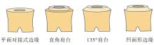
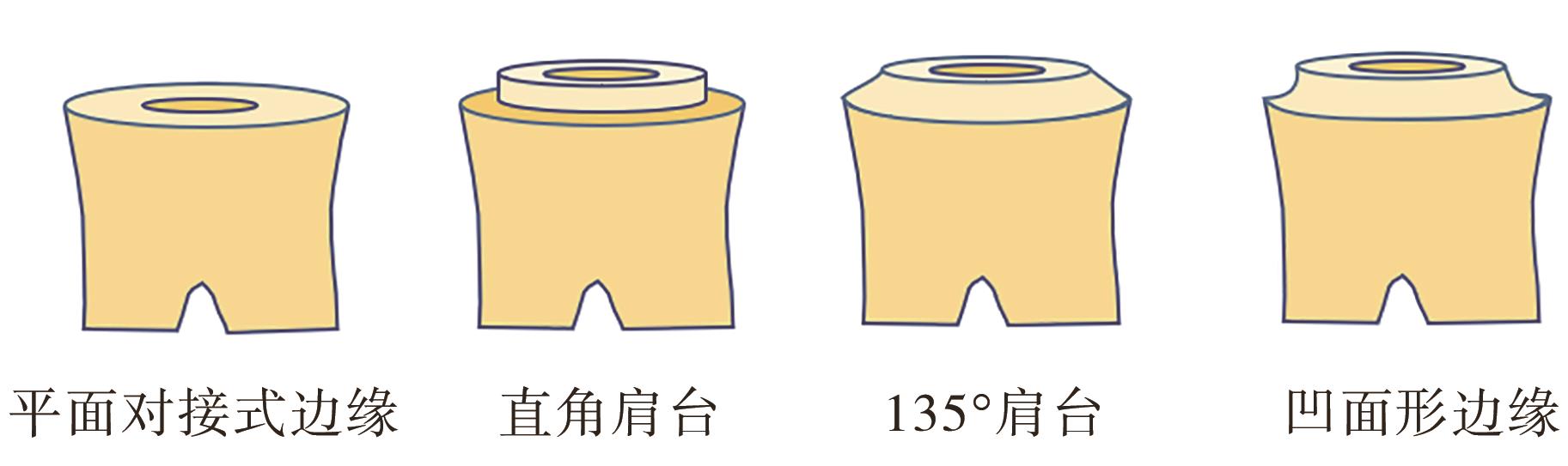
| 34 | 王慧媛, 付强, 张春光, 等. 边缘形式对大面积缺损第一磨牙髓腔固位冠应力分布的影响[J]. 口腔医学研究, 2015, 31(11): 1121-1124. |
| Wang HY, Fu Q, Zhang CG, et al. Research on the biomechanical effects of restoration method on first molar with significant loss of coronal structure[J]. J Oral Sci Res, 2015, 31(11): 1121-1124. | |
| 35 | 郭靖, 王潇宇, 李学盛, 等. 不同边缘设计的髓腔固位冠修复下颌前磨牙的应力分析[J]. 南方医科大学学报, 2016, 36(2): 200-204. |
| Guo J, Wang XY, Li XS, et al. Influence of different designs of marginal preparation on stress distribution in the mandibular premolar restored with endocrown[J]. J South Med Univ, 2016, 36(2): 200-204. |
| [1] | 杨岩朵,陈红,许祖达,赵媛. 生物陶瓷材料iRoot BP Plus与三氧化矿物聚合物在活髓切断术中疗效对比的Meta分析[J]. 国际口腔医学杂志, 2024, 51(2): 176-186. |
| [2] | 颜愈佳,邹玲. 生物陶瓷类根管封闭剂的研究进展[J]. 国际口腔医学杂志, 2022, 49(5): 578-585. |
| [3] | 黎敏,华成舸,蒋丽. 提高氧化锆陶瓷粘接性能新技术的研究进展[J]. 国际口腔医学杂志, 2021, 48(4): 485-490. |
| [4] | 李米雪子,张琛. 椅旁计算机辅助设计/计算机辅助制作髓腔固位冠修复根管治疗后磨牙的临床考量[J]. 国际口腔医学杂志, 2021, 48(3): 274-279. |
| [5] | 于婉琦,周延民,赵静辉. 口腔种植体新材料的研究现状[J]. 国际口腔医学杂志, 2019, 46(4): 488-496. |
| [6] | 侯晔坡,高杰. Er:YAG激光照射对牙科陶瓷材料粘接影响的研究进展[J]. 国际口腔医学杂志, 2019, 46(1): 68-72. |
| [7] | 张雅蓉, 刘洋, 张玲, 于海洋. 不同切端设计的上前牙瓷贴面受载能力的定量研究[J]. 国际口腔医学杂志, 2017, 44(3): 301-303. |
| [8] | 姚陈敏, 周丽群, 黄翠. 前牙磨耗牙色修复材料的选择[J]. 国际口腔医学杂志, 2017, 44(3): 363-367. |
| [9] | 曹国庆, 王林霞, 杜莉平. 有限元法在桩核冠修复研究中的应用[J]. 国际口腔医学杂志, 2017, 44(2): 209-213. |
| [10] | 刘伟 陈西文 朱智敏. 前牙氧化锆全瓷翼板粘接桥的短期临床观察[J]. 国际口腔医学杂志, 2014, 41(5): 530-535. |
| [11] | 李励芸 魏文佳 孟翔峰. 长期水储存对玻璃陶瓷与牙本质间树脂粘接界面的影响[J]. 国际口腔医学杂志, 2013, 40(4): 436-439. |
| [12] | 景页 孟翔峰. 自粘接树脂水门汀与二氧化锆陶瓷间粘接耐久性的研究[J]. 国际口腔医学杂志, 2013, 40(3): 301-304. |
| [13] | 林艺华1 宋晓萌2 张玮3. 3种树脂加强型玻璃离子与氧化锆陶瓷粘接性能的研究[J]. 国际口腔医学杂志, 2013, 40(3): 305-308. |
| [14] | 浩志超1综述 孟玉坤2审校. 牙科用氧化锆陶瓷的低温时效及其影响因素[J]. 国际口腔医学杂志, 2012, 39(4): 494-497. |
| [15] | 王传勇综述 李伟 蒋丽审校. 生物陶瓷表面蛋白吸附的研究进展[J]. 国际口腔医学杂志, 2012, 39(4): 550-553. |
|