国际口腔医学杂志 ›› 2023, Vol. 50 ›› Issue (2): 186-194.doi: 10.7518/gjkq.2023035
汪牡丹( ),宋东哲,黄定明()
Wang Mudan(),Song Dongzhe,Huang Dingming.()
摘要:
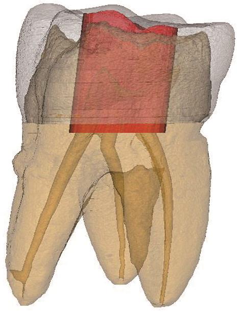
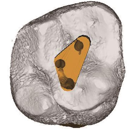
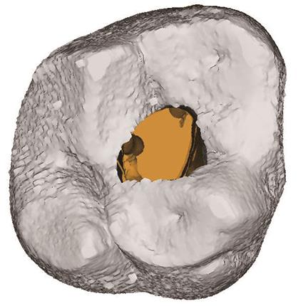
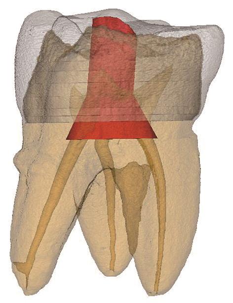
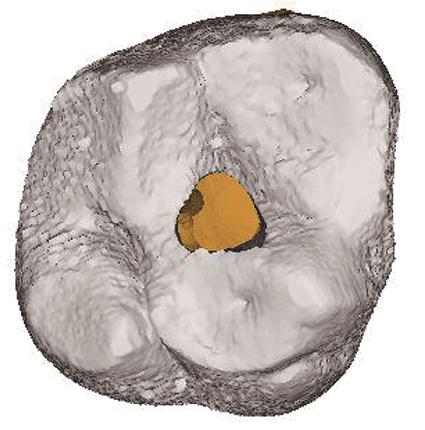
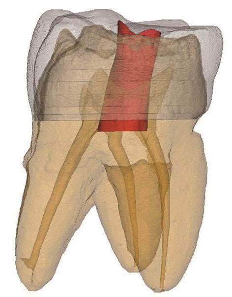
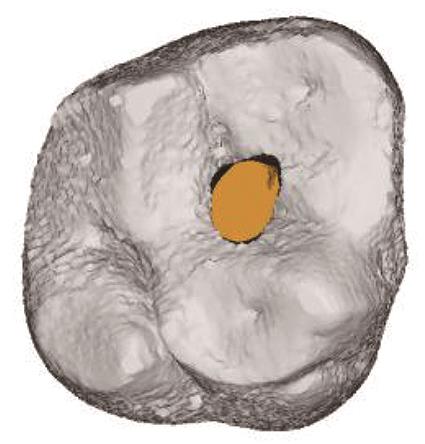
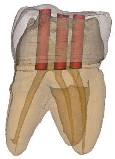
根管治疗术是目前治疗牙髓根尖周疾病最常用且有效的手段之一。根管治疗过程会导致牙体组织损失,可能会降低患牙的抗折性。开髓洞型的设计与冠部牙体组织的磨除量密切相关。如何在保证根管治疗的有效开展下进行微创开髓,减少冠部牙体组织损失,以提高术后患牙的抗折性能,是本领域的研究热点。与强调直线入路的传统开髓洞型相比,以保守开髓洞型为代表的微创开髓洞型能否增强患牙的抗折性存在争议。本文从不同开髓洞型设计要点、根管治疗术后患牙抗折性能的研究现状入手,对相关研究进行文献回顾和综述。综合研究结果,可以认为微创开髓洞型减少了冠部牙体组织损失量,降低了应力集中;但过分微创的开髓洞型未能实现患牙抗折性能的提升,可能是因为微创设计破坏了直线通路所致。本文为微创开髓洞型在根管治疗中的应用提供了新思路,设计新的微创开髓洞型以兼顾冠部牙体组织的保存与直线通路的建立,或许是未来的研究方向。
中图分类号:
| 1 | 周学东, 陈智, 岳林. 牙体牙髓病学[M]. 5版. 北京: 人民卫生出版, 2020. |
| Zhou XD, Chen Z, Yue L. Prosthodontics[M]. 5th ed. Beijing: People’s Medical Publishing House, 2020. | |
| 2 | Liao WC, Chen CH, Pan YH, et al. Vertical root fracture in non-endodontically and endodontically treated teeth: current understanding and future challenge[J]. J Pers Med, 2021, 11(12): 1375. |
| 3 | Gutmann J. Minimally invasive dentistry (Endodontics)[J]. J Conserv Dent, 2013, 16(4): 282. |
| 4 | Gluskin AH, Peters CI, Peters OA. Minimally invasive endodontics: challenging prevailing paradigms[J]. Br Dent J, 2014, 216(6): 347-353. |
| 5 | Shabbir J, Zehra T, Najmi N, et al. Access cavity preparations: classification and literature review of traditional and minimally invasive endodontic access cavity designs[J]. J Endod, 2021, 47(8): 1229-1244. |
| 6 | Silva EJNL, Pinto KP, Ferreira CM, et al. Current status on minimal access cavity preparations: a critical analysis and a proposal for a universal nomenclature[J]. Int Endod J, 2020, 53(12): 1618-1635. |
| 7 | Wang Q, Liu YX, Wang ZH, et al. Effect of access cavities and canal enlargement on biomechanics of endodontically treated teeth: a finite element analysis[J]. J Endod, 2020, 46(10): 1501-1507. |
| 8 | 高羽轩, 张岚, 周学东, 等. 直线通路微创开髓洞型对上颌第一前磨牙力学性能影响的有限元分析[J]. 中华口腔医学杂志, 2022, 57(1): 52-59. |
| Gao YX, Zhang L, Zhou XD, et al. Effect of straight-line minimally invasive access cavity on the mechanical properties of maxillary first premolars: a finite element analysis[J]. Chin J Stomatol, 2022, 57(1): 52-59. | |
| 9 | Santosh SS, Ballal S, Natanasabapathy V. Influence of minimally invasive access cavity designs on the fracture resistance of endodontically treated mandi-bular molars subjected to thermocycling and dynamic loading[J]. J Endod, 2021, 47(9): 1496-1500. |
| 10 | Silva EJNL, Lima CO, Barbosa AFA, et al. Preser-ving dentine in minimally invasive access cavities does not strengthen the fracture resistance of restored mandibular molars[J]. Int Endodontic J, 2021, 54(6): 966-974. |
| 11 | Patel S, Bhuva B, Bose R. Present status and future directions: vertical root fractures in root filled teeth[J]. Int Endod J, 2022, 55(): 804-826. |
| 12 | Silva EJNL, Versiani MA, Souza EM, et al. Minimally invasive access cavities: does size really matter[J]. Int Endod J, 2021, 54(2): 153-155. |
| 13 | Plotino G, Grande NM, Isufi A, et al. Fracture strength of endodontically treated teeth with diffe-rent access cavity designs[J]. J Endod, 2017, 43(6): 995-1000. |
| 14 | Saber SM, Hayaty DM, Nawar NN, et al. The effect of access cavity designs and sizes of root canal pre-parations on the biomechanical behavior of an en-dodontically treated mandibular first molar: a finite element analysis[J]. J Endod, 2020, 46(11): 1675-1681. |
| 15 | Jiang QZ, Huang YT, Tu XR, et al. Biomechanical properties of first maxillary molars with different endodontic cavities: a finite element analysis[J]. J Endod, 2018, 44(8): 1283-1288. |
| 16 | Zhang YY, Liu YX, She YH, et al. The effect of en-dodontic access cavities on fracture resistance of first maxillary molar using the extended finite element method[J]. J Endod, 2019, 45(3): 316-321. |
| 17 | Sabeti M, Kazem M, Dianat O, et al. Impact of access cavity design and root canal taper on fracture resistance of endodontically treated teeth: an ex vivo investigation[J]. J Endod, 2018, 44(9): 1402-1406. |
| 18 | Augusto CM, Barbosa AFA, Guimarães CC, et al. A laboratory study of the impact of ultraconservative access cavities and minimal root canal tapers on the ability to shape canals in extracted mandibular molars and their fracture resistance[J]. Int Endod J, 2020, 53(11): 1516-1529. |
| 19 | Neelakantan P, Khan K, Hei Ng GP, et al. Does the orifice-directed dentin conservation access design debride pulp chamber and mesial root canal systems of mandibular molars similar to a traditional access design[J]. J Endod, 2018, 44(2): 274-279. |
| 20 | Corsentino G, Pedullà E, Castelli L, et al. Influence of access cavity preparation and remaining tooth substance on fracture strength of endodontically treated teeth[J]. J Endod, 2018, 44(9): 1416-1421. |
| 21 | Liu YX, Liu H, Fan B. Influence of cavity designs on fracture behavior of a mandibular first premolar with a severely curved h-shaped canal[J]. J Endod, 2021, 47(6): 1000-1006. |
| 22 | Fu YJ, Zhang L, Gao Y, et al. A comparison of vo-lume of tissue removed and biomechanical analysis of different access cavity designs in 2-rooted mandibular first molars: a multisample 3-dimensional finite element analysis[J]. J Endod, 2022, 48(3): 362-369. |
| 23 | 陈新民, 赵云凤. 口腔生物力学[M]. 北京: 科学出版社, 2010. |
| Chen XM, Zhao YF. Dental biomechanics[M]. Beijing: Science Press, 2010. | |
| 24 | Welch-Phillips A, Gibbons D, Ahern DP, et al. What is finite element analysis[J]. Clin Spine Surg, 2020, 33(8): 323-324. |
| 25 | Kim SY, Kim BS, Kim H, et al. Occlusal stress distribution and remaining crack propagation of a cracked tooth treated with different materials and designs: 3D finite element analysis[J]. Dent Mater, 2021, 37(4): 731-740. |
| 26 | 刘子嫣, 赵凌, 杨丽媛, 等. 开髓方式与全冠修复对上颌中切牙应力分布影响的三维有限元分析[J]. 华西口腔医学杂志, 2019, 37(6): 642-647. |
| Liu ZY, Zhao L, Yang LY, et al. Three-dimensional finite element analysis of different endodontic access methods and full crown restoration in the ma-xillary central incisor[J]. West China J Stomatol, 2019, 37(6): 642-647. | |
| 27 | Yuan KY, Niu CG, Xie Q, et al. Comparative evaluation of the impact of minimally invasive preparation vs. conventional straight-line preparation on tooth biomechanics: a finite element analysis[J]. Eur J Oral Sci, 2016, 124(6): 591-596. |
| 28 | Jalali P, Allen C, Meyer C, et al. Stress distribution in a tooth treated through minimally invasive access compared to one treated through traditional access: a finite element analysis study[J]. J Conserv Dent, 2018, 21(5): 505. |
| 29 | Wan BY, Shahmoradi M, Zhang ZP, et al. Modelling of stress distribution and fracture in dental occlusal fissures[J]. Sci Rep, 2019, 9(1): 4682. |
| 30 | Lai H, Lin X, Zhang Y, et al. Effect of different en-dodontic access preparations on the biomechanical behavior of lithium disilicate and resin nanoceramic onlay restorations: an in vitro and 3D finite element analysis study[J]. J Prosthet Dent, 2022: S0022-3913(22)00006-3. |
| 31 | Assif D, Nissan J, Gafni Y, et al. Assessment of the resistance to fracture of endodontically treated molars restored with amalgam[J]. J Prosthet Dent, 2003, 89(5): 462-465. |
| 32 | Moore B, Verdelis K, Kishen A, et al. Impacts of contracted endodontic cavities on instrumentation efficacy and biomechanical responses in maxillary molars[J]. J Endod, 2016, 42(12): 1779-1783. |
| 33 | Rover G, Belladonna FG, Bortoluzzi EA, et al. Influence of access cavity design on root canal detection, instrumentation efficacy, and fracture resistance assessed in maxillary molars[J]. J Endod, 2017, 43(10): 1657-1662. |
| 34 | Silva AA, Belladonna FG, Rover G, et al. Does ultraconservative access affect the efficacy of root canal treatment and the fracture resistance of two-roo-ted maxillary premolars[J]. Int Endod J, 2020, 53(2): 265-275. |
| 35 | Xia J, Wang WD, Li ZM, et al. Impacts of contrac-ted endodontic cavities compared to traditional en-dodontic cavities in premolars[J]. BMC Oral Health, 2020, 20(1): 250. |
| 36 | Krishan R, Paqué F, Ossareh A, et al. Impacts of conservative endodontic cavity on root canal instrumentation efficacy and resistance to fracture assessed in incisors, premolars, and molars[J]. J Endod, 2014, 40(8): 1160-1166. |
| 37 | Silva EJNL, Rover G, Belladonna FG, et al. Impact of contracted endodontic cavities on fracture resistance of endodontically treated teeth: a systematic review of in vitro studies[J]. Clin Oral Investig, 2018, 22(1): 109-118. |
| [1] | 陆磊,王鑫,康泽标,谢富强. 计算机辅助导航手术在复杂颌面部骨折中的应用新进展[J]. 国际口腔医学杂志, 2023, 50(6): 696-703. |
| [2] | 高宇天,苏勤. 酸性氧化电位水在根管治疗中的研究与应用[J]. 国际口腔医学杂志, 2023, 50(4): 401-406. |
| [3] | 汤芝伟,高莺. 靶向牙髓显微外科技术的应用与进展[J]. 国际口腔医学杂志, 2022, 49(6): 678-683. |
| [4] | 王璐璇,侯本祥. 根管内氢氧化钙残留对根管治疗的影响[J]. 国际口腔医学杂志, 2022, 49(3): 367-372. |
| [5] | 戢晓,景钫淇,李雅,薛晶. 根管预备顺序的数据模拟优化研究[J]. 国际口腔医学杂志, 2022, 49(1): 37-47. |
| [6] | 何蓉,刘学军,周宇琨. 光子引导的光声流效应在根管荡洗中应用的系统评价[J]. 国际口腔医学杂志, 2021, 48(6): 644-655. |
| [7] | 邢桂琪,郭林溪,苏勤. 根管治疗后疾病的综合评估和治疗决策[J]. 国际口腔医学杂志, 2021, 48(5): 579-584. |
| [8] | 刘昱晨,田敏,牛丽娜,方明. 粘接固定桥存留率的影响因素及提高对策[J]. 国际口腔医学杂志, 2021, 48(5): 585-591. |
| [9] | 彭玮琪,高原,徐欣. 髓腔通路设计的微创理念及其研究进展[J]. 国际口腔医学杂志, 2021, 48(4): 433-438. |
| [10] | 胡文杰. 牙槽嵴保存术的临床实施问题探讨[J]. 国际口腔医学杂志, 2021, 48(3): 249-259. |
| [11] | 李米雪子,张琛. 椅旁计算机辅助设计/计算机辅助制作髓腔固位冠修复根管治疗后磨牙的临床考量[J]. 国际口腔医学杂志, 2021, 48(3): 274-279. |
| [12] | 季梦真,漆美瑶,杜珂芯,全淑琪,张煜强,郑庆华. 开髓洞型对全冠修复后隐裂牙抗力影响的三维有限元研究[J]. 国际口腔医学杂志, 2021, 48(1): 41-49. |
| [13] | 谭凯璇,李帆,张利娟,李姗姗,卢洁,张颖,杨芳. 根管再治疗并发皮下气肿1例[J]. 国际口腔医学杂志, 2020, 47(5): 563-566. |
| [14] | 石海涛,黄金霞,潘剑. 内镜技术在上颌窦异物取出术中的应用进展[J]. 国际口腔医学杂志, 2020, 47(4): 452-457. |
| [15] | 唐蓓,赵文俊,王虎,郑广宁,游梦. 根管超填导致下牙槽神经损伤2例[J]. 国际口腔医学杂志, 2020, 47(3): 293-296. |
|