国际口腔医学杂志 ›› 2026, Vol. 53 ›› Issue (5): 615-623.doi: 10.7518/gjkq.2026628
• 专家笔谈 •
刘璐玮( ),严斌()
Luwei Liu(),Bin Yan()
摘要:
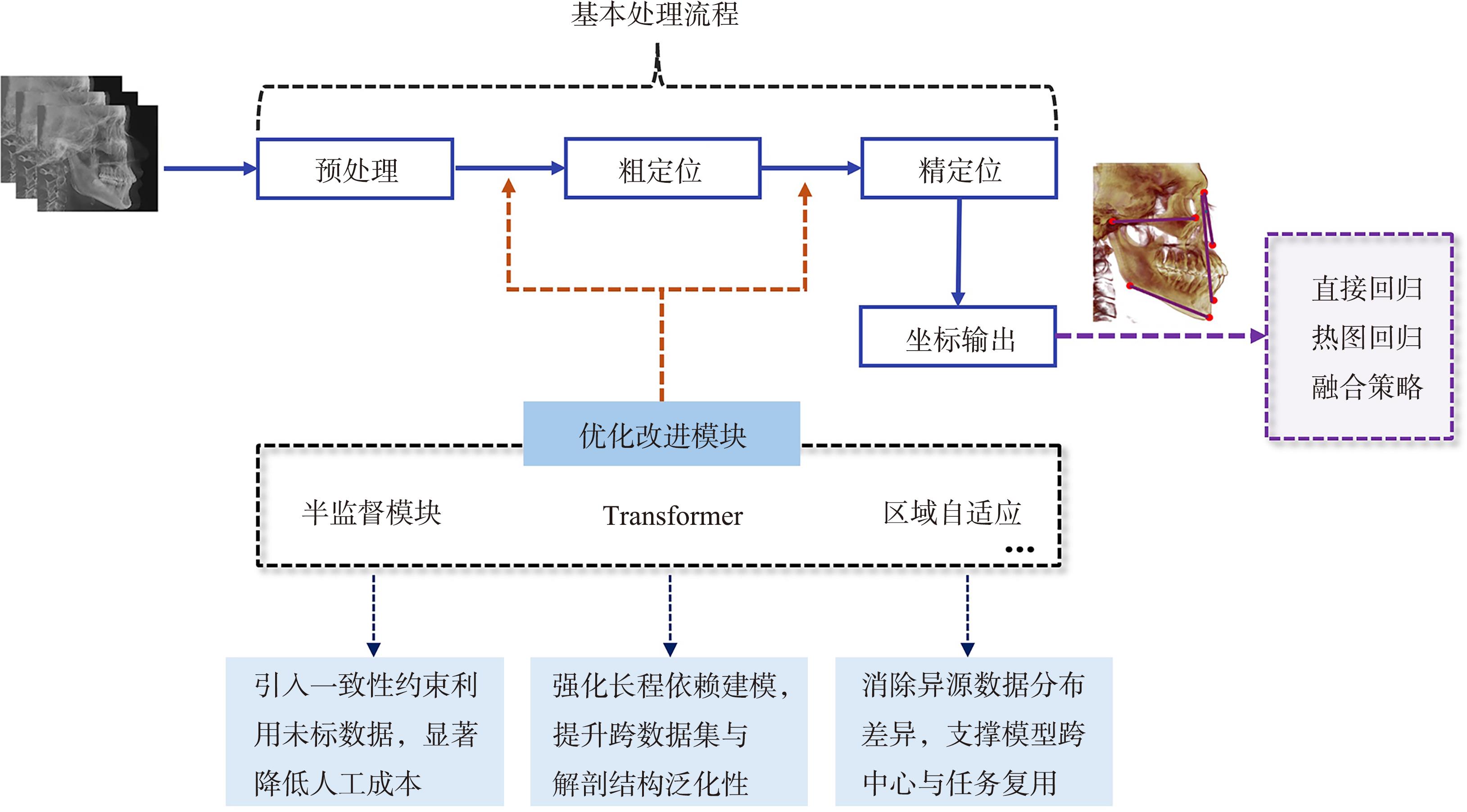
近年来,深度学习技术在医学影像分析领域取得了显著进展,颅颌面软硬组织自动定点及头影测量作为三维测量与分析的核心环节,在提高测量效率、降低观察者差异及支持个体化诊疗方面展现出重要潜力。本文回顾二维自动头影测量的发展历程,重点叙述基于深度学习的三维颅颌面软硬组织自动定点及头影测量方法的研究进展,分析三维自动定点在测量规范、数据依赖、泛化能力与临床可解释性等方面面临的主要挑战,对未来临床数据库建设、测量标准体系构建以及多模态多任务模型在临床决策支持中的应用前景进行展望,为三维颅颌面自动定点及头影测量技术的进一步研究与临床转化提供参考。
中图分类号:
| [1] | Durão AR, Pittayapat P, Rockenbach MIB, et al. Validity of 2D lateral cephalometry in orthodontics: a systematic review[J]. Prog Orthod, 2013, 14: 31. |
| [2] | LeCun Y, Bengio Y, Hinton G. Deep learning[J]. Nature, 2015, 521(7553): 436-444. |
| [3] | Broadbent BH. A new X-ray technique and its application to orthodontia[J]. Angle Orthod, 1931, 1(2): 45-66. |
| [4] | Proffit WR, Fields HW, Sarver DM. Contemporary orthodontics[M]. 4th ed. New York: Elsevier Heal-th Sciences, 2007. |
| [5] | Fracchia DE, Bignotti D, Lai S, et al. Reproducibility of AI in cephalometric landmark detection: a preliminary study[J]. Diagnostics, 2025, 15(19): 2521. |
| [6] | Scarfe WC, Farman AG, Sukovic P. Clinical applications of cone-beam computed tomography in dental practice[J]. J Can Dent Assoc, 2006, 72(1): 75-80. |
| [7] | Kapila S, Conley RS, Harrell WE Jr. The current status of cone beam computed tomography imaging in orthodontics[J]. Dentomaxillofac Radiol, 2011, 40(1): 24-34. |
| [8] | Weinberg SM, Naidoo S, Govier DP, et al. Anthropometric precision and accuracy of digital three-dimensional photogrammetry[J]. J Craniofacial Surg, 2006, 17(3): 477-483. |
| [9] | Nahm KY, Kim Y, Choi YS, et al. Accurate registration of cone-beam computed tomography scans to 3-dimensional facial photographs[J]. Am J Orthod Dentofacial Orthop, 2014, 145(2): 256-264. |
| [10] | Bollen AM. Cephalometry in orthodontics: 2D and 3D[J]. Am J Orthod Dentofacial Orthop, 2019, 156(1): 161. |
| [11] | Dot G, Schouman T, Chang S, et al. Automatic 3-dimensional cephalometric landmarking via deep lear-ning[J]. J Dent Res, 2022, 101(11): 1380-1387. |
| [12] | Gwilliam JR, Cunningham SJ, Hutton T. Reprodu-cibility of soft tissue landmarks on three-dimensional facial scans[J]. Eur J Orthod, 2006, 28(5): 408-415. |
| [13] | Perrotti G, Baccaglione G, Clauser T, et al. Total face approach (TFA) 3D cephalometry and superimposition in orthognathic surgery: evaluation of the vertical dimensions in a consecutive series[J]. Me-thods Protoc, 2021, 4(2): 36. |
| [14] | Hsu SS, Gateno J, Bell RB, et al. Accuracy of a computer-aided surgical simulation protocol for orthognathic surgery: a prospective multicenter study[J]. J Oral Maxillofac Surg, 2013, 71(1): 128-142. |
| [15] | Yang GJ, Xie F, Chen FH, et al. A comparative study of traditional and computer-aided surgical simulation guides in orthognathic correction of bimaxillary protrusion[J]. J Craniofacial Surg, 2025, 36(2): 553-557. |
| [16] | Song C, Jeong Y, Huh H, et al. Multi-scale 3D ce-phalometric landmark detection based on direct regression with 3D CNN architectures[J]. Diagnostics, 2024, 14(22): 2605. |
| [17] | Serafin M, Baldini B, Cabitza F, et al. Accuracy of automated 3D cephalometric landmarks by deep learning algorithms: systematic review and Meta-analysis[J]. Radiol Med, 2023, 128(5): 544-555. |
| [18] | Liu B, Liu C, Xiong Y, et al. Accuracy and reliability of 3D cephalometric landmark detection with deep learning[J]. Eur J Med Res, 2025, 30(1): 1000. |
| [19] | Blum FMS, Möhlhenrich SC, Raith S, et al. Evaluation of an artificial intelligence-based algorithm for automated localization of craniofacial landmarks[J]. Clin Oral Invest, 2023, 27(5): 2255-2265. |
| [20] | Lee JH, Yu HJ, Kim MJ, et al. Automated cephalometric landmark detection with confidence regions using Bayesian convolutional neural networks[J]. BMC Oral Health, 2020, 20: 270. |
| [21] | Tang HM, Liu S, Shi YX, et al. Automatic segmentation and landmark detection of 3D CBCT images using semi supervised learning for assisting orthognathic surgery planning[J]. Sci Rep, 2025, 15: 8814. |
| [22] | Tao LR, Zhang X, Yang Y, et al. Craniomaxillofacial landmarks detection in CT scans with limited labeled data via semi-supervised learning[J]. Heliyon, 2024, 10(14): e34583. |
| [23] | Lu G, Shu HZ, Bao H, et al. CMF-Net: craniomaxillofacial landmark localization on CBCT images u-sing geometric constraint and transformer[J]. Phys Med Biol, 2023, 68(9): 095020. |
| [24] | Bao H, Zhang KJ, Yu CH, et al. Evaluating the accuracy of automated cephalometric analysis based on artificial intelligence[J]. BMC Oral Heal, 2023, 23: 191. |
| [25] | Jiang Y, Jiang CY, Shi B, et al. Automatic identification of hard and soft tissue landmarks in cone-beam computed tomography via deep learning with diversity datasets: a methodological study[J]. BMC Oral Health, 2025, 25: 505. |
| [26] | Tanikawa C, Nakamura H, Mimura T, et al. Deve-lopment of artificial intelligence-supported automa-tic three-dimensional surface cephalometry[J]. Orthod Craniofacial Res, 2025, 28(4): 636-646. |
| [27] | Sahlsten J, Järnstedt J, Jaskari J, et al. Deep learning for 3D cephalometric landmarking with heterogeneous multi-center CBCT dataset[J]. PLoS One, 2024, 19(6): e0305947. |
| [28] | Deng HH, Liu Q, Chen A, et al. Clinical feasibility of deep learning-based automatic head CBCT image segmentation and landmark detection in computer-aided surgical simulation for orthognathic surgery[J]. Int J Oral Maxillofac Surg, 2023, 52(7): 793-800. |
| [29] | Tao L, Li M, Zhang X, et al. Automatic craniomaxillofacial landmarks detection in CT images of individuals with dentomaxillofacial deformities by a two-stage deep learning model[J]. BMC Oral Heal-th, 2023, 23(1): 876. |
| [30] | Nishimoto S, Saito T, Ishise H, et al. Three-dimensional craniofacial landmark detection in series of CT slices using multi-phased regression networks[J]. Diagnostics, 2023, 13(11): 1930. |
| [31] | Gillot M, Miranda F, Baquero B, et al. Automatic landmark identification in cone-beam computed tomography[J]. Orthod Craniofacial Res, 2023, 26(4): 560-567. |
| [32] | Berends B, Bielevelt F, Schreurs R, et al. Fully automated landmarking and facial segmentation on 3D photographs[J]. Sci Rep, 2024, 14(1): 6463. |
| [33] | Al-Baker B, Ayoub A, Ju XY, et al. Patch-based convolutional neural networks for automatic landmark detection of 3D facial images in clinical settings[J]. Eur J Orthod, 2024, 46(6): cjae056. |
| [34] | Chong YM, Du FZ, Ma XD, et al. Automated anatomical landmark detection on 3D facial images u-sing U-NET-based deep learning algorithm[J]. Quant Imaging Med Surg, 2024, 14(3): 2466-2474. |
| [35] | Wu YW, Zhao ZC, Lu JL, et al. A personalized automated system of 3D facial soft tissue landmarks annotation based on deep learning and computer vision[J]. BMC Oral Health, 2025, 26(1): 6. |
| [36] | Baksi S, Freezer S, Matsumoto T, et al. Accuracy of an automated method of 3D soft tissue landmark detection[J]. Eur J Orthod, 2021, 43(6): 622-630. |
| [37] | Weingart JV, Schlager S, Metzger MC, et al. Automated detection of cephalometric landmarks using deep neural patchworks[J]. Dentomaxillofac Radiol, 2023, 52(6): 20230059. |
| [38] | Jindanil T, Burlacu-Vatamanu OE, Baldini B, et al. Automated orofacial virtual patient creation using two cohorts of MSCT vs. CBCT scans[J]. Head Face Med, 2025, 21(1): 21. |
| [39] | Wang RH, Ho CT, Lin HH, et al. Three-dimensional cephalometry for orthognathic planning: normative data and analyses[J]. J Formos Med Assoc, 2020, 119(1 Pt 2): 191-203. |
| [40] | Pittayapat P, Limchaichana-Bolstad N, Willems G, et al. Three-dimensional cephalometric analysis in orthodontics: a systematic review[J]. Orthod Craniofac Res, 2014, 17(2): 69-91. |
| [41] | Muhammad D, Bendechache M. Unveiling the black box: a systematic review of Explainable Artificial Intelligence in medical image analysis[J]. Comput Struct Biotechnol J, 2024, 24: 542-560. |
| [42] | Mathew MG, Jose S, Yadav S, et al. AI in dentistry: a legal minefield[J]. Br Dent J, 2025, 238(11): 838. |
| [1] | 周文媛,范娟,熊再道,朱林,余泽正,王璐,金龙,张盼盼,顾永春. 卷积神经网络ResNet-101和Inception-v3对口腔曲面体层片中下颌第一磨牙远舌根变异的诊断[J]. 国际口腔医学杂志, 2026, 53(3): 328-334. |
| [2] | 林超英,张岚,黄定明. 人工智能在根管治疗中的研究进展[J]. 国际口腔医学杂志, 2025, 52(5): 572-578. |
| [3] | 周小洁,侯本祥. 基于深度学习技术诊断龋病方法的研究进展[J]. 国际口腔医学杂志, 2025, 52(5): 579-585. |
| [4] | 汪云毅,朱珠,张峰. 人工智能在头影测量自动定点算法上的研究进展[J]. 国际口腔医学杂志, 2024, 51(5): 630-641. |
| [5] | 陈新月,潘晓予,杨燕,贾媛媛,陈亮. 卷积神经网络在牙体牙髓病学中的应用进展[J]. 国际口腔医学杂志, 2024, 51(4): 483-488. |
| [6] | 刘力嘉,毛婧,龙欢,蒲亚龙,王军. 二维与三维头影测量自动定点的研究进展[J]. 国际口腔医学杂志, 2022, 49(1): 100-108. |
| [7] | 施丹妮,杨鑫,吴建勇. 锥形束CT三维头影测量参考坐标系的研究进展[J]. 国际口腔医学杂志, 2021, 48(4): 398-404. |
| [8] | 王耀骏, 严斌, 王林. 牙周膜本构模型的研究进展[J]. 国际口腔医学杂志, 2017, 44(5): 538-543. |
| [9] | 曹聪聪, 李精韬, 郑谦. 腭隐裂患者颅颌面生长发育的研究进展[J]. 国际口腔医学杂志, 2017, 44(4): 390-392. |
| [10] | 宿洪丽. 口腔正畸学临床教学方法改革探讨[J]. 国际口腔医学杂志, 2017, 44(1): 41-44. |
|
||