国际口腔医学杂志 ›› 2026, Vol. 53 ›› Issue (4): 607-614.doi: 10.7518/gjkq.2026223
• 综述 • 上一篇
赵蕾1( ),石永乐2,刘杨1,林翠1,李春燕1,任家银3()
Lei Zhao1(),Yongle Shi2,Yang Liu1,Cui Lin1,Chunyan Li1,Jiayin Ren3()
摘要:
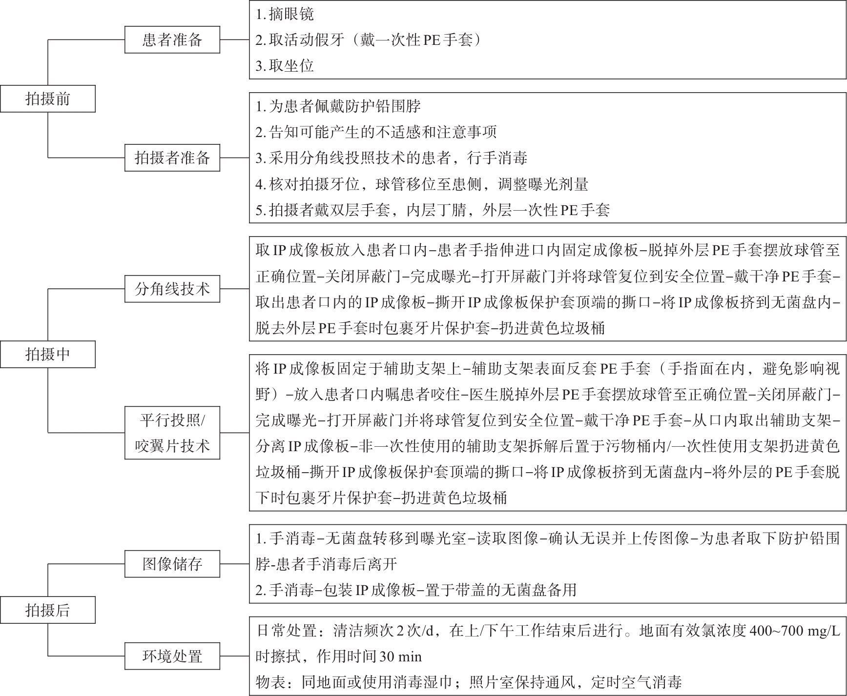
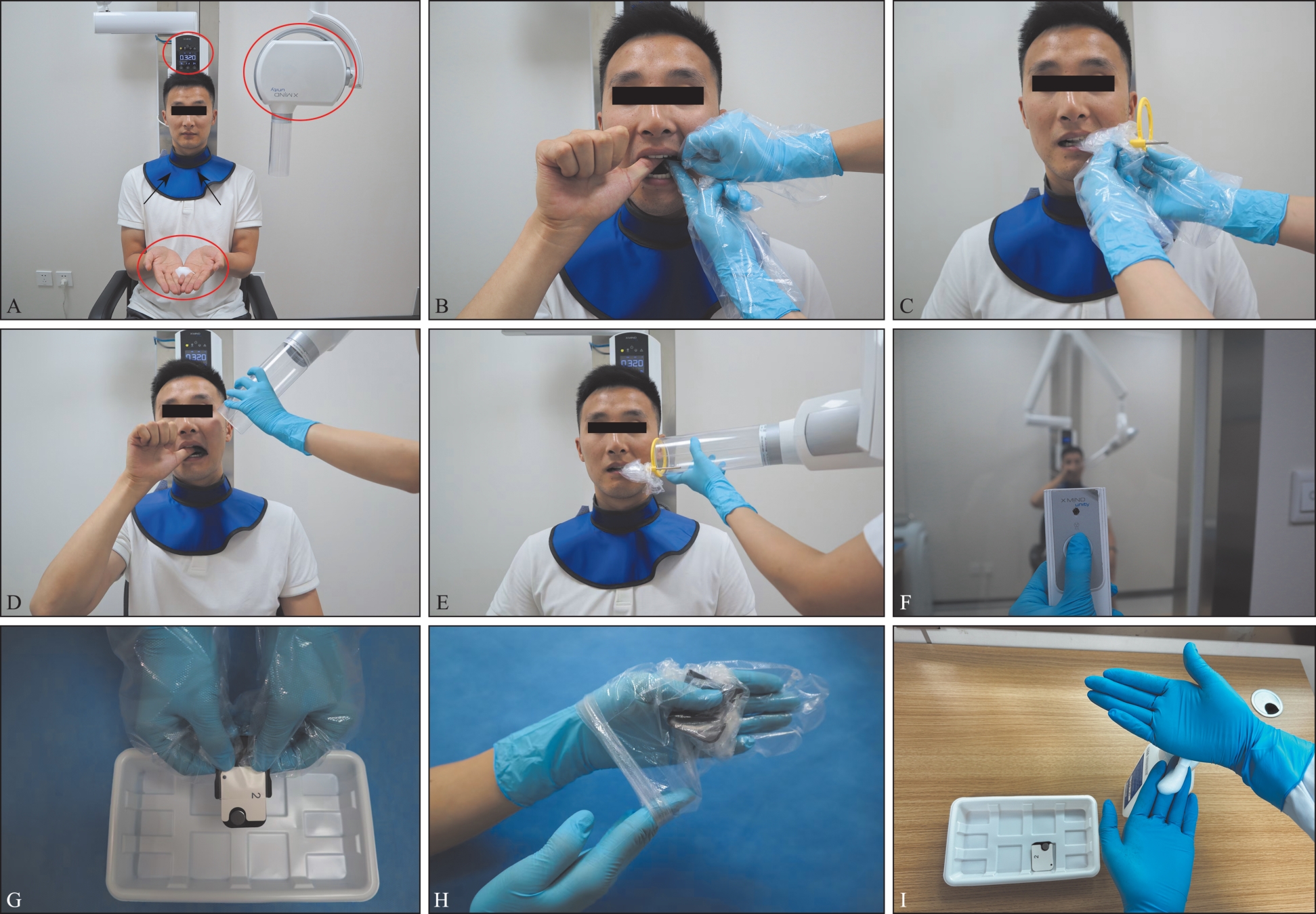
在口腔诊疗中,口内片拥有经济、便捷、辐射剂量小,分辨率较高等优势,是临床诊疗中应用最广泛的辅助检查。但是,在口内片拍摄环节,存在诸多与感染相关问题,如:口内片特殊的拍摄方式使环境被血液、唾液污染;部分患者口内存在开放性伤口或以口腔症状为首发的特殊隐匿性感染;拍摄者拍摄流程不规范;器械、拍摄环境处置不正确等。这些综合因素使拍摄环节存在交叉感染的高风险,但在临床工作中经常被忽视。因此,本文综述了数字化口内X线片拍摄技术中的医院感染风险现状,总结国内外文献,结合临床经验,优化了口内片拍摄标准流程、制定了高风险患者拍摄策略以及器械和环境的消毒处置规范,以期能有效降低口内片拍摄过程中的感染风险。
中图分类号:
| [1] | 赵蕾, 游梦, 王凯利. 数字化分角线投照根尖片质量影像因素分析[J]. 影像技术, 2023, 35(2): 13-17. |
| Zhao L, You M, Wang KL. Factors related to image quality of digital periapical film using bisecting-angle technique[J]. Image Technol, 2023, 35(2): 13-17. | |
| [2] | MacDonald DS, Colosi DC, Mupparapu M, et al. Guidelines for oral and maxillofacial imaging: COVID-19 considerations[J]. Oral Surg Oral Med Oral Pathol Oral Radiol, 2021, 131(1): 99-110. |
| [3] | 赵蕾, 游梦, 王凯利, 等. 传染病疫情下口腔影像检查的防控与建议[J]. 当代护士(中旬刊), 2021, 28(11): 118-120. |
| Zhao L, You M, Wang KL, et al. Recommendations for prevention and control of infectious diseases du-ring oral imaging examination[J]. Modern Nurse, 2021, 28(11): 118-120. | |
| [4] | Pinheiro SL, Martoni SC, Ogera RR. Assessment of microbial contamination of radiographic equipment and materials during intraoral imaging procedures[J]. Minerva Stomatol, 2012, 61(5): 197-203. |
| [5] | Mossa-Basha M, Meltzer CC, Kim DC, et al. Ra-diology department preparedness for COVID-19: radiology scientific expert panel[J]. Radiology, 2020, 296(2): E106-E112. |
| [6] | Sewerin I. Gagging in dental radiography[J]. Oral Surg Oral Med Oral Pathol, 1984, 58(6): 725-728. |
| [7] | 潘剑, 曹昊天, 刘济远, 等. 口腔医护人员传染病职业暴露危险因素及防护[J]. 国际口腔医学杂志,2020, 47(3): 366-372. |
| Pan J, Cao HT, Liu JY, et al. Occupational exposure risk factors and protection for dental health care workers from contagion[J]. Int J Stomatol, 2020, 47(3): 366-372. | |
| [8] | Katsouda M, Coolidge T, Simos G, et al. Factors associated with gagging during radiographic and in-traoral photographic examinations in 4-12-year-old children[J]. Eur Arch Paediatr Dent, 2021, 22(2): 129-137. |
| [9] | Wenzel A, Møystad A. Work flow with digital intraoral radiography: a systematic review[J]. Acta Odon-tol Scand, 2010, 68(2): 106-114. |
| [10] | Hokett SD, Honey JR, Ruiz F, et al. Assessing the effectiveness of direct digital radiography barrier sheaths and finger cots[J]. J Am Dent Assoc, 2000, 131(4): 463-467. |
| [11] | 张灶钦, 耿继武, 许志强, 等. 广东省44家口腔诊所牙科X射线诊断放射防护现状及常见问题探讨[J]. 中国辐射卫生, 2022, 31(6): 704-707, 712. |
| Zhang ZQ, Geng JW, Xu ZQ, et al. Current situation and discussion on common problems of radiation protection for dental X-ray diagnostic project in 44 dental clinics in Guangdong Province, China[J]. Chin J Radiol Health, 2022, 31(6): 704-707, 712. | |
| [12] | Reis LA, Gómez La-Rotta EI, Diniz PB, et al. Occupational exposure to potentially infectious biological material among physicians, dentists, and nurses at a university[J]. Saf Health Work, 2019, 10(4): 445-451. |
| [13] | Malta CP, Damasceno NN, Ribeiro RA, et al. Mi-crobiological contamination in digital radiography: evaluation at the radiology clinic of an educational institution[J]. Acta Odontol Latinoam, 2016, 29(3): 239-247. |
| [14] | 金桂明. 医用X射线设备对医院建筑的要求[J]. 中国医院建筑与装备, 2019, 20(2): 79-80. |
| Jin GM. Requirements of medical X-ray equipment for hospital buildings[J]. Technol Equip, 2019, 20(2): 79-80. | |
| [15] | Hunter A, Kalathingal S, Shrout M, et al. The effectiveness of a pre-procedural mouthrinse in reducing bacteria on radiographic phosphor plates[J]. Ima-ging Sci Dent, 2014, 44(2): 149-154. |
| [16] | Charuakkra A, Prapayasatok S, Janhom A, et al. Infection control and patient discomfort with an alternative plastic barrier in intraoral digital radiography[J]. Dentomaxillofac Radiol, 2017, 46(2): 20160253. |
| [17] | Mupparapu M. Editorial: dental practitioners’ role in the assessment and containment of coronavirus disease (COVID-19): evolving recommendations from the centers for disease control[J]. Quintessence Int, 2020, 51(5): 349-350. |
| [18] | Stoopler ET, Murdoch-Kinch CA. American Dental Association specialty recognition of oral medicine: implications for the dental profession[J]. J Am Dent Assoc, 2020, 151(7): 472-473. |
| [19] | 中华人民共和国国家卫生健康委员会. 医院感染管理办法[R/OL]. [2026-07-06]. |
| a27b03f9e8fd6/files/%E5%8C%BB%E9%99%A2% | |
| E6%84%9F%E6%9F%93%E7%AE%A1%E7%90 | |
| %86%E5%8A%9E%E6%B3%95.pdf. | |
| National Health Commission of the People's Republic of China. Measures for the administration of | |
| nosocomial infection[R/OL]. [2026-07-06]. | |
| ca 49 b08dda 27b03f9e8fd6/files/%E 5%8C%BB%E9%99%A2%E6%84%9F%E6%9F%93%E7%AE%A | |
| 1 %E 7%90%86%E5%8 A% 9E%E6%B3%95.pdf. | |
| [20] | 刘翠梅. 《口腔器械消毒灭菌技术操作规范》释义[J]. 中华医院感染学杂志, 2017, 27(15): 3382-3384, 3396. |
| Liu CM. Interpretation of “technical regulations for disinfection and sterilization of dental instruments”[J]. Chin J Nosocomiol, 2017, 27(15): 3382-3384, 3396. | |
| [21] | Tyan K, Levin A, Avalos-Pacheco A, et al. Conside-rations for the selection and use of disinfectants against SARS-CoV-2 in a health care setting[J]. Open Forum Infect Dis, 2020, 7(9): ofaa396. |
| [22] | 赵蕾, 宋宇, 王凯利. 多重耐药菌规范化管理在放射科应用后的效果评价[J]. 当代护士(上旬刊), 2020, 27(6): 185-187. |
| Zhao L, Song Y, Wang KL. Effect of evaluation of standardized management of multidrug-resistant bacteria in radiology department[J]. Today Nurse, 2020, 27(6): 185-187. | |
| [23] | 医疗机构环境表面清洁与消毒管理规范WS/T 512—2016[J]. 中国感染控制杂志, 2017, 16(4): 388-392. |
| Regulation for cleaning and disinfection management of environmental surface in healthcare. WS/T512—2016[J]. Chin J Infect Control, 2017, 16(4): 388-392. | |
| [24] | 唐倩, 胡姚佳, 朱丽辉. 消毒湿巾临床应用研究进展[J]. 中国消毒学杂志, 2021, 38(3): 221-225. |
| Tang Q, Hu YJ, Zhu LH. Research progress on the clinical application of disinfectant wipes[J]. Chin J Disinfect, 2021, 38(3): 221-225. | |
| [25] | 吴睿, 许斯, 张艳, 等. 重症监护病房高频接触表面清洁消毒干预效果评价[J]. 中华医院感染学杂志, 2018, 28(19): 3026-3028. |
| Wu R, Xu S, Zhang Y, et al. Effect of interventions on cleaning and disinfection of high-frequency contact surfaces in intensive care unit[J]. Chin J Nosocomiol, 2018, 28(19): 3026-3028. | |
| [26] | 朱金婷, 何惠英. 浅谈口腔诊疗环境医院感染防控措施[J]. 继续医学教育, 2021, 35(8): 74-76. |
| Zhu JT, He HY. Hospital infection prevention and control in the oral environment[J]. Continu Med Edu, 2021, 35(8): 74-76. | |
| [27] | 孙岩, 王少星, 赵宝莲, 等. 口腔诊疗中潜在感染风险与防控措施[J]. 现代口腔医学杂志, 2023, 37(3): 206-209. |
| Sun Y, Wang SX, Zhao BL, et al. Potential infection risks in dental treatments and prevention measures[J]. J Modern Stomatol, 2023, 37(3): 206-209. |
| [1] | 吴思佳,舒畅,王洋,王媛,邓淑丽,王慧明. 根管内感染控制对年轻恒牙牙髓再生治疗的影响及研究进展[J]. 国际口腔医学杂志, 2023, 50(4): 388-394. |
| [2] | 王铝亚,张竞心,林洁. 口腔术后聚维酮碘与氯己定控制感染效果的系统评价分析[J]. 国际口腔医学杂志, 2023, 50(4): 438-444. |
| [3] | 林洁,刘帆. 基于多酶清洗剂控制的口腔专科器械清洗消毒模式的探索[J]. 国际口腔医学杂志, 2022, 49(3): 324-327. |
| [4] | 王璐璇,侯本祥. 根管内氢氧化钙残留对根管治疗的影响[J]. 国际口腔医学杂志, 2022, 49(3): 367-372. |
| [5] | 易俭如,罗梦奇,尹一佳,刘治清,柳茜,石永乐,杨征,刘帆,韩向龙. 新型冠状病毒肺炎流行期降低口腔诊疗中气溶胶传播风险的策略[J]. 国际口腔医学杂志, 2020, 47(3): 362-365. |
| [6] | 曾淑蓉,张莉,王丽萍,刘治清. 加强口腔医院消毒供应中心管理控制医院感染[J]. 国际口腔医学杂志, 2008, 35(S1): -. |
| [7] | 章小缓综述 凌均棨审校. 口腔综合治疗台水路中的生物膜与污染控制[J]. 国际口腔医学杂志, 2008, 35(5): 546-546~549. |
|
||