国际口腔医学杂志 ›› 2026, Vol. 53 ›› Issue (3): 388-400.doi: 10.7518/gjkq.2026106
• 综述 • 上一篇
陈浩哲( ),杨茂莹,祝颂松,姜楠()
Haozhe Chen(),Maoying Yang,Songsong Zhu,Nan Jiang()
摘要:
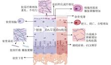
关节腔注射治疗作为骨关节炎(OA)的一种治疗方式,其有效应用要求药物能够深入渗透软骨,因此软骨的渗透性成为了药物作用的关键。然而OA状态下软骨固有的低渗结构发生变化,进一步影响了关节腔注射的药物渗透。本文综述了包括透明软骨和纤维软骨在内的不同类型软骨及不同软骨区域的结构特性以及OA引起的软骨变化对药物扩散可能产生的影响,并以此为基础进一步综述了增强药物渗透能力的关节内药物递送策略及存在的问题,旨在为 OA 治疗领域的研究与临床实践提供新的思路,推动关节内药物递送系统的迭代升级与研发工作。
中图分类号:
| [1] | Huang HR, Lou ZJ, Zheng SM, et al. Intra-articular drug delivery systems for osteoarthritis therapy: shifting from sustained release to enhancing penetration into cartilage[J]. Drug Deliv, 2022, 29(1): 767-791. |
| [2] | Katz JN, Arant KR, Loeser RF. Diagnosis and treatment of hip and knee osteoarthritis: a review[J]. JAMA, 2021, 325(6): 568-578. |
| [3] | Hunziker EB, Lippuner K, Shintani N. How best to preserve and reveal the structural intricacies of cartilaginous tissue[J]. Matrix Biol, 2014, 39: 33-43. |
| [4] | Bajpayee AG, Grodzinsky AJ. Cartilage-targeting drug delivery: can electrostatic interactions help[J]. Nat Rev Rheumatol, 2017, 13(3): 183-193. |
| [5] | Bergholt MS, St-Pierre JP, Offeddu GS, et al. Raman spectroscopy reveals new insights into the zo-nal organization of native and tissue-engineered articular cartilage[J]. ACS Cent Sci, 2016, 2(12): 885-895. |
| [6] | Buchanan JL. Types of fibrocartilage[J]. Clin Podiatr Med Surg, 2022, 39(3): 357-361. |
| [7] | Mérida-Velasco JR, Rodríguez-Vázquez JF, Mérida-Velasco JA, et al. Development of the human temporomandibular joint[J]. Anat Rec, 1999, 255(1): 20-33. |
| [8] | Fischenich KM, Wahlquist JA, Wilmoth RL, et al. Human articular cartilage is orthotropic where microstructure, micromechanics, and chemistry vary with depth and split-line orientation[J]. Osteoarthritis Cartilage, 2020, 28(10): 1362-1372. |
| [9] | Önnerfjord P, Khabut A, Reinholt FP, et al. Quantitative proteomic analysis of eight cartilaginous tissues reveals characteristic differences as well as similarities between subgroups[J]. J Biol Chem, 2012, 287(23): 18913-18924. |
| [10] | Nissinen MT, Hänninen N, Prakash M, et al. Functional and structural properties of human patellar articular cartilage in osteoarthritis[J]. J Biomech, 2021, 126: 110634. |
| [11] | Cederlund AA, Aspden RM. Walking on water: revisiting the role of water in articular cartilage biomechanics in relation to tissue engineering and rege-nerative medicine[J]. J R Soc Interface, 2022, 19(193): 20220364. |
| [12] | Wei LW, Pan QQ, Teng JY, et al. Intra-articular administration of PLGA resveratrol sustained-release nanoparticles attenuates the development of rat osteoarthritis[J]. Mater Today Bio, 2023, 24: 100884. |
| [13] |
Sardroud HA, Dos Santos Rosa G, Dust W, et al. Comparison study on hyaline cartilage versus fibrocartilage formation in a pig model by using 3D-bioprinted hydrogel and hybrid constructs[J]. Biofabrication, 2024, 17(1). doi: 10.1088/1758-5090/ad88a6 .
doi: 10.1088/1758-5090/ad88a6 |
| [14] | Scarcia L, Pileggi M, Camilli A, et al. Degenerative disc disease of the spine: from anatomy to pathophysiology and radiological appearance, with morphological and functional considerations[J]. J Pers Med, 2022, 12(11): 1810. |
| [15] | Schwer J, Galbusera F, Ignatius A, et al. Non-invasive regional parameter identification of degenera-ted human meniscus[J]. Comput Biol Med, 2024, 182: 109230. |
| [16] | Waghorne J, Bonomo FP, Rabbani A, et al. On the characteristics of natural hydraulic dampers: an i-mage-based approach to study the fluid flow beha-viour inside the human meniscal tissue[J]. Acta Biomater, 2024, 175: 157-169. |
| [17] | Perni S, Prokopovich P. Rheometer enabled study of cartilage frequency-dependent properties[J]. Sci Rep, 2020, 10(1): 20696. |
| [18] | Morejon A, Norberg CD, De Rosa M, et al. Compressive properties and hydraulic permeability of human meniscus: relationships with tissue structure and composition[J]. Front Bioeng Biotechnol, 2021, 8: 622552. |
| [19] | Jaumard NV, Welch WC, Winkelstein BA. Spinal facet joint biomechanics and mechanotransduction in normal, injury and degenerative conditions[J]. J Biomech Eng, 2011, 133(7): 071010. |
| [20] | Mélou C, Pellen-Mussi P, Jeanne S, et al. Osteoarthritis of the temporomandibular joint: a narrative overview[J]. Medicina (Kaunas), 2022, 59(1): 8. |
| [21] | Fan XW, Wu XX, Trevisan Franca De Lima L, et al. The deterioration of calcified cartilage integrity reflects the severity of osteoarthritis—a structural, molecular, and biochemical analysis[J]. FASEB J, 2022, 36(2): e22142. |
| [22] | Huang Y, Chen C, Wang FY, et al. Observation of solute transport between articular cartilage and subchondral bone in live mice[J]. Cartilage, 2021, 13(): 398S-407S. |
| [23] | Liu N, Jiang JL, Liu TC, et al. Compositional, structural, and biomechanical properties of three diffe-rent soft tissue-hard tissue insertions: a comparative review[J]. ACS Biomater Sci Eng, 2024, 10(5): 2659-2679. |
| [24] | Evans LAE, Pitsillides AA. Structural clues to arti-cular calcified cartilage function: a descriptive review of this crucial interface tissue[J]. J Anat, 2022, 241(4): 875-895. |
| [25] | Pouran B, Raoof A, Matthijs de Winter DA, et al. Topographic features of nano-pores within the osteochondral interface and their effects on transport properties-a 3D imaging and modeling study[J]. J Biomech, 2021, 123: 110504. |
| [26] | Wang WY, Ye RX, Xie WQ, et al. Roles of the calcified cartilage layer and its tissue engineering reconstruction in osteoarthritis treatment[J]. Front Bioeng Biotechnol, 2022, 10: 911281. |
| [27] | Mahmoudian A, Lohmander LS, Jafari H, et al. Towards classification criteria for early-stage knee osteoarthritis: a population-based study to enrich for progressors[J]. Semin Arthritis Rheum, 2021, 51(1): 285-291. |
| [28] | Chery DR, Han B, Zhou Y, et al. Decorin regulates cartilage pericellular matrix micromechanobiology[J]. Matrix Biol, 2021, 96: 1-17. |
| [29] | Moo EK, Ebrahimi M, Sibole SC, et al. The intrinsic quality of proteoglycans, but not collagen fibres, degrades in osteoarthritic cartilage[J]. Acta Biomater, 2022, 153: 178-189. |
| [30] | Ebrahimi M, Finnilä MAJ, Turkiewicz A, et al. Elastic, dynamic viscoelastic and model-derived fibril-reinforced poroelastic mechanical properties of normal and osteoarthritic human femoral condyle cartilage[J]. Ann Biomed Eng, 2021, 49(9): 2622-2634. |
| [31] | Ebrahimi M, Ojanen S, Mohammadi A, et al. Elastic, viscoelastic and fibril-reinforced poroelastic material properties of healthy and osteoarthritic human tibial cartilage[J]. Ann Biomed Eng, 2019, 47(4): 953-966. |
| [32] | Hamilton M, Wang JX, Dhar P, et al. Controlled-release hydrogel microspheres to deliver multipotent stem cells for treatment of knee osteoarthritis[J]. Bioengineering (Basel), 2023, 10(11): 1315. |
| [33] | Morici L, Allémann E, Rodríguez-Nogales C, et al. Cartilage-targeted drug nanocarriers for osteoarthritis therapy[J]. Int J Pharm, 2024, 666: 124843. |
| [34] | Sun ZW, Sun YL, Lu T, et al. A swelling-based biphasic analysis on the quasi-static biomechanical behaviors of healthy and degenerative intervertebral discs[J]. Comput Methods Programs Biomed, 2023, 235: 107513. |
| [35] | Gupta S, Xiao R, Fainor M, et al. Level dependent alterations in human facet cartilage mechanics and bone morphometry with spine degeneration[J]. J Orthop Res, 2023, 41(3): 674-683. |
| [36] | Almarza AJ, Athanasiou KA. Design characteristics for the tissue engineering of cartilaginous tissues[J]. Ann Biomed Eng, 2004, 32(1): 2-17. |
| [37] | Hu N, Qiu JW, Xu B, et al. The role of cartilage stem/progenitor cells in cartilage repair in osteoarthritis[J]. Curr Stem Cell Res Ther, 2023, 18(7): 892-903. |
| [38] | Zou YC, Huang PC, Lin HY, et al. The dynamic progression of temporomandibular joint osteoarthritis-like lesions elicited by mandibular shift in a rat mo-del[J]. Ann Anat Anat Anz, 2024, 255: 152301. |
| [39] | Yan JF, Qin WP, Xiao BC, et al. Pathological calcification in osteoarthritis: an outcome or a disease ini-tiator[J]. Biol Rev Camb Philos Soc, 2020, 95(4): 960-985. |
| [40] | Wang XZ, Wu Q, Zhang R, et al. Stage-specific and location-specific cartilage calcification in osteoarthritis development[J]. Ann Rheum Dis, 2023, 82(3): 393-402. |
| [41] | Bernabei I, So A, Busso N, et al. Cartilage calcification in osteoarthritis: mechanisms and clinical relevance[J]. Nat Rev Rheumatol, 2023, 19(1): 10-27. |
| [42] | Heath S, Han Y, Hua R, et al. Assessment of glycosaminoglycan content in bone using Raman spectroscopy[J]. Bone, 2023, 171: 116751. |
| [43] | Hassan CR, Lee W, Komatsu DE, et al. Evaluation of nucleus pulposus fluid velocity and pressure alte-ration induced by cartilage endplate sclerosis using a poro-elastic finite element analysis[J]. Biomech Model Mechanobiol, 2021, 20(1): 281-291. |
| [44] | Zeng WN, Zhang Y, Wang D, et al. Intra-articular injection of kartogenin-enhanced bone marrow-derived mesenchymal stem cells in the treatment of knee osteoarthritis in a rat model[J]. Am J Sports Med, 2021, 49(10): 2795-2809. |
| [45] | Hagag UI, Halfaya FM, Al-Muzafar HM, et al. Impacts of mesenchymal stem cells and hyaluronic a-cid on inflammatory indicators and antioxidant defense in experimental ankle osteoarthritis[J]. World J Orthop, 2024, 15(11): 1056-1074. |
| [46] | Xiao LK, Cui JR, Sun Z, et al. Therapeutic potential of nanotechnology-based approaches in osteoarthritis[J]. Front Pharmacol, 2022, 13: 920824. |
| [47] | Morgese G, Cavalli E, Rosenboom JG, et al. Cyclic polymer grafts that lubricate and protect damaged cartilage[J]. Angew Chem Int Ed, 2018, 57(6): 1621-1626. |
| [48] | Xiong W, Han ZY, Ding SL, et al. In situ remode-ling of efferocytosis via lesion-localized microspheres to reverse cartilage senescence[J]. Adv Sci (Weinh), 2024, 11(19): e2400345. |
| [49] | Yang QF, Liu GH, Chen GH, et al. Novel injectable adhesive hydrogel loaded with exosomes for holistic repair of hemophilic articular cartilage defect[J]. Bioact Mater, 2024, 42: 85-111. |
| [50] | Tuppurainen J, Paakkari P, Jäntti J, et al. Revealing detailed cartilage function through nanoparticle diffusion imaging: a computed tomography & finite e-lement study[J]. Ann Biomed Eng, 2024, 52(9): 2584-2595. |
| [51] | Bajpayee AG, Wong CR, Bawendi MG, et al. Avidin as a model for charge driven transport into cartilage and drug delivery for treating early stage post-traumatic osteoarthritis[J]. Biomaterials, 2014, 35(1): 538-549. |
| [52] | Bajpayee AG, Scheu M, Grodzinsky AJ, et al. A rabbit model demonstrates the influence of cartilage thickness on intra-articular drug delivery and retention within cartilage[J]. J Orthop Res, 2015, 33(5): 660-667. |
| [53] | Bajpayee AG, Scheu M, Grodzinsky AJ, et al. Electrostatic interactions enable rapid penetration, enhanced uptake and retention of intra-articular injec-ted avidin in rat knee joints[J]. J Orthop Res, 2014, 32(8): 1044-1051. |
| [54] | Mehta S, Boyer TL, Akhtar S, et al. Sustained intra-cartilage delivery of interleukin-1 receptor antagonist using cationic peptide and protein-based carriers[J]. Osteoarthritis Cartilage, 2023, 31(6): 780-792. |
| [55] | Gupta SS, Mishra V, Das Mukherjee M, et al. Amino acid derived biopolymers: recent advances and biomedical applications[J]. Int J Biol Macromol, 2021, 188: 542-567. |
| [56] | Vedadghavami A, Hakim B, He TF, et al. Cationic peptide carriers enable long-term delivery of insulin-like growth factor-1 to suppress osteoarthritis-induced matrix degradation[J]. Arthritis Res Ther, 2022, 24(1): 172. |
| [57] | He TF, Zhang CZ, Vedadghavami A, et al. Multi-arm Avidin nano-construct for intra-cartilage deli-very of small molecule drugs[J]. J Control Release, 2020, 318: 109-123. |
| [58] | Formica FA, Barreto G, Zenobi-Wong M. Cartilage-targeting dexamethasone prodrugs increase the efficacy of dexamethasone[J]. J Control Release, 2019, 295: 118-129. |
| [59] | Ali NA, Morsi NM, Badr-Eldin SM, et al. Diacerein-loaded surface modified iron oxide microparticles (SMIOMPs): an emerging magnetic system for management of osteoarthritis via intra-articular injection[J]. Front Bioeng Biotechnol, 2024, 12: 1439085. |
| [60] | Rogina A, Pušić M, Štefan L, et al. Characterization of chitosan-based scaffolds seeded with sheep nasal chondrocytes for cartilage tissue engineering[J]. Ann Biomed Eng, 2021, 49(6): 1572-1586. |
| [61] | Kim Y, Klutz AM, Jacobson KA. Systematic investigation of polyamidoamine dendrimers surface-modified with poly(ethylene glycol) for drug delivery applications: synthesis, characterization, and evaluation of cytotoxicity[J]. Bioconjug Chem, 2008, 19(8): 1660-1672. |
| [62] | Geiger BC, Wang S, Padera RF Jr, et al. Cartilage-penetrating nanocarriers improve delivery and efficacy of growth factor treatment of osteoarthritis[J]. Sci Transl Med, 2018, 10(469): eaat8800. |
| [63] | Lin JQ, Hu WC, Gao T, et al. ε-Poly-l-lysine as an efficient cartilage penetrating and residing drug carrier with high intraarticular injection safety for trea-ting osteoarthritis[J]. Chem Eng J, 2022, 430: 133018. |
| [64] | Feng K, Xie XT, Yuan J, et al. Reversing the surface charge of MSC-derived small extracellular vesicles by εPL-PEG-DSPE for enhanced osteoarthritis treatment[J]. J Extracell Vesicles, 2021, 10(13): e12160. |
| [65] | Vedadghavami A, Wagner EK, Mehta S, et al. Cartilage penetrating cationic peptide carriers for applications in drug delivery to avascular negatively charged tissues[J]. Acta Biomater, 2019, 93: 258-269. |
| [66] | Lynn DM, Langer R. Degradable poly(β-amino esters): synthesis, characterization, and self-assembly with plasmid DNA[J]. J Am Chem Soc, 2000, 122(44): 10761-10768. |
| [67] | Perni S, Prokopovich P. Poly-beta-amino-esters nano-vehicles based drug delivery system for cartilage[J]. Nanomedicine, 2017, 13(2): 539-548. |
| [68] | Saeedi T, Prokopovich P. Screening of poly-beta amino ester coated emulsion of ketorolac for cartilage delivery[J]. J Mater Chem B, 2024, 12(24): 5930-5939. |
| [69] | Cipollaro L, Trucillo P, Bragazzi NL, et al. Liposomes for intra-articular analgesic drug delivery in orthopedics: state-of-art and future perspectives. Insights from a systematic mini-review of the literature[J]. Medicina (Kaunas), 2020, 56(9): 423. |
| [70] | Kim HR, Cho HB, Lee S, et al. Fusogenic liposomes encapsulating mitochondria as a promising delivery system for osteoarthritis therapy[J]. Biomaterials, 2023, 302: 122350. |
| [71] | Ebada HM, Nasra MM, Nassra RA, et al. Cationic nanocarrier of rhein based on hydrophobic ion pai-ring approach as intra-articular targeted regenerative therapy for osteoarthritis[J]. Colloids Surf B Biointerfaces, 2022, 211: 112285. |
| [72] | Zong LJ, Wang Q, Sun HY, et al. Intra-articular injection of PLGA/polydopamine core-shell nanoparticle attenuates osteoarthritis progression[J]. ACS Appl Mater Interfaces, 2024, 16(17): 21450-21462. |
| [73] | Jiang T, Kan HM, Rajpura K, et al. Development of targeted nanoscale drug delivery system for osteoarthritic cartilage tissue[J]. J Nanosci Nanotechnol, 2018, 18(4): 2310-2317. |
| [74] | Zhang H, Wu SL, Chen WK, et al. Bone/cartilage targeted hydrogel: strategies and applications[J]. Bioact Mater, 2022, 23: 156-169. |
| [75] | Rothenfluh DA, Bermudez H, O’Neil CP, et al. Biofunctional polymer nanoparticles for intra-articular targeting and retention in cartilage[J]. Nat Mater, 2008, 7(3): 248-254. |
| [76] | Pi YB, Zhang X, Shi JJ, et al. Targeted delivery of non-viral vectors to cartilage in vivo using a chondrocyte-homing peptide identified by phage display[J]. Biomaterials, 2011, 32(26): 6324-6332. |
| [77] | Shao ZX, Zhang X, Pi YB, et al. Polycaprolactone electrospun mesh conjugated with an MSC affinity peptide for MSC homing in vivo [J]. Biomaterials, 2012, 33(12): 3375-3387. |
| [78] | Hughes C, Faurholm B, Dell’Accio F, et al. Human single-chain variable fragment that specifically targets arthritic cartilage[J]. Arthritis Rheum, 2010, 62(4): 1007-1016. |
| [79] | Hu Q, Chen Q, Yan XY, et al. Chondrocyte affinity peptide modified PAMAM conjugate as a nanoplatform for targeting and retention in cartilage[J]. Nanomedicine (Lond), 2018, 13(7): 749-767. |
| [80] | Ebada HMK, Nasra MMA, Nassra RA, et al. Chondroitin sulfate-functionalized lipid nanoreservoirs: a novel cartilage-targeting approach for intra-articular delivery of cassic acid for osteoarthritis treatment[J]. Drug Deliv, 2022, 29(1): 652-663. |
| [81] | Wu HP, Zhang L, Dong XK, et al. Targeted delivery of berberine using bionic nanomaterials for Atherosclerosis therapy[J]. Biomed Pharmacother, 2024, 178: 117135. |
| [82] | Chen PF, Liu X, Gu CH, et al. A plant-derived natural photosynthetic system for improving cell anabolism[J]. Nature, 2022, 612(7940): 546-554. |
| [83] | Brown S, Pistiner J, Adjei IM, et al. Nanoparticle properties for delivery to cartilage: the implications of disease state, synovial fluid, and off-target uptake[J]. Mol Pharm, 2019, 16(2): 469-479. |
| [84] | Laver-Rudich Z, Silbermann M. Cartilage surface charge. A possible determinant in aging and osteoarthritic processes[J]. Arthritis Rheum, 1985, 28(6): 660-670. |
| [85] | Price FM, Levick JR, Mason RM. Glycosaminoglycan concentration in synovium and other tissues of rabbit knee in relation to synovial hydraulic resistance[J]. J Physiol, 1996, 495(Pt 3): 803-820. |
| [86] | von Mentzer U, Selldén T, Råberg L, et al. Synovial fluid profile dictates nanoparticle uptake into cartila-ge—implications of the protein corona for novel arthritis treatments[J]. Osteoarthritis Cartilage, 2022, 30(10): 1356-1364. |
| [87] | Vedadghavami A, He TF, Zhang CZ, et al. Charge-based drug delivery to cartilage: hydrophobic and not electrostatic interactions are the dominant cause of competitive binding of cationic carriers in synovial fluid[J]. Acta Biomater, 2022, 151: 278-289. |
| [88] | Andersson E, Tykesson E, Lohmander LS, et al. Quantification of chondroitin sulfate, hyaluronic acid and N-glycans in synovial fluid—a technical performance study[J]. Osteoarthr Cartil Open, 2023, 5(3): 100380. |
| [89] | Duman H, Eker F, Akdaşçi E, et al. Silver nanoparticles: a comprehensive review of synthesis methods and chemical and physical properties[J]. Nanomaterials (Basel), 2024, 14(18): 1527. |
| [90] | Warren MR, Vedadghavami A, Bhagavatula S, et al. Effects of polycationic drug carriers on the electromechanical and swelling properties of cartilage[J]. Biophys J, 2022, 121(18): 3542-3561. |
| [91] | Sangsuwan R, Yik JHN, Owen M, et al. Intra-articular injection of flavopiridol-loaded microparticles for treatment of post-traumatic osteoarthritis[J]. Acta Biomater, 2022, 149: 347-358. |
| [1] | 郭怡婧,杜思雨,陈亚冰,王雷. 辛伐他汀前药在牙周炎治疗中的研究进展[J]. 国际口腔医学杂志, 2026, 53(2): 239-246. |
| [2] | 韦秀湘,李昊. 新型组织黏合剂在口腔领域的研究进展[J]. 国际口腔医学杂志, 2025, 52(2): 154-160. |
| [3] | 罗启培,张新春. 智能响应型水凝胶及其在药物控释中的应用[J]. 国际口腔医学杂志, 2025, 52(1): 123-132. |
| [4] | 温星悦, 赵骏宇, 赵崇钧, 王贵欣, 黄睿洁. 壳聚糖治疗牙周病的研究进展[J]. 国际口腔医学杂志, 2024, 51(4): 416-424. |
| [5] | 梁屹,裴锡波,万乾炳. 光响应水凝胶在生物医学领域应用的研究进展[J]. 国际口腔医学杂志, 2022, 49(1): 12-18. |
| [6] | 杨文英1 张文丽1 罗应伟2. 颞下颌关节骨关节炎动物模型的研究进展[J]. 国际口腔医学杂志, 2015, 42(6): 677-680. |
| [7] | 阴健1 廖爽1 张伟华2 何嘉3 王航1. 关节腔内注射氨基葡萄糖治疗兔颞下颌关节骨关节炎的组织学研究[J]. 国际口腔医学杂志, 2014, 41(1): 36-39. |
| [8] | 代康 焦凯综述 王美青审校. 细胞因子在骨关节炎软骨病变中的作用[J]. 国际口腔医学杂志, 2012, 39(4): 491-493. |
| [9] | 朱桂全综述 梁新华审校. 低氧诱导因子- 1α在骨关节炎发病机制中的研究进展[J]. 国际口腔医学杂志, 2008, 35(3): 325-325~328. |
| [10] | 肖进,谷志远,谢志坚. 软骨细胞的细胞骨架研究[J]. 国际口腔医学杂志, 2005, 32(01): 25-27. |
|
||