国际口腔医学杂志 ›› 2024, Vol. 51 ›› Issue (2): 164-171.doi: 10.7518/gjkq.2024029
陈韫欣1( ),李舒舒2,黄梓澄1,孔卫东2()
Yunxin Chen1(),Shushu Li2,Zicheng Huang1,Weidong Kong2()
摘要:
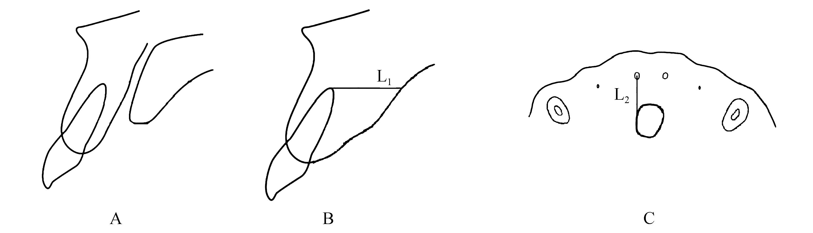
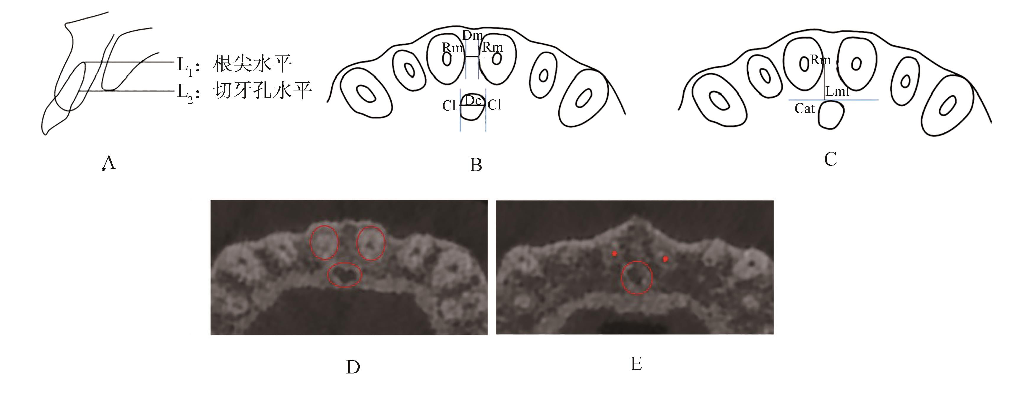
上中切牙的位置在面部美学中有十分重要的意义。在正畸治疗过程中,为获得理想的矫正效果,上中切牙会根据实际情况进行三维方向上的移动。以往观点认为上中切牙移动的范围只受到上颌骨唇、腭侧骨皮质的限制,但随着锥形束计算机体层成像(CBCT)的应用,发现在上颌骨内存在着解剖形态不规则的切牙管,其外侧壁由致密的皮质骨围绕而成,若上中切牙与之发生碰撞不仅会导致接触部位的牙根吸收,还可导致骨开窗、骨开裂,引发牙周问题,甚至还可能压迫鼻腭管神经等。因此,上中切牙三维移动的边界与切牙管的形态及位置关系密切,应引起重视,以避免临床工作中对上中切牙的牙根造成不必要伤害。本文就切牙管对上中切牙三维移动的影响做一综述。
中图分类号:
| 1 | 林久祥, 陈莉莉, 韩冰, 等. 健康正畸为本美学正畸为鉴——健康矫治理念的构建与传动矫治技术研发应用[J]. 北京大学学报(医学版), 2022, 54(5): 837-841. |
| Lin JX, Chen LL, Han B, et al. Healthy orthodontics is the reference of aesthetic orthodontics-construction of healthy orthodontics concept and development and application of transmission orthodontics[J]. J Peking Uni (Health Sci), 2022, 54(5): 837-841. | |
| 2 | Feller L, Khammissa RA, Thomadakis G, et al. Apical external root resorption and repair in orthodontic tooth movement: biological events[J]. Biomed Res Int, 2016, 2016: 4864195. |
| 3 | Kim JH, Oka K, Jin ZW, et al. Fetal development of the incisive canal, especially of the delayed closure due to the nasopalatine duct: a study using serial sections of human fetuses[J]. Anat Rec (Hoboken), 2017, 300(6): 1093-1103. |
| 4 | Lake, Iwanaga J, Kikuta S, et al. The incisive canal: a comprehensive review[J]. Cureus, 2018, 10(7): e3069. |
| 5 | Bornstein MM, Balsiger R, Sendi P, et al. Morphology of the nasopalatine canal and dental implant surgery: a radiographic analysis of 100 consecutive patients using limited cone-beam computed tomography[J]. Clin Oral Implants Res, 2011, 22(3): 295-301. |
| 6 | Milanovic P, Selakovic D, Vasiljevic M, et al. Morphological characteristics of the nasopalatine canal and the relationship with the anterior maxillary bone-a cone beam computed tomography study[J]. Diagnostics, 2021, 11(5): 915. |
| 7 | Mardinger O, Namani-Sadan N, Chaushu G, et al. Morphologic changes of the nasopalatine canal rela-ted to dental implantation: a radiologic study in different degrees of absorbed maxillae[J]. J Periodontol, 2008, 79(9): 1659-1662. |
| 8 | Etoz M, Sisman Y. Evaluation of the nasopalatine canal and variations with cone-beam computed tomography[J]. Surg Radiol Anat, 2014, 36(8): 805-812. |
| 9 | 王扬, 姜曚, 袁艺航, 等. 鼻腭管解剖形态多样性的锥形束CT研究[J]. 临床口腔医学杂志, 2015, 31(12): 710-714. |
| Wang Y, Jiang M, Yuan YH, et al. Analysis of anatomical variations of nasopalatine canal using cone beam computed tomography[J]. J Clin Stomatol, 2015, 31(12): 710-714. | |
| 10 | 吴连俊, 姚李韬, 张彩霞, 等. 切牙管形态学特征的CBCT测量分析[J]. 口腔医学研究, 2020, 36(7): 693-697. |
| Wu LJ, Yao LT, Zhang CX, et al. Morphological characteristics of incisive canal by cone beam computed tomography[J]. J Oral Sci Res, 2020, 36(7): 693-697. | |
| 11 | Mraiwa N, Jacobs R, van Cleynenbreugel J, et al. The nasopalatine canal revisited using 2D and 3D CT imaging[J]. Dentomaxillofac Radiol, 2004, 33(6): 396-402. |
| 12 | Xu YH, Xie JY. Comparison of the effects of mini-implant and traditional anchorage on patients with maxillary dentoalveolar protrusion[J]. Angle Orthod, 2017, 87(2): 320-327. |
| 13 | Horiuchi A, Hotokezaka H, Kobayashi K. Correlation between cortical plate proximity and apical root resorption[J]. Am J Orthod Dentofac Orthop, 1998, 114(3): 311-318. |
| 14 | Cho EA, Kim SJ, Choi YJ, et al. Morphologic eva-luation of the incisive canal and its proximity to the maxillary central incisors using computed tomography images[J]. Angle Orthod, 2016, 86(4): 571-576. |
| 15 | 徐海洋, 张卫兵. 骨性Ⅱ类成年患者上中切牙根尖区牙槽骨量的CBCT研究[J]. 口腔医学, 2019, 39(6): 510-513, 529. |
| Xu HY, Zhang WB. A CBCT study of the alveolar bone in the maxillary central incisor root apex of the skeletal Class Ⅱ adults[J]. Stomatology, 2019, 39(6): 510-513, 529. | |
| 16 | 徐海洋, 张卫兵. 骨性Ⅱ类成年患者上颌中切牙牙根与切牙管三维位置关系的研究[J]. 中华口腔正畸学杂志, 2019, 26(4): 203-208. |
| Xu HY, Zhang WB. A study of three-dimensional relationship between the maxillary central incisor roots and the incisive canal in skeletal Class Ⅱ malocclusion in adults[J]. Chin J Orthod, 2019, 26(4): 203-208. | |
| 17 | Chan E, Darendeliler MA. Physical properties of root cementum: part 7. Extent of root resorption under areas of compression and tension[J]. Am J Orthod Dentofac Orthop, 2006, 129(4): 504-510. |
| 18 | Chung CJ, Choi YJ, Kim KH. Approximation and contact of the maxillary central incisor roots with the incisive canal after maximum retraction with temporary anchorage devices: report of 2 patients[J]. Am J Orthod Dentofac Orthop, 2015, 148(3): 493-502. |
| 19 | Pan Y, Chen S. Contact of the incisive canal and upper central incisors causing root resorption after retraction with orthodontic mini-implants: a CBCT study[J]. Angle Orthod, 2019, 89(2): 200-205. |
| 20 | Proffit WR, Fields HW, Sarver DM. 当代口腔正畸学[M]. 王林, 译. 北京: 人民军医出版社, 2014: 670-671. |
| Proffit WR, Fields HW, Sarver DM. Contemporary orthodontics[M]. Wang Lin, Trans. Beijing: People’s Military Medical Press, 2014: 670-671. | |
| 21 | Chung CJ, Nguyen T, Lee JH, et al. Incisive canal remodelling following maximum anterior retraction reduces apical root resorption[J]. Orthod Craniofac Res, 2021, 24(): 59-65. |
| 22 | Khurana S, Parasher P, Mukherjee P, et al. Cone beam computed tomographic-based retrospective study on Newark population for the assessment of distance between incisive canal and maxillary central incisors: clinical implications[J]. Indian J Dent Res, 2020, 31(2): 175-179. |
| 23 |
倪洁丽, 秦金炜, 张阳. 成年骨性Ⅱ类错 患者上颌中切牙牙根与切牙管的位置关系研究[J]. 南京医科大学学报(自然科学版), 2021, 41(1): 103-108. 患者上颌中切牙牙根与切牙管的位置关系研究[J]. 南京医科大学学报(自然科学版), 2021, 41(1): 103-108.
|
| Ni JL, Qin JW, Zhang Y. Study on the relationship between the roots of maxillary central incisors and the incisive canal in skeletal Ⅱ adults[J]. J Nanjing Med Uni (Nat Sci), 2021, 41(1): 103-108. | |
| 24 | 郑怡, 谢敏, 韦理英. 骨性Ⅱ类均角成年患者上颌中切牙与切牙管位置关系的锥形束计算机断层扫描研究[J]. 广西医科大学学报, 2021, 38(8): 1541-1545. |
| Zheng Y, Xie M, Wei LY. A cone-beam CT study on position relationship between upper central incisors and incisive canal of adults with skeletal class Ⅱ ave-rage-angle pattern malocclusion[J]. J Guangxi Med Uni, 2021, 38(8): 1541-1545. | |
| 25 | Al-Rokhami RK, Sakran KA, Alhammadi MS, et al. Proximity of upper central incisors to incisive canal among subjects with maxillary dentoalveolar protrusion in various facial growth patterns[J]. Angle Orthod, 2022, 92(4): 529-536. |
| 26 | Owens EG, Goodacre CJ, Loh PL, et al. A multicenter interracial study of facial appearance. Part 1: a comparison of extraoral parameters[J]. Int J Pros-thodont, 2002, 15(3): 273-282. |
| 27 | Imamura T, Uesugi S, Ono T. Unilateral maxillary central incisor root resorption after orthodontic treatment for Angle Class Ⅱ , division 1 malocclusion with significant maxillary midline deviation: a possible correlation with root proximity to the incisive canal[J]. Korean J Orthod, 2020, 50(3): 216-226. |
| 28 | Nakada T, Motoyoshi M, Horinuki E, et al. Cone-beam computed tomography evaluation of the association of cortical plate proximity and apical root resorption after orthodontic treatment[J]. J Oral Sci, 2016, 58(2): 231-236. |
| 29 | Bidra AS, Uribe F, Taylor TD, et al. The relationship of facial anatomic landmarks with midlines of the face and mouth[J]. J Prosthet Dent, 2009, 102(2): 94-103. |
| 30 | 钱煦, 李小兵. 正畸治疗中上前牙的美学因素[J]. 国际口腔医学杂志, 2008, 35(S1): 328-330. |
| Qian X, Li XB. Anterior dental esthetics in ortho-dontic treatment[J]. Int J Stomatol, 2008, 35(S1): 328-330. | |
| 31 | 颜冬, 施雨佳, 葛悦, 等. 切牙管与上颌中切牙位置关系的定量研究[J]. 口腔医学, 2021, 41(7): 627-630, 639. |
| Yan D, Shi YJ, Ge Y, et al. Quantitative study of the relationship between the incisive canal and maxillary central incisors[J]. Stomatology, 2021, 41(7): 627-630, 639. | |
| 32 | Jia X, Hu W, Meng H. Relationship of central incisor implant placement to the ridge configuration anterior to the nasopalatine canal in dentate and partially edentulous individuals: a comparative study[J]. Peerj, 2015, 3: e1315. |
| 33 | Tian YL, Liu F, Sun HJ, et al. Alveolar bone thickness around maxillary central incisors of different inclination assessed with cone-beam computed tomography[J]. Korean J Orthod, 2015, 45(5): 245-252. |
| 34 | Fernández-Alonso A, Suárez-Quintanilla JA, Muinelo-Lorenzo J, et al. Three-dimensional study of nasopalatine canal morphology: a descriptive retrospective analysis using cone-beam computed tomography[J]. Surg Radiol Anat, 2014, 36(9): 895-905. |
| 35 | Li W, Chen F, Zhang F, et al. Volumetric measurement of root resorption following molar mini-screw implant intrusion using cone beam computed tomo-graphy[J]. PLoS One, 2013, 8(4): e60962. |
| 36 | 吴碧蓉, 骆英, 王晖. 种植支抗治疗露龈笑过程中切牙牙根吸收的CBCT检测[J]. 口腔医学, 2014, 34(7): 517-519. |
| Wu BR, Luo Y, Wang H. Measurement of incisor root resorption by CBCT in treatment of gummy smile using implant anchorage[J]. Stomatology, 2014, 34(7): 517-519. | |
| 37 | Aras I, Tuncer AV. Comparison of anterior and posterior mini-implant-assisted maxillary incisor intrusion: root resorption and treatment efficiency[J]. Angle Orthod, 2016, 86(5): 746-752. |
| 38 | Thakur AR, Burde K, Guttal K, et al. Anatomy and morphology of the nasopalatine canal using cone-beam computed tomography[J]. Imaging Sci Dent, 2013, 43(4): 273-281. |
| 39 | Soumya P, Koppolu P, Pathakota KR, et al. Maxillary incisive canal characteristics: a radiographic study using cone beam computerized tomography[J]. Radiol Res Pract, 2019, 2019: 6151253. |
| 40 | Costa EDD, Nejaim Y, Martins LAC, et al. Morphological evaluation of the nasopalatine canal in patients with different facial profiles and ages[J]. J Oral Maxillofac Surg, 2019, 77(4): 721-729. |
| 41 | 袁艺航, 张成晓雪, 王扬, 等. 成都正常人群上颌前牙区鼻腭管相关解剖结构的锥形束CT研究[J]. 国际口腔医学杂志, 2017, 44(5): 566-572. |
| Yuan YH, Zhang CXX, Wang Y, et al. Cone-beam computed tomography evaluation of nasopalatine canal anatomy at maxillary anterior region in Chengdu normal population[J]. Int J Stomatol, 2017, 44(5): 566-572. | |
| 42 | 戴静桃, 李平, 李安, 等. 切牙管与上颌中切牙牙根位置关系的CBCT研究[J]. 中国美容医学, 2014, 23(22): 1904-1908. |
| Dai JT, Li P, Li A, et al. Study on the positional relation of incisive canal and maxillary central incisor root by cone-beam computed tomography[J]. Chin J Aesthetic Med, 2014, 23(22): 1904-1908. | |
| 43 | Chatriyanuyoke P, Lu CI, Suzuki Y, et al. Nasopalatine canal position relative to the maxillary central incisors: a cone beam computed tomography assessment[J]. J Oral Implantol, 2012, 38(6): 713-717. |
| 44 | Artzi Z, Nemcovsky CE, Bitlitum I, et al. Displacement of the incisive foramen in conjunction with implant placement in the anterior maxilla without jeo-pardizing vitality of nasopalatine nerve and vessels: a novel surgical approach[J]. Clin Oral Implants Res, 2000, 11(5): 505-510. |
| 45 | Yu JH, Nguyen T, Kim YI, et al. Morphologic changes of the incisive canal and its proximity to maxillary incisor roots after anterior tooth movement[J]. Am J Orthod Dentofac Orthop, 2022, 161(3): 396-403.e1. |
| [1] | 斯佳萍,吕林,王思婕,周宇,陈小燕. 不同类型的辅弓在正畸前牙压低中的应用与研究进展[J]. 国际口腔医学杂志, 2024, 51(2): 241-248. |
| [2] | 王宁祥,刘帅,林良缘,吴娟. 多发性特发性根颈部吸收的研究进展[J]. 国际口腔医学杂志, 2021, 48(3): 362-366. |
| [3] | 周懿婕,宋光泰. 年轻恒牙挫入性损伤的处理策略[J]. 国际口腔医学杂志, 2021, 48(2): 135-140. |
| [4] | 赵玉洁,管晓燕,李小兰,陈琦君,王倩,刘建国. 巨噬细胞极化参与正畸牙移动的研究进展[J]. 国际口腔医学杂志, 2020, 47(4): 478-483. |
| [5] | 李寒月,夏露露,华先明. 牙周加速成骨正畸临床应用效果的研究进展[J]. 国际口腔医学杂志, 2020, 47(2): 206-211. |
| [6] | 陈雪,李纾. 牙颈部外吸收[J]. 国际口腔医学杂志, 2019, 46(5): 516-521. |
| [7] | 高鑫,曾融生. 骨保护素在口腔领域的研究进展[J]. 国际口腔医学杂志, 2019, 46(3): 316-319. |
| [8] | 崔跃, 姜欢, 胡敏. 破骨细胞蛋白酪氨酸磷酸酶与正畸移动牙牙根吸收的关系[J]. 国际口腔医学杂志, 2017, 44(1): 87-91. |
| [9] | 刘林1 王生瑜2综述 袁小平1审校. 甲状旁腺素及其相关蛋白对正畸牙根吸收后修复相关细胞的作用[J]. 国际口腔医学杂志, 2012, 39(3): 376-379. |
| [10] | 陆史俊1综述 王林2 王震东2审校. 前牙压低技术在深覆牙合患者矫治中的应用进展[J]. 国际口腔医学杂志, 2011, 38(6): 674-676. |
| [11] | 姜世同1 王作君2 安忠军1 焦广军1 姜良坤1 罗圆圆1. 倒位埋伏阻生上中切牙的临床矫治探索[J]. 国际口腔医学杂志, 2011, 38(5): 515-517. |
| [12] | 王智, 靳淑梅综述 张君审校. 牙根吸收的原因与机制[J]. 国际口腔医学杂志, 2010, 37(01): 101-101~105. |
| [13] | 王娇翠综述 赵玮审校. 乳牙根生理性吸收的研究现状[J]. 国际口腔医学杂志, 2009, 36(3): 300-302. |
| [14] | 左志刚综述 胡敏审校. 正畸治疗导致牙根吸收的影响因素和诊断评估[J]. 国际口腔医学杂志, 2009, 36(1): 111-111~113. |
| [15] | 谢永建,麦理想,张晟,郭慧,王洋,王大为. 实验性牙根吸收组织中基质金属蛋白酶- 1 及其抑制剂- 1 的定位表达[J]. 国际口腔医学杂志, 2008, 35(5): 481-481~484,605. |
|