国际口腔医学杂志 ›› 2023, Vol. 50 ›› Issue (3): 293-301.doi: 10.7518/gjkq.2023028
李沛然( ),毕瑞野,王旻,王瑞宇,刘尧,姜楠,曹品银,祝颂松()
Li Peiran(),Bi Ruiye,Wang Min,Wang Ruiyu,Liu Yao,Jiang Nan,Cao Pinyin,Zhu Songsong.()
摘要:
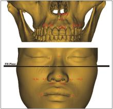
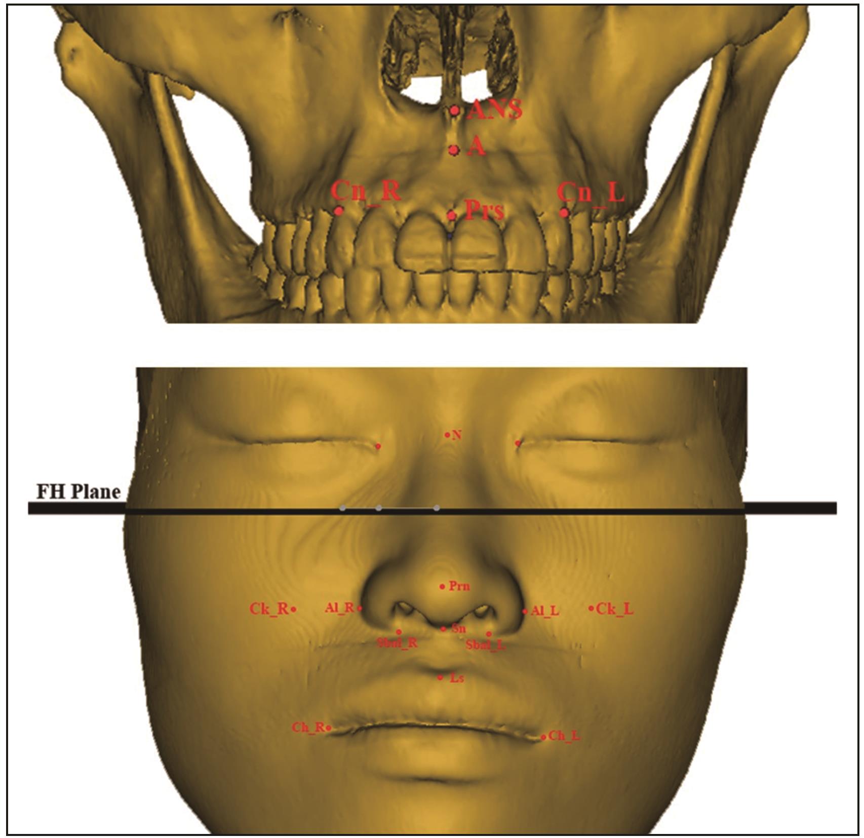
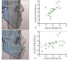
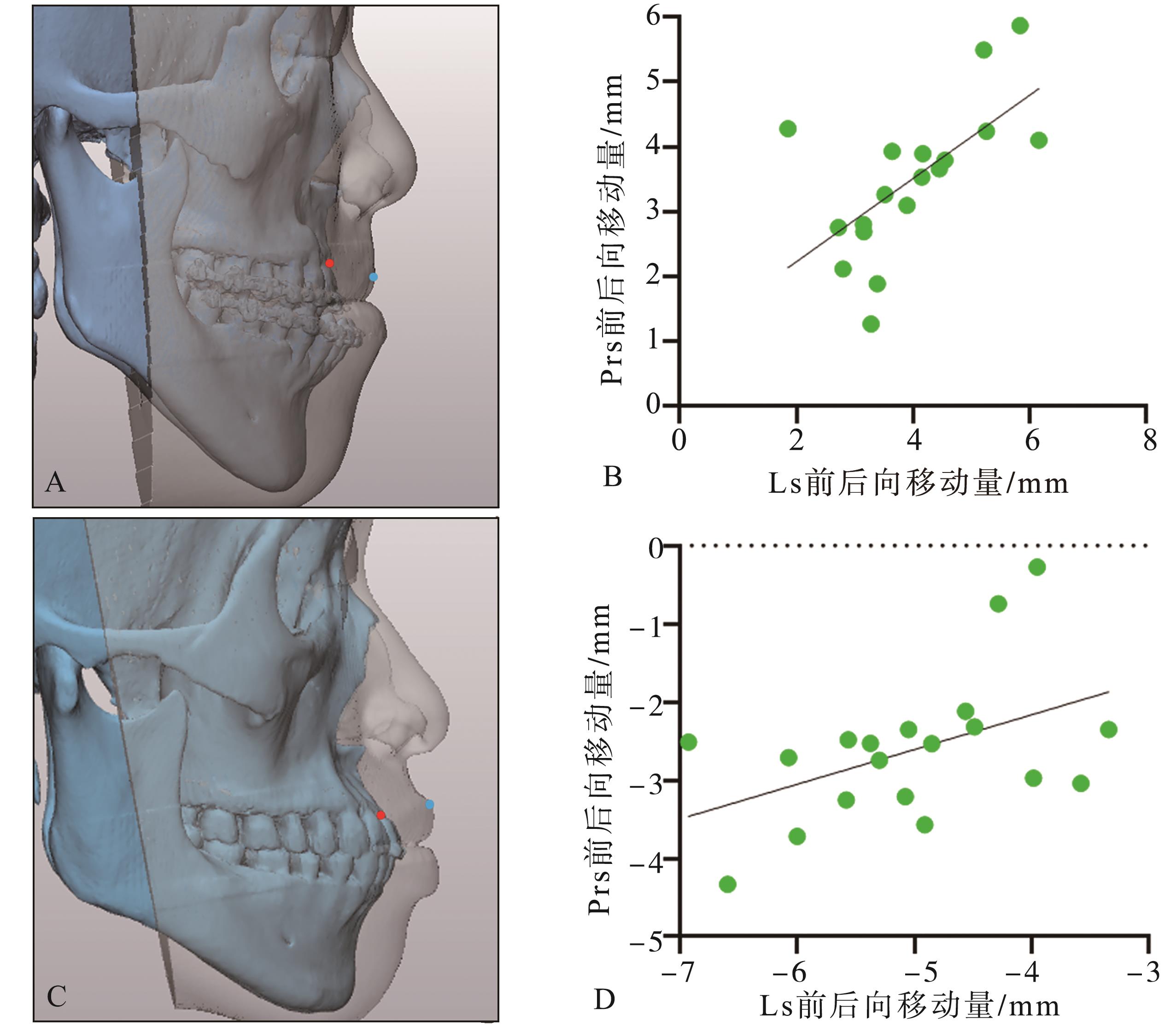
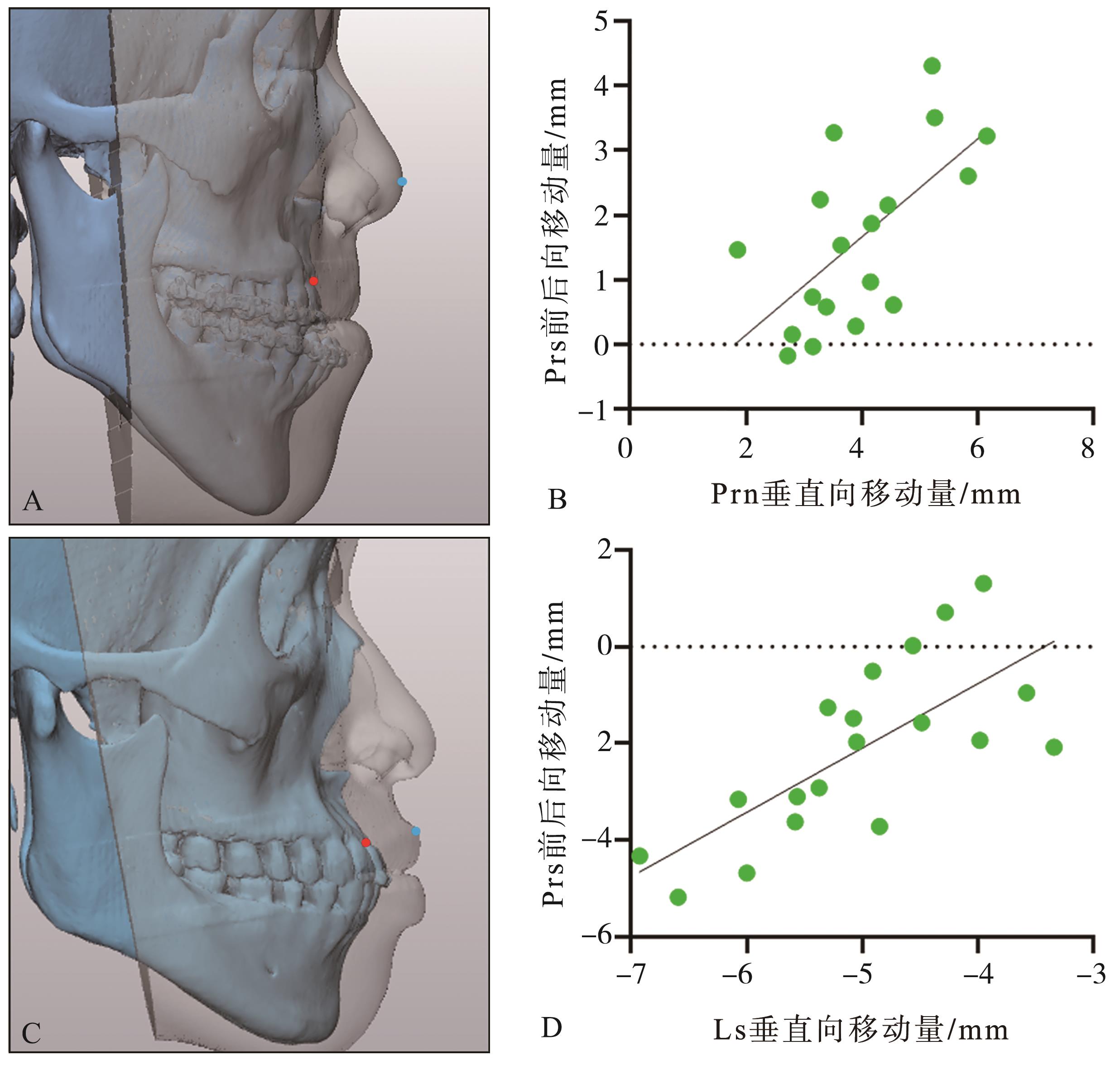
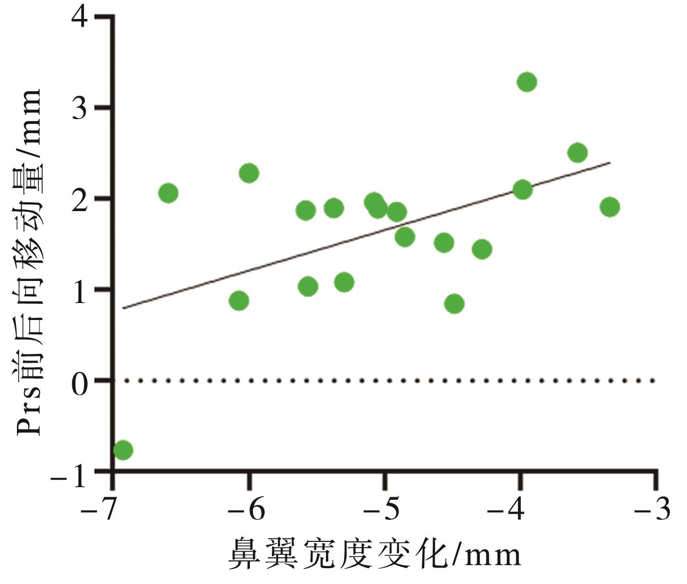
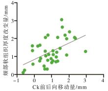
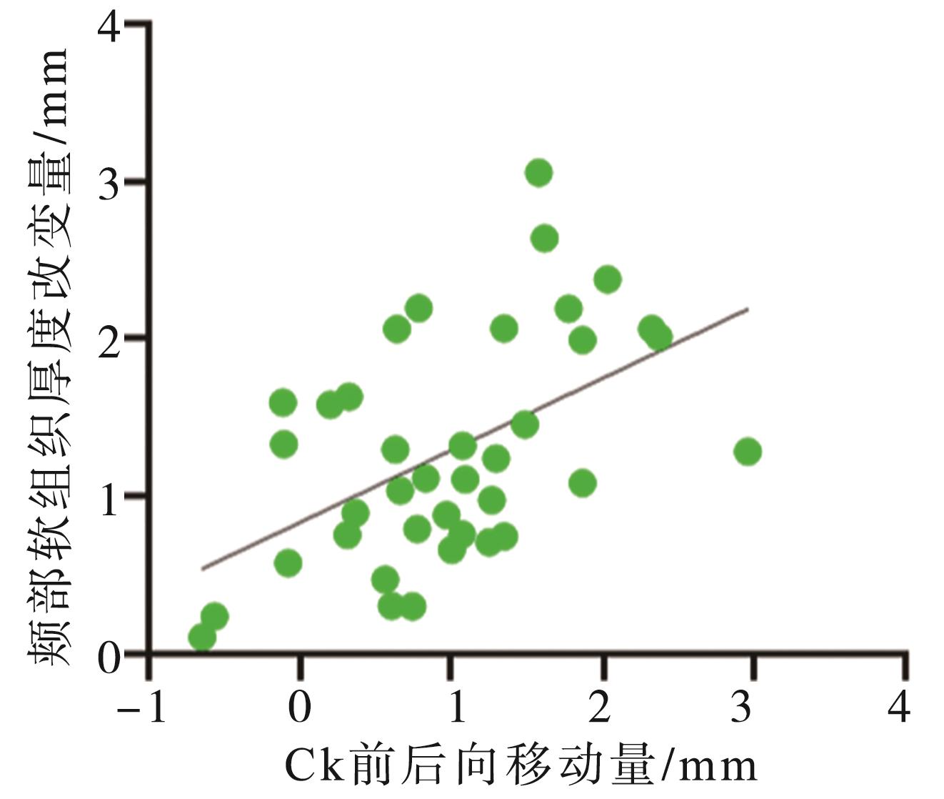
目的 测量不同上颌骨正颌术式前后鼻唇区域软组织变化,分析可能导致变化的因素。 方法 2017—2021年于四川大学华西口腔医院正颌及关节外科住院行正颌手术的患者37例,其中因上颌后缩行上颌骨Le Fort Ⅰ型骨切开前徙术(LFIA)的患者18例,因上颌前突行上颌骨前份根尖下截骨后退术(AMOS)的患者19例。收集其术前即刻和术后6~12个月的螺旋CT影像,通过Mimics和3-Matic软件进行三维重建和匹配,测量软硬组织标志点变化。使用GraphPad Prism软件进行统计学分析,显著性水平α=0.05。 结果 2种术式术后最主要的软组织变化均发生在上唇区域且改变方向与骨组织移动方向相同。在前后向软组织改变上,LFIA组软硬组织改变的比例为0.628,而AMOS组为0.465。颊部和鼻尖软组织在2种术式后均发生了前移,其中AMDS组中颊部软组织前移的量与软组织厚度的改变具有显著正相关性。在垂直向软组织变化上,LFIA组鼻尖点发生上抬且其距离与颌骨前徙距离具有显著正相关性,AMOS组上唇缘点发生下降且其距离与颌骨后退距离具有显著正相关性。在水平向软组织变化上,鼻翼宽度在行2种术式后均出现增宽,但在AMOS组中鼻翼宽度增宽量与后退距离呈显著负相关。 结论 AMOS和LFIA术后的鼻唇区域软组织改变与颌骨移动量及方向具有显著相关性。上唇前后向改变与颌骨移动距离具有显著正相关性,鼻翼宽度改变与上颌骨后退距离具有相关性。以上相关性的发现可能帮助临床医生更准确地判断鼻唇区域的软硬组织改变。
中图分类号:
| 1 | Posnick JC. Orthognathic surgery: principles and practice[M]. Saunders: St. Louis, Missouri, 2014: 61-68. |
| 2 | Lo LJ, Weng JL, Ho CT, et al. Three-dimensional region-based study on the relationship between soft and hard tissue changes after orthognathic surgery in patients with prognathism[J]. PLoS One, 2018, 13(8): e0200589. |
| 3 | Aydil B, Özer N, Marşan G. Bimaxillary surgery in Class Ⅲ malocclusion: soft and hard tissue changes[J]. J Craniomaxillofac Surg, 2013, 41(3): 254-257. |
| 4 | Becker OE, Avelar RL, Dolzan ADON, et al. Soft and hard tissue changes in skeletal Class Ⅲ patients treated with double-jaw orthognathic surgery-maxillary advancement and mandibular setback[J]. Int J Oral Maxillofac Surg, 2014, 43(2): 204-212. |
| 5 | Park JH, Jung HD, Cha JY, et al. Hard and soft tissue changes and long-term stability after vertical height reduction genioplasty using biodegradable fixation[J]. Int J Oral Maxillofac Surg, 2019, 48(8): 1051-1056. |
| 6 | Park JY, Kim MJ, Hwang SJ. Soft tissue profile changes after setback genioplasty in orthognathic surgery patients[J]. J Cranio Maxillofac Surg, 2013, 41(7): 657-664. |
| 7 | Kim KA, Chang YJ, Lee SH, et al. Three-dimensional soft tissue changes according to skeletal changes after mandibular setback surgery by using cone-beam computed tomography and a structured light scanner[J]. Prog Orthod, 2019, 20(1): 25. |
| 8 | Bhagat SK, Kannan S, Babu MRR, et al. Soft tissue changes following combined anterior segmental bimaxillary orthognathic procedures[J]. J Maxillofac Oral Surg, 2019, 18(1): 93-99. |
| 9 | Okudaira M, Kawamoto T, Ono T, et al. Soft-tissue changes in association with anterior maxillary osteotomy: a pilot study[J]. Oral Maxillofac Surg, 2008, 12(3): 131-138. |
| 10 | Storms AS, Miclotte A, Grosjean L, et al. Short-term hard and soft tissue changes after mandibular advancement surgery in Class Ⅱ patients: a retrospective cephalometric study[J]. Eur J Orthod, 2017, 39(5): 567-576. |
| 11 | Jung J, Lee CH, Lee JW, et al. Three dimensional evaluation of soft tissue after orthognathic surgery[J]. Head Face Med, 2018, 14(1): 21. |
| 12 | Vasudavan S, Jayaratne YSN, Padwa BL. Nasolabial soft tissue changes after Le Fort Ⅰ advancement[J]. J Oral Maxillofac Surg, 2012, 70(4): e270-e277. |
| 13 | Worasakwutiphong S, Chuang YF, Chang HW, et al. Nasal changes after orthognathic surgery for patients with prognathism and Class Ⅲ malocclusion: analysis using three-dimensional photogrammetry[J]. J Formos Med Assoc, 2015, 114(2): 112-123. |
| 14 | Akan B, Gökçe G, Karadede Ünal B, et al. Assessment of soft tissue changes after LeFort Ⅰ advancement[J]. Eur Arch Otorhinolaryngol, 2021, 278(3): 813-819. |
| 15 | Freihofer HP Jr. The lip profile after correction of retromaxillism in cleft and non-cleft patients[J]. J Maxillofac Surg, 1976, 4: 136-141. |
| 16 | Kau CH, Cronin AJ, Richmond S. A three-dimensional evaluation of postoperative swelling following orthognathic surgery at 6 months[J]. Plast Reconstr Surg, 2007, 119(7): 2192-2199. |
| 17 | Tiwari R, Chakravarthi PS, Kattimani VS, et al. A perioral soft tissue evaluation after orthognathic surgery using three-dimensional computed tomography scan[J]. Open Dent J, 2018, 12: 366-376. |
| 18 | Cevidanes LH, Motta A, Proffit WR, et al. Cranial base superimposition for 3-dimensional evaluation of soft-tissue changes[J]. Am J Orthod Dentofacial Orthop, 2010, 137(4 ): S120-S129. |
| 19 | Dong Y, Zhao YM, Bai SZ, et al. Three-dimensional anthropometric analysis of Chinese faces and its application in evaluating facial deformity[J]. J Oral Maxillofac Surg, 2011, 69(4): 1195-1206. |
| 20 | Farkas LG. Anthropometry of the head and face[M]. New York: Raven Press, 1994. |
| 21 | Ferrario VF, Sforza C, Serrao G, et al. Soft tissue facial growth and development as assessed by the three-dimensional computerized mesh diagram ana-lysis[J]. Am J Orthod Dentofacial Orthop, 1999, 116(2): 215-228. |
| 22 | Soncul M, Bamber MA. Evaluation of facial soft tissue changes with optical surface scan after surgical correction of Class Ⅲ deformities[J]. J Oral Maxillofac Surg, 2004, 62(11): 1331-1340. |
| 23 | Alkadhi RM, Finkelman MD, Trotman CA, et al. The role of lip thickness in upper lip response to sagittal change of incisor position[J]. Orthod Craniofac Res, 2019, 22(1): 53-57. |
| 24 | Hodgkinson D, Firth FA, Farella M. Effect of incisor retraction on facial aesthetics[J]. J Orthod, 2019, 46(): 49-53. |
| 25 | Kim M, Lee DY, Lim YK, et al. Three-dimensional evaluation of soft tissue changes after mandibular setback surgery in Class Ⅲ malocclusion patients according to extent of mandibular setback, vertical skeletal pattern, and genioplasty[J]. Oral Surg Oral Med Oral Pathol Oral Radiol Endodontology, 2010, 109(5): e20-e32. |
| 26 | Kim SC, Kwon JG, Jeong WS, et al. Three-dimensional photogrammetric analysis of facial soft-to-hard tissue ratios after bimaxillary surgery in facial asymmetry patients with and without sturge-weber syndrome[J]. Ann Plast Surg, 2018, 81(2): 178-185. |
| 27 | Tseng YC, Chen HJ, Cheng JH, et al. Appearance on face reading (cheek line) after orthognathic surgery[J]. Br J Oral Maxillofac Surg, 2018, 56(5): 394-400. |
| 28 | Louis PJ, Austin RB, Waite PD, et al. Soft tissue changes of the upper lip associated with maxillary advancement in obstructive sleep apnea patients[J]. J Oral Maxillofac Surg, 2001, 59(2): 151-156. |
| 29 | Collins PC, Epker BN. The alar base cinch: a technique for prevention of alar base flaring secondary to maxillary surgery[J]. Oral Surg Oral Med Oral Pathol, 1982, 53(6): 549-553. |
| 30 | Tian JH, Wei DH, Zhao YJ, et al. Labial soft tissue contour dynamics following immediate implants and immediate provisionalization of single maxillary incisors: a 1-year prospective study[J]. Clin Implant Dent Relat Res, 2019, 21(3): 492-502. |
| [1] | 樊永杰,董婷婷. 内蒙古地区蒙古族和汉族成人面部软组织侧貌角度对比研究[J]. 国际口腔医学杂志, 2022, 49(6): 648-656. |
| [2] | 罗恩. 人工智能正颌外科的探索与临床初步应用[J]. 国际口腔医学杂志, 2022, 49(2): 125-131. |
| [3] | 王悦,文冰,邓梦婷,李建平. 低能量激光治疗对种植体周围组织愈合的研究进展[J]. 国际口腔医学杂志, 2021, 48(6): 725-730. |
| [4] | 王立冬,马文,付帅,张长彬,崔庆赢,梁燕,黎明. 不同方法制作正颌手术数字化牙合板的研究及精确性分析[J]. 国际口腔医学杂志, 2021, 48(2): 156-164. |
| [5] | 王涛. 外科优先序列治疗——正颌外科的发展热点之一及其误区[J]. 国际口腔医学杂志, 2020, 47(5): 497-505. |
| [6] | SupriyaShakya,张鑫,王剑. 种植盾构术的研究进展[J]. 国际口腔医学杂志, 2020, 47(1): 109-114. |
| [7] | 房方方,常雅琴,董迎春,秦莎莎,陈斌. 异种胶原蛋白基质与自体结缔组织移植瓣牙周软组织增量效果的系统评价与Meta分析[J]. 国际口腔医学杂志, 2019, 46(2): 135-141. |
| [8] | 刘玲玲,刘树泰. 上颌腭侧软组织厚度的测量方法及影响因素[J]. 国际口腔医学杂志, 2019, 46(2): 234-237. |
| [9] | 张旭,李妍熹,李涵识,魏洁雅,鄢鑫语,郑玮,李宇. 正畸软组织侧貌改变预测的研究进展[J]. 国际口腔医学杂志, 2019, 46(1): 105-111. |
| [10] | 米梦梦,夏海斌,王敏. 釉基质蛋白衍生物在口腔种植中的研究进展[J]. 国际口腔医学杂志, 2018, 45(5): 522-526. |
| [11] | 樊永杰, 阿荣高娃. 三维摄影术测量蒙古族正常成人面部软组织的研究[J]. 国际口腔医学杂志, 2018, 45(3): 291-294. |
| [12] | 万双全, 邓飞龙. 上皮下结缔组织瓣在种植软组织缺陷中的应用[J]. 国际口腔医学杂志, 2018, 45(1): 68-73. |
| [13] | 陈静, 黄晓峰. 激光在口腔正畸临床应用中的进展[J]. 国际口腔医学杂志, 2017, 44(6): 712-716. |
| [14] | 范盛梓, 谢志刚. 牙龈生物型对种植牙美学影响的研究进展[J]. 国际口腔医学杂志, 2017, 44(5): 580-582. |
| [15] | 戴智, 侯敏, 张春香. 渐进性髁突吸收致下颌后缩正颌外科的治疗进展[J]. 国际口腔医学杂志, 2017, 44(3): 359-362. |
|