国际口腔医学杂志 ›› 2020, Vol. 47 ›› Issue (4): 413-417.doi: 10.7518/gjkq.2020066
钱慧芬1,2,林云红3,吴美莹1,代自超1,李星星1( )
)
Qian Huifen1,2,Lin Yunhong3,Wu Meiying1,Dai Zichao1,Li Xingxing1()
摘要:
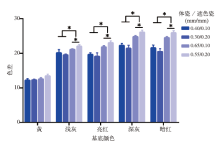
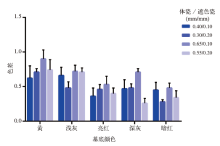
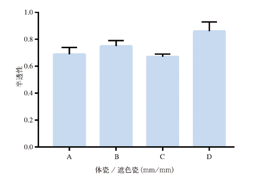
目的 分析不同瓷层厚度和变色基底对瓷贴面修复四环素牙的色彩学性能和半透性的影响。方法 用粉浆涂塑烧结法制作瓷试件:体瓷/遮色瓷厚度分别为0.40 mm/0.10 mm、0.30 mm/0.20 mm、0.65 mm/0.10 mm和0.55 mm/ 0.20 mm。利用分光光度计测量试件在模拟四环素牙背景及黑白背景下的L*、a*、b*值,计算试件在模拟四环素牙背景上与背景颜色的色差ΔE001,与试件在白背景上色差ΔE002以及试件的半透性TP值。结果 1)除黄色基底外,其余基底上总厚度0.75 mm的2组色差ΔE001显著大于0.50 mm组,差异有统计学意义(P<0.01)。2)不同背景下,不同厚度瓷试件的色差ΔE002均<1.25,且差异无统计学意义(P>0.01)。3)不同厚度瓷试件TP值均<1,且差异无统计学意义(P>0.01)。结论 对于偏黄、偏红和偏灰的四环素牙,选用0.50 mm厚IPS d.SIGN瓷贴面(0.10 mm的遮色瓷与0.40 mm的体瓷)可实现目标色的良好匹配,但其通透性与天然牙相比较差。
中图分类号:
| [1] |
Kürklü D, Azer SS, Yilmaz B, et al. Porcelain thick-ness and cement shade effects on the colour and translucency of porcelain veneering materials[J]. J Dent, 2013,41(11):1043-1050.
doi: 10.1016/j.jdent.2013.08.017 pmid: 24004966 |
| [2] | Bagis B, Turgut S. Optical properties of current cera-mics systems for laminate veneers[J]. J Dent, 2013,41(Suppl 3):e24-e30. |
| [3] | 杜瑞钿, 李彦, 范丹妮, 等. 烤瓷贴面临床效果相关影响因素的回顾性研究[J]. 口腔颌面修复学杂志, 2014,15(1):15-20. |
| Du RT, Li Y, Fan DN, et al. A retrospective study on the contributing risk factors for clinical failures of porcelain laminate veneers[J]. Chin J Prosthod, 2014,15(1):15-20. | |
| [4] |
Igiel C, Weyhrauch M, Mayer B, et al. Effects of ceramic layer thickness, cement color, and abutment tooth color on color reproduction of feldspathic veneers[J]. Int J Esthet Dent, 2018,13(1):110-119.
pmid: 29379907 |
| [5] |
Chaiyabutr Y, Kois JC, Lebeau D, et al. Effect of abutment tooth color, cement color, and ceramic thickness on the resulting optical color of a CAD/CAM glass-ceramic lithium disilicate-reinforced crown[J]. J Prosthet Dent, 2011,105(2):83-90.
doi: 10.1016/S0022-3913(11)60004-8 pmid: 21262405 |
| [6] |
Skyllouriotis AL, Yamamoto HL, Nathanson D. Masking properties of ceramics for veneer restora-tions[J]. J Prosthet Dent, 2017,118(4):517-523.
doi: 10.1016/j.prosdent.2016.12.003 pmid: 28341059 |
| [7] |
Sari T, Ural C, Yüzbasioglu E, et al. Color match of a feldspathic ceramic CAD-CAM material for ultra-thin laminate veneers as a function of substrate shade, restoration color, and thickness[J]. J Prosthet Dent, 2018,119(3):455-460.
doi: 10.1016/j.prosdent.2017.02.022 pmid: 28552290 |
| [8] | 潘祁. 模拟四环素牙瓷贴面修复体的色彩学实验研究[D]. 西安: 第四军医大学, 2010. |
| Pan Q. Color assessment on the veneer restorations of mimic-tetracycline pigm entation teeth[D]. Xi’an: The Fourth Military Medical University, 2010. | |
| [9] | 阮丹平, 钱程辉, 张修银, 等. 四环素着色前牙色度学分布特征的检测分析[J]. 上海口腔医学, 2006,15(6):567-570. |
| Ruan DP, Qian CH, Zhang XY, et al. Clinical ana-lysis of colour distribution in tetracycline teeth[J]. Shanghai J Stomatol, 2006,15(6):567-570. | |
| [10] | 钱海馨, 张修银, 杨丹苓. IPS-EMPRESS Ⅱ全瓷修复变色牙的临床效果[J]. 国际口腔医学杂志, 2011,38(3):274-276. |
| Qian HX, Zhang XY, Yang DL. Evaluate color change of discolored tooth after restored with IPS-EMPRSS Ⅱceramics[J]. Int J Stomatol, 2011,38(3):274-276. | |
| [11] |
Chen JH, Shi CX, Wang M, et al. Clinical evaluation of 546 tetracycline-stained teeth treated with porce-lain laminate veneers[J]. J Dent, 2005,33(1):3-8.
doi: 10.1016/j.jdent.2004.06.008 pmid: 15652162 |
| [12] |
Sánchez AR, Rogers RS 3rd, Sheridan PJ. Tetracyc-line and other tetracycline-derivative staining of the teeth and oral cavity[J]. Int J Dermatol, 2004,43(10):709-715.
doi: 10.1111/j.1365-4632.2004.02108.x pmid: 15485524 |
| [13] |
Botelho MG, Chan AWK, Newsome PRH, et al. A randomized controlled trial of home bleaching of tetracycline-stained teeth[J]. J Dent, 2017,67:29-35.
doi: 10.1016/j.jdent.2017.05.003 pmid: 28478214 |
| [14] | 金恩龙, 吴大宏, 闫亮, 等. IPS e.max铸瓷贴面与VITA VM9烤瓷贴面的比较[J]. 中国组织工程研究, 2018,22(22):3474-3479. |
| Jin EL, Wu DH, Yan L, et al. IPS e.max ceramic veneers versus VITA VM9 porcelain laminate veneers[J]. Chin J Tissue Eng Res, 2018,22(22):3474-3479. | |
| [15] |
Shono NN, Al Nahedh HN. Contrast ratio and mas-king ability of three ceramic veneering materials[J]. Oper Dent, 2012,37(4):406-416.
doi: 10.2341/10-237-L pmid: 22339384 |
| [16] |
Turgut S, Bagis B, Ayaz EA. Achieving the desired colour in discoloured teeth, using leucite-based CAD-CAM laminate systems[J]. J Dent, 2014,42(1):68-74.
doi: 10.1016/j.jdent.2013.10.018 pmid: 24239927 |
| [17] | Al Hamad KQ, Qadan MM, Al Wahadni AM. Spe-ctrophotometric analysis of the influence of metal alloy choice, opaque thickness, and repeated firing on the shade of metal ceramic restorations[J]. J Esthet Restor Dent, 2016,28(Suppl 1):S56-S67. |
| [18] | 裴延平, 陈吉华, 常青, 等. 不同金属基底和遮色瓷厚度对金瓷修复体色彩的影响[J]. 华西口腔医学杂志, 2005,23(2):133-135. |
| Pei YP, Chen JH, Chang Q, et al. Effects of varying the opaque thickness and the type of metal ceramic alloy on color[J]. West China J Stomatol, 2005,23(2):133-135. | |
| [19] | Paravina RD, Ghinea R, Herrera LJ, et al. Color dif-ference thresholds in dentistry[J]. J Esthet Restor Dent, 2015,27(Suppl 1):S1-S9. |
| [20] | Ghinea R, Pérez MM, Herrera LJ, et al. Color difference thresholds in dental ceramics[J]. J Dent, 2010,38(Suppl 2):e57-e64. |
| [21] |
Salas M, Lucena C, Herrera LJ, et al. Translucency thresholds for dental materials[J]. Dent Mater, 2018,34(8):1168-1174.
doi: 10.1016/j.dental.2018.05.001 pmid: 29764698 |
| [1] | 黄依欢,李委航,马典,陈瑾,钱捷,李旭东. IPS e.maxCAD和Lava Ultimate在 贴面修复中的有限元分析[J]. 国际口腔医学杂志, 2023, 50(4): 423-432. 贴面修复中的有限元分析[J]. 国际口腔医学杂志, 2023, 50(4): 423-432. |
| [2] | 沈冬妮,施莹,傅柏平. 后牙牙合贴面修复的研究进展[J]. 国际口腔医学杂志, 2021, 48(3): 287-291. |
| [3] | 张婧婷,潘旭东,张文云. 遮色层厚度对聚醚醚酮-Crea.lign修复体颜色的影响[J]. 国际口腔医学杂志, 2020, 47(4): 418-423. |
| [4] | 黄满英,付云. 牙龈生物型的测量方法[J]. 国际口腔医学杂志, 2019, 46(2): 171-176. |
| [5] | 刘玲玲,刘树泰. 上颌腭侧软组织厚度的测量方法及影响因素[J]. 国际口腔医学杂志, 2019, 46(2): 234-237. |
| [6] | 张停停,宗娟娟. 自体结缔组织移植术的研究现状[J]. 国际口腔医学杂志, 2019, 46(1): 89-93. |
| [7] | 靳志亨,李清. 核瓷和饰瓷厚度变化对全瓷修复体抗弯强度影响的研究进展[J]. 国际口腔医学杂志, 2019, 46(1): 94-98. |
| [8] | 魏煦, 杨芸, 张凯, 孙方方. 热压铸瓷面瓷贴面的临床效果评估[J]. 国际口腔医学杂志, 2017, 44(6): 696-700. |
| [9] | 张雅蓉, 刘洋, 张玲, 于海洋. 不同切端设计的上前牙瓷贴面受载能力的定量研究[J]. 国际口腔医学杂志, 2017, 44(3): 301-303. |
| [10] | 罗志强, 叶钟泰. 新疆地区不同人群上颌窦外侧骨壁厚度的锥形束CT测量分析[J]. 国际口腔医学杂志, 2017, 44(1): 55-58. |
| [11] | 陈丽娟1 孟庆飞2 万延俊1. 不同设计类型的IPS Empress铸瓷贴面前牙美学修复的疗效观察[J]. 国际口腔医学杂志, 2016, 43(5): 511-514. |
| [12] | 高原,徐佳蕾,杨倩,黄定明,周学东. 根管内分离器械的处理评估与取出策略[J]. 国际口腔医学杂志, 2016, 43(3): 249-259. |
| [13] | 陆轩,陈小冬,邢文忠,李振春. 全瓷贴面修复的临床效果评估[J]. 国际口腔医学杂志, 2015, 42(2): 170-172. |
| [14] | 谢雨菲 陆佩珺 胡铮 冯静 沈刚. 颧牙槽嵴区域骨密质厚度的锥形束CT测量分析[J]. 国际口腔医学杂志, 2014, 41(3): 281-285. |
| [15] | 刘天爽1 樊成2 李振春3 陈小冬3. 不同切端全瓷贴面的破坏力学分析[J]. 国际口腔医学杂志, 2012, 39(5): 565-567. |
|