国际口腔医学杂志 ›› 2021, Vol. 48 ›› Issue (2): 180-186.doi: 10.7518/gjkq.2021041
丁张帆( ),郭陟永,苗诚,李春洁,宣鸣,王晓毅,张壮()
Ding Zhangfan(),Guo Zhiyong,Miao Cheng,Li Chunjie,Xuan Ming,Wang Xiaoyi,Zhang Zhuang()
摘要:
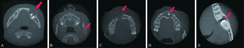
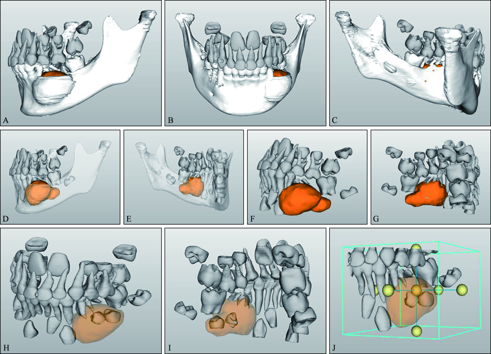
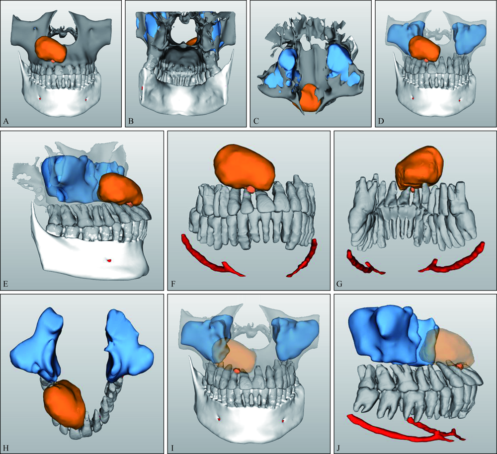
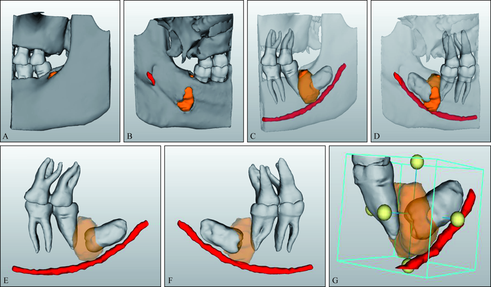
基于锥形束CT(CBCT)的三维重建是通过CBCT影像学技术获得人体组织结构的二维图像,然后运用计算机处理原始数据,提取兴趣区域,并进行三维重建,从而获得三维图像的技术。目前基于CBCT的三维重建在口腔临床上的应用较少。本文通过总结应用CBCT三维可视化技术辅助诊治5例颌骨囊性病变的方法和疗效,探讨该技术在颌骨囊性病变手术中的临床应用价值。
中图分类号:
| [1] |
Liedke GS, Spin-Neto R, Vizzotto MB, et al. Diagnostic accuracy of cone beam computed tomography sections with various thicknesses for detecting misfit between the tooth and restoration in metal-restored teeth[J]. Oral Surg Oral Med Oral Pathol Oral Radiol, 2015,120(2):e131-e137.
doi: 10.1016/j.oooo.2015.05.004 pmid: 26166035 |
| [2] |
Jiang K, Che C, Ding Z, et al. Precision diagnosis and antidiastole on supernumerary cusp of tooth by CBCT[J]. Surg Radiol Anat, 2016,38(9):1099-1104.
doi: 10.1007/s00276-016-1636-7 pmid: 26861010 |
| [3] |
Junqueira CH, Janson G, Junqueira MH, et al. Compa-rison between full face and hemifacial CBCT cephalograms in clinically symmetrical patients: a pilot study[J]. Dental Press J Orthod, 2015,20(2):83-89.
doi: 10.1590/2176-9451.20.2.083-089.oar pmid: 25992992 |
| [4] |
Matherne RP, Angelopoulos C, Kulild JC, et al. Use of cone-beam computed tomography to identify root canal systems in vitro[J]. J Endod, 2008,34(1):87-89.
doi: 10.1016/j.joen.2007.10.016 pmid: 18155501 |
| [5] | 朱敏, 林梓桐, 文珊辉, 等. CBCT根管形态三维容积重建可视化技术的研究[J]. 口腔医学研究, 2015,31(6):601-603. |
| Zhu M, Lin ZT, Wen SH, et al. Visualization of root canal morphology with volume rendering technique based on CBCT images[J]. J Oral Sci Res, 2015,31(6):601-603. | |
| [6] | 常旖旎, 鲁雯, 聂生东. 医学图像三维可视化技术及其应用[J]. 中国医学物理学杂志, 2012,29(2):3254-3258. |
| Chang YN, Lu W, Nie SD. Research on 3-D visuali-zation technology and application of medical image[J]. Chin J Med Phys, 2012,29(2):3254-3258. | |
| [7] | 王猛, 孔繁之. 医学图像三维可视化技术及其新进展[J]. 医学影像学杂志, 2015,25(6):1095-1097. |
| Wang M, Kong FZ. Research on 3D visualization technology of medical image[J]. J Med Imaging, 2015,25(6):1095-1097. | |
| [8] | 孔戈, 郭春岚, 李珍. CBCT髓室底3D重建在牙髓根尖周疾病中的应用[J]. 北京口腔医学, 2017,25(1):33-35. |
| Kong G, Guo CL, Li Z. Application of three-dimensional reconstruction of the floor of pulp cham-ber by CBCT in endodontics[J]. Beijing J Stomatol, 2017,25(1):33-35. | |
| [9] |
Hilgenfeld T, Juerchott A, Deisenhofer UK, et al. In vivo accuracy of tooth surface reconstruction based on CBCT and dental MRI-A clinical pilot study[J]. Clin Oral Implants Res, 2019,30(9):920-927.
doi: 10.1111/clr.13498 pmid: 31257638 |
| [10] |
Zhou Y, Li JP, Lv WC, et al. Three-dimensional CBCT images registration method for TMJ based on reconstructed condyle and skull base[J]. Dentomaxillofac Radiol, 2018,47(5):20170421.
doi: 10.1259/dmfr.20170421 pmid: 29595332 |
| [11] |
Kayaoglu G, Peker I, Gumusok M, et al. Root and canal symmetry in the mandibular anterior teeth of patients attending a dental clinic: CBCT study[J]. Braz Oral Res, 2015,29(1):1-7.
doi: 10.1590/1807-3107BOR-2015.vol29.0092errata pmid: 26398110 |
| [12] |
Giri S, Tripathi A, Patil R, et al. Analysis of bite marks in food stuffs by CBCT 3D-reconstruction[J]. J Oral Biol Craniofacial Res, 2019,9(1):24-27.
doi: 10.1016/j.jobcr.2018.08.006 |
| [13] |
Schlueter B, Kim KB, Oliver D, et al. Cone beam computed tomography 3D reconstruction of the mandibular condyle[J]. Angle Orthod, 2008,78(5):880-888.
doi: 10.2319/072007-339.1 pmid: 18298200 |
| [14] |
Guyader E, Savéan J, Clodic C, et al. Three-dimensional reconstruction of the temporal bone: comparison of in situ, CT, and CBCT measurements[J]. Eur Ann Otorhinolaryngol Head Neck Dis, 2018,135(6):393-398.
doi: 10.1016/j.anorl.2018.08.013 pmid: 30220575 |
| [1] | 杨雨楠,刘鹏,王虎,游梦. 上颌窦黏膜增厚的锥形束CT影像分析[J]. 国际口腔医学杂志, 2023, 50(3): 302-307. |
| [2] | 吴文智,冯达兴,陈垂壮,周丽鹃. 海口地区下颌第一恒磨牙近中中央根管发生率及相关因素[J]. 国际口腔医学杂志, 2022, 49(4): 420-425. |
| [3] | 叶泽林,刘璐,龙虎,游梦. 弯曲前牙的影像评价及治疗的研究进展[J]. 国际口腔医学杂志, 2022, 49(2): 173-181. |
| [4] | 田浩楠,林敏,谢丛蔓,任嫒姝. 上颌腭侧阻生尖牙与寰椎后桥相关性的锥形束CT研究[J]. 国际口腔医学杂志, 2021, 48(5): 536-540. |
| [5] | 施丹妮,杨鑫,吴建勇. 锥形束CT三维头影测量参考坐标系的研究进展[J]. 国际口腔医学杂志, 2021, 48(4): 398-404. |
| [6] | 王立冬,马文,付帅,张长彬,崔庆赢,梁燕,黎明. 不同方法制作正颌手术数字化牙合板的研究及精确性分析[J]. 国际口腔医学杂志, 2021, 48(2): 156-164. |
| [7] | 王奔,许喆桢,韦曦. 数字化微创技术在牙髓根尖周病学中的应用与进展[J]. 国际口腔医学杂志, 2021, 48(1): 110-118. |
| [8] | 唐蓓,赵文俊,王虎,郑广宁,游梦. 根管超填导致下牙槽神经损伤2例[J]. 国际口腔医学杂志, 2020, 47(3): 293-296. |
| [9] | 章婷婷,胡常红,彭燕,周文翘,张慧聪,刘蝶. 300例不同年龄段有牙颌人群上唇软组织侧貌的锥形束CT三维测量分析[J]. 国际口腔医学杂志, 2020, 47(2): 182-188. |
| [10] | 王春林,刘从华,宋思吟,周丽淑,林丽佳. 运用锥形束CT诊断上下颌横向发育不调的研究进展[J]. 国际口腔医学杂志, 2020, 47(1): 121-124. |
| [11] | 黎祺, 黄少宏. 岭南地区广府民系人群下颌第二恒磨牙牙根和根管形态的锥形束CT研究[J]. 国际口腔医学杂志, 2019, 46(6): 640-649. |
| [12] | 曹焜,李家锋,孙玉华,鲍强,卢秋宁,唐巍. 下颌下窝的锥形束CT影像分析[J]. 国际口腔医学杂志, 2019, 46(2): 209-212. |
| [13] | 孟怡彤,张晓东. 成人个别正常颌上气道不同软件三维测量的比较研究[J]. 国际口腔医学杂志, 2018, 45(6): 690-694. |
| [14] | 徐迅, 黄建生, 甘泽坤, 罗震. 上颌第一磨牙区腭侧骨板的锥形束CT测量结果及其临床意义[J]. 国际口腔医学杂志, 2017, 44(6): 686-690. |
| [15] | 袁艺航, 张成晓雪, 王扬, 何双双, 宋雪娟, 王虎. 成都正常人群上颌前牙区鼻腭管相关解剖结构的锥形束CT研究[J]. 国际口腔医学杂志, 2017, 44(5): 566-572. |
|