国际口腔医学杂志 ›› 2020, Vol. 47 ›› Issue (1): 121-124.doi: 10.7518/gjkq.2020031
• 综述 • 上一篇
王春林,刘从华( ),宋思吟,周丽淑,林丽佳
Wang Chunlin,Liu Conghua(),Song Siyin,Zhou Lishu,Lin Lijia
摘要:
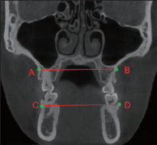
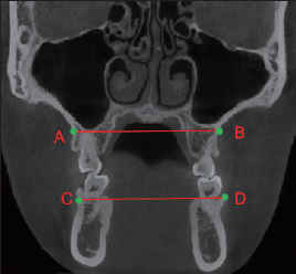
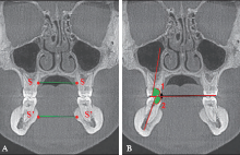
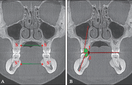
上下颌横向发育不调是导致错畸形的重要因素,临床上常表现为后牙反、牙列拥挤、腭盖高拱、Wilson曲线过深、上颌磨牙颊倾、下颌磨牙舌倾等。本文就近年来上下颌横向发育不调在锥形束CT上的诊断方法、诊断标准的研究现状作一综述。
中图分类号:
| [1] | 白丁, 赵志河 . 口腔正畸策略、控制与技巧[M]. 北京: 人民卫生出版社, 2015. |
| Bai D, Zhao ZH. Advanced strategy with positive control in orthodontics[M]. Beijing: People’s Medical Publishing House, 2015. | |
| [2] | Sawchuk D, Currie K, Vich ML , et al. Diagnostic methods for assessing maxillary skeletal and dental transverse deficiencies: a systematic review[J]. Korean J Orthod, 2016,46(5):331-342. |
| [3] | 高雪梅, 杨凯, 曾祥龙 . 儿童呼吸方式与颌面生长发育的关系[J]. 中华医学杂志, 2005,85(44):3105-3106. |
| Gao XM, Yang K, Zeng XL . A study on the relation-ship between maxillofacial development and brea-thing pattern in children[J]. Natl Med J China, 2005,85(44):3105-3106. | |
| [4] | Miner RM, Al Qabandi S, Rigali PH , et al. Cone-beam computed tomography transverse analyses. Part 2: measures of performance[J]. Am J Orthod Dentofacial Orthop, 2015,148(2):253-263. |
| [5] | de Oliveira MA Jr, Pereira MD, Hino CT , et al. Prediction of transverse maxillary dimension using orthodontic models[J]. J Craniofac Surg, 2008,19(6):1465-1471. |
| [6] | Sawchuk D, Currie K, Vich ML , et al. Diagnostic methods for assessing maxillary skeletal and dental transverse deficiencies: a systematic review[J]. Korean J Orthod, 2016,46(5):331-342. |
| [7] | Major PW, Johnson DE, Hesse KL , et al. Effect of head orientation on posterior anterior cephalometric landmark identification[J]. Angle Orthod, 1996,66(1):51-60. |
| [8] | Lee KM, Hwang HS, Cho JH . Comparison of transverse analysis between posteroanterior cephalogram and cone-beam computed tomography[J]. Angle Orthod, 2014,84(4):715-719. |
| [9] | Andrews LF . The 6-elements orthodontic philosophy: treatment goals, classification, and rules for treating[J]. Am J Orthod Dentofac Orthop, 2015,148(6):883-887. |
| [10] | Tamburrino RK, Boucher NS, Vanarsdall RL , et al. The transverse dimension: diagnosis and relevance to functional occlusion[J]. RWISO J, 2010,2(1):11-20. |
| [11] | Katona TR .An engineering analysis of dental occlusion principles[J]. Am J Orthod Dentofacial Orthop, 2009, 135(6): 696.e1-696.e8,697. |
| [12] | Ricketts RM . Perspectives in the clinical application of cephalometrics. The first fifty years[J]. Angle Orthod, 1981,51(2):115-150. |
| [13] | Miner RM, Al Qabandi S, Rigali PH , et al. Cone-beam computed tomography transverse analysis. Part Ⅰ: normative data[J]. Am J Orthod Dentofacial Orthop, 2012,142(3):300-307. |
| [14] | Hwang S, Song J, Lee J , et al. Three-dimensional evaluation of dentofacial transverse widths in adults with different sagittal facial patterns[J]. Am J Orthod Dentofacial Orthop, 2018,154(3):365-374. |
| [15] | Tyan S, Park HS, Janchivdorj M , et al. Three-dimensional analysis of molar compensation in patients with facial asymmetry and mandibular prognathism[J]. Angle Orthod, 2016,6(3):421-430. |
| [16] | Shewinvanakitkul W, Hans MG, Narendran S , et al. Measuring buccolingual inclination of mandibular canines and first molars using CBCT[J]. Orthod Craniofac Res, 2011,14(3):168-174. |
| [17] | Ahn J, Kim SJ, Lee JY , et al. Transverse dental compensation in relation to sagittal and transverse skeletal discrepancies in skeletal class Ⅲ patients[J]. Am J Orthod Dentofacial Orthop, 2017,151(1):148-156. |
| [18] | Vanarsdall RL Jr . Transverse dimension and long-term stability[J]. Semin Orthod, 1999,5(3):171-180. |
| [19] | Hwang S, Noh Y, Choi YJ , et al. Dentofacial transverse development in Koreans according to skeletal maturation: a cross-sectional study[J]. Korean J Orthod, 2018,48(1):39-47. |
| [20] | Yehya Mostafa R, Bous RM, Hans MG , et al. Effects of Case Western Reserve University’s transverse analysis on the quality of orthodontic treatment[J]. Am J Orthod Dentofacial Orthop, 2017,152(2):178-192. |
| [21] | Podesser B, Williams S, Bantleon HP , et al. Quantitation of transverse maxillary dimensions using computed tomography: a methodological and reproducibility study[J]. Eur J Orthod, 2004,26(2):209-215. |
| [1] | 杨雨楠,刘鹏,王虎,游梦. 上颌窦黏膜增厚的锥形束CT影像分析[J]. 国际口腔医学杂志, 2023, 50(3): 302-307. |
| [2] | 吴文智,冯达兴,陈垂壮,周丽鹃. 海口地区下颌第一恒磨牙近中中央根管发生率及相关因素[J]. 国际口腔医学杂志, 2022, 49(4): 420-425. |
| [3] | 叶泽林,刘璐,龙虎,游梦. 弯曲前牙的影像评价及治疗的研究进展[J]. 国际口腔医学杂志, 2022, 49(2): 173-181. |
| [4] | 田浩楠,林敏,谢丛蔓,任嫒姝. 上颌腭侧阻生尖牙与寰椎后桥相关性的锥形束CT研究[J]. 国际口腔医学杂志, 2021, 48(5): 536-540. |
| [5] | 施丹妮,杨鑫,吴建勇. 锥形束CT三维头影测量参考坐标系的研究进展[J]. 国际口腔医学杂志, 2021, 48(4): 398-404. |
| [6] | 丁张帆,郭陟永,苗诚,李春洁,宣鸣,王晓毅,张壮. 基于锥形束CT的三维可视化技术在颌骨囊性病变手术中的应用[J]. 国际口腔医学杂志, 2021, 48(2): 180-186. |
| [7] | 王奔,许喆桢,韦曦. 数字化微创技术在牙髓根尖周病学中的应用与进展[J]. 国际口腔医学杂志, 2021, 48(1): 110-118. |
| [8] | 唐蓓,赵文俊,王虎,郑广宁,游梦. 根管超填导致下牙槽神经损伤2例[J]. 国际口腔医学杂志, 2020, 47(3): 293-296. |
| [9] | 章婷婷,胡常红,彭燕,周文翘,张慧聪,刘蝶. 300例不同年龄段有牙颌人群上唇软组织侧貌的锥形束CT三维测量分析[J]. 国际口腔医学杂志, 2020, 47(2): 182-188. |
| [10] | 黎祺, 黄少宏. 岭南地区广府民系人群下颌第二恒磨牙牙根和根管形态的锥形束CT研究[J]. 国际口腔医学杂志, 2019, 46(6): 640-649. |
| [11] | 曹焜,李家锋,孙玉华,鲍强,卢秋宁,唐巍. 下颌下窝的锥形束CT影像分析[J]. 国际口腔医学杂志, 2019, 46(2): 209-212. |
| [12] | 孟怡彤,张晓东. 成人个别正常颌上气道不同软件三维测量的比较研究[J]. 国际口腔医学杂志, 2018, 45(6): 690-694. |
| [13] | 徐迅, 黄建生, 甘泽坤, 罗震. 上颌第一磨牙区腭侧骨板的锥形束CT测量结果及其临床意义[J]. 国际口腔医学杂志, 2017, 44(6): 686-690. |
| [14] | 袁艺航, 张成晓雪, 王扬, 何双双, 宋雪娟, 王虎. 成都正常人群上颌前牙区鼻腭管相关解剖结构的锥形束CT研究[J]. 国际口腔医学杂志, 2017, 44(5): 566-572. |
| [15] | 吴杉杉, 张茹, 侯本祥. 钙化根管的诊断与治疗[J]. 国际口腔医学杂志, 2017, 44(3): 279-283. |
|