Int J Stomatol ›› 2026, Vol. 53 ›› Issue (6): 810-820.doi: 10.7518/gjkq.2026147
• Expert Forum • Previous Articles
Siying Liu( ),Jie Gao()
CLC Number:
| [1] | 王旭东, 魏弘朴, 李彪. 从“经验外科”到“精准外科”:精准正颌外科体系的建立与临床应用[J]. 华西口腔医学杂志, 2023, 41(5): 491-501. |
| Wang XD, Wei HP, Li B. From “Empirical Surgery” to “Precision Surgery”: establishment and clinical application of precision orthognathic surgery system[J]. West China J Stomatol, 2023, 41(5): 491-501. | |
| [2] | Posnick JC, Makan S, Bostock D, et al. Primary maxillary deficiency dentofacial deformities: occlusion and facial esthetic surgical outcomes[J]. J Oral Maxillofac Surg, 2018, 76(9): 1966-1982. |
| [3] | Li JTW, Leung YY. Digital diagnostics and treatment planning for orthognathic surgery[J]. Aust Dent J, 2025, 70(S1): S82-S92. |
| [4] | Bench RW. The visual treatment objective: ortho-dontic's most effective treatment planning tool[J]. Proc Found Orthod Res, 1971: 165-194. |
| [5] | 闫佳恕, 李彪, 王旭东. 数字化正颌外科技术在临床实践中面临的挑战[J]. 口腔疾病防治, 2024, 32(5): 395-400. |
| Yan JS, Li B, Wang XD. Challenges of computer-assisted orthognathic surgery in clinical application[J]. J Prev Treat Stomatol Dis, 2024, 32(5): 395-400. | |
| [6] | Peck S, Peck L. Selected aspects of the art and science of facial esthetics[J]. Semin Orthod, 1995, 1(2): 105-126. |
| [7] | Oh HS, Korn EL, Zhang XY, et al. Correlations between cephalometric and photographic measurements of facial attractiveness in Chinese and US patients after orthodontic treatment[J]. Am J Orthod Dentofacial Orthop, 2009, 136(6): 762.e1-762.e14. |
| [8] | Andrews LF. The six keys to normal occlusion[J]. Am J Orthod, 1972, 62(3): 296-309. |
| [9] | Andrews LF. The 6-elements orthodontic philosophy: treatment goals, classification, and rules for treating[J]. Am J Orthod Dentofacial Orthop, 2015, 148(6): 883-887. |
| [10] | Andrews WA, Andrews LF. The six elements or-thodontic philosophy: goals, diagnosis, classification, and treatment[M]. Meridian: Dental Press, 2025. |
| [11] | 张珂, 白丁. Andrews口颌面协调六要素在侧貌美学中的应用[J]. 国际口腔医学杂志, 2010, 37(2): 236-239. |
| Zhang K, Bai D. Application of Andrews' six elements of orofacial harmony in profile esthetics[J]. Int J Stomatol, 2010, 37(2): 236-239. | |
| [12] | Lundström A, Lundström F, Lebret LM, et al. Natural head position and natural head orientation: basic considerations in cephalometric analysis and research[J]. Eur J Orthod, 1995, 17(2): 111-120. |
| [13] | Lo LJ, Weng JL, Ho CT, et al. Three-dimensional region-based study on the relationship between soft and hard tissue changes after orthognathic surgery in patients with prognathism[J]. PLoS One, 2018, 13(8): e0200589. |
| [14] | San Miguel Moragas J, Oth O, Büttner M, et al. A systematic review on soft-to-hard tissue ratios in orthognathic surgery part Ⅱ: chin procedures[J]. J Cranio Maxillofac Surg, 2015, 43(8): 1530-1540. |
| [15] | Knoops PGM, Borghi A, Breakey RWF, et al. Three-dimensional soft tissue prediction in orthognathic surgery: a clinical comparison of Dolphin, ProPlan CMF, and probabilistic finite element modelling[J]. Int J Oral Maxillofac Surg, 2019, 48(4): 511-518. |
| [16] | Dammling C, Powell K, Repasky T, et al. Validation of Andrews analysis in the virtual environment[J]. J Oral Maxillofac Surg, 2023, 81(8): 979-987. |
| [17] | Resnick CM, Daniels KM, Vlahos M. Does Andrews facial analysis predict esthetic sagittal maxillary position[J]. Oral Surg Oral Med Oral Pathol Oral Radiol, 2018, 125(4): 376-381. |
| [18] | Resnick CM, Kim S, Yorlets RR, et al. Evaluation of Andrews' analysis as a predictor of ideal sagittal maxillary positioning in orthognathic surgery[J]. J Oral Maxillofac Surg, 2018, 76(10): 2169-2176. |
| [19] | 王策. 应用Andrews六要素要素Ⅱ评估骨性Ⅱ类患者上下颌骨矢状向位置特征的研究[D]. 沈阳: 中国医科大学, 2022. |
| Wang C. Study on the application of Andrews six element Ⅱ to evaluate the sagittal positional characte-ristics of the upper and lower jaws in patients with skeletal class Ⅱ[D]. Shenyang: China Medical University, 2022. | |
| [20] | 杨妍, 梁源, 金钫, 等. 中国骨性错𬌗畸形人群基于眉间点确定上颌理想前界线的可行性研究[J]. 中华口腔正畸学杂志, 2025, 32(3): 146-150. |
| Yang Y, Liang Y, Jin F, et al. Feasibility of using the glabella to determine the ideal sagittal position of the maxilla in Chinese patients with skeletal malocclusion[J]. Chin J Orthod, 2025, 32(3): 146-150. | |
| [21] | Miao P, Gao J, Lu ZY, et al. Cone beam computed tomography imaging of sagittal positions of the mandibular prominence and maxillary central incisors in adult Chinese Han men as an aesthetic profile determinant[J]. Medicine, 2020, 99(43): e22778. |
| [22] | Perrett DI, May KA, Yoshikawa S. Facial shape and judgements of female attractiveness[J]. Nature, 1994, 368(6468): 239-242. |
| [23] | 杨妍, 曹猛, 刘思颖, 等. 中国汉族正常𬌗人群GALL线与眉间点矢状向关系的研究[J]. 中华口腔正畸学杂志, 2017, 24(3): 128-132. |
| Yang Y, Cao M, Liu SY, et al. The relationship between GALL Line and glabella in sagittal direction of Han Chinese with individual normal occlusion[J]. Chin J Orthod, 2017, 24(3): 128-132. | |
| [24] | Sandoval C, Díaz A, Manríquez G. Relationship between craniocervical posture and skeletal class: a statistical multivariate approach for studying Class Ⅱ and Class Ⅲ malocclusions[J]. Cranio, 2021, 39(2): 133-140. |
| [25] | Slaimayeh AAS. 自然头位下头影测量稳定参考平面研究[D]. 成都: 四川大学, 2021. |
| Slaimayeh AAS. Study on stable reference plane of cephalometric measurement under natural head position[D]. Chengdu: Sichuan University, 2021. | |
| [26] | Andrews WA. AP relationship of the maxillary central incisors to the forehead in adult white females[J]. Angle Orthod, 2008, 78(4): 662-669. |
| [27] | 王晓婷, 林立. Andrews六要素与上颌矢状向定位的研究进展[J]. 中华口腔正畸学杂志, 2024, 31(3): 148-152. |
| Wang XT, Lin L. Research proceeding of Andrews' six elements and the sagittal position of maxilla[J]. Chin J Orthod, 2024, 31(3): 148-152. | |
| [28] | Bertot AA, Dammling CW, Souccar NM, et al. A cross-sectional study examining Andrews' analysis in Caucasian and African American subjects[J]. J Oral Maxillofac Surg, 2024, 82(12): 1528-1536. |
| [29] | Farkas LG, Katic MJ, Forrest CR. International anthropometric study of facial morphology in various ethnic groups/races[J]. J Craniofac Surg, 2005, 16(4): 615-646. |
| [30] | Suhk J, Park J, Nguyen AH. Nasal analysis and ana-tomy: anthropometric proportional assessment in asians-aesthetic balance from forehead to chin, part Ⅰ[J]. Semin Plast Surg, 2015, 29(4): 219-225. |
| [31] | 何冰. 广东地区正常𬌗人群Andrews侧貌分析研究[D]. 广州: 南方医科大学, 2013. |
| He B. Andrews lateral facial analysis study on normal occlusion population in Guangdong region[D]. Guangzhou: Southern Medical University, 2013. | |
| [32] | He DQ, Gu Y, Sun YN. Evaluation of aesthetic anteroposterior position of maxillary incisors in patients with extraction treatment using facial refe-rence lines[J]. J Int Med Res, 2019, 47(7): 2951-2960. |
| [33] | Zou BS, Zhou Y, Lowe AA, et al. Changes in anteroposterior position and inclination of the maxillary incisors after surgical-orthodontic treatment of ske-letal class Ⅲ malocclusions[J]. J Cranio Maxillofac Surg, 2015, 43(10): 1986-1993. |
| [34] | An JS, Jeong W, Sonnesen L, et al. Effects of presurgical mandibular incisor decompensation on long-term outcomes of Class Ⅲ surgical orthodontic treatment[J]. J Clin Med, 2021, 10(13): 2870. |
| [35] | Seker ED, Sunal Akturk E, Yilmaz HN, et al. Occlusal plane rotation and orthodontic decompensation: influence on the outcome of surgical correction of Class Ⅲ malocclusion[J]. J Orofac Orthop, 2023, 84(6): 373-383. |
| [36] | Liang YQ, Zhang RC, Wang S, et al. Evaluation of a safe decompensation range for mandibular incisors during presurgical orthodontic treatment for severe skeletal Class Ⅲ malocclusion: a cone-beam computed tomography study[J]. Am J Orthod Dentofacial Orthop, 2026, 169(4): 440-457.e2. |
| [37] | 贺红. 正畸牙移动中牙周界限的探讨[J]. 中华口腔医学杂志, 2021, 56(10): 966-970. |
| He H. Periodontal limits of orthodontic tooth movement[J]. Chin J Stomatol, 2021, 56(10): 966-970. |
|
||
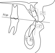
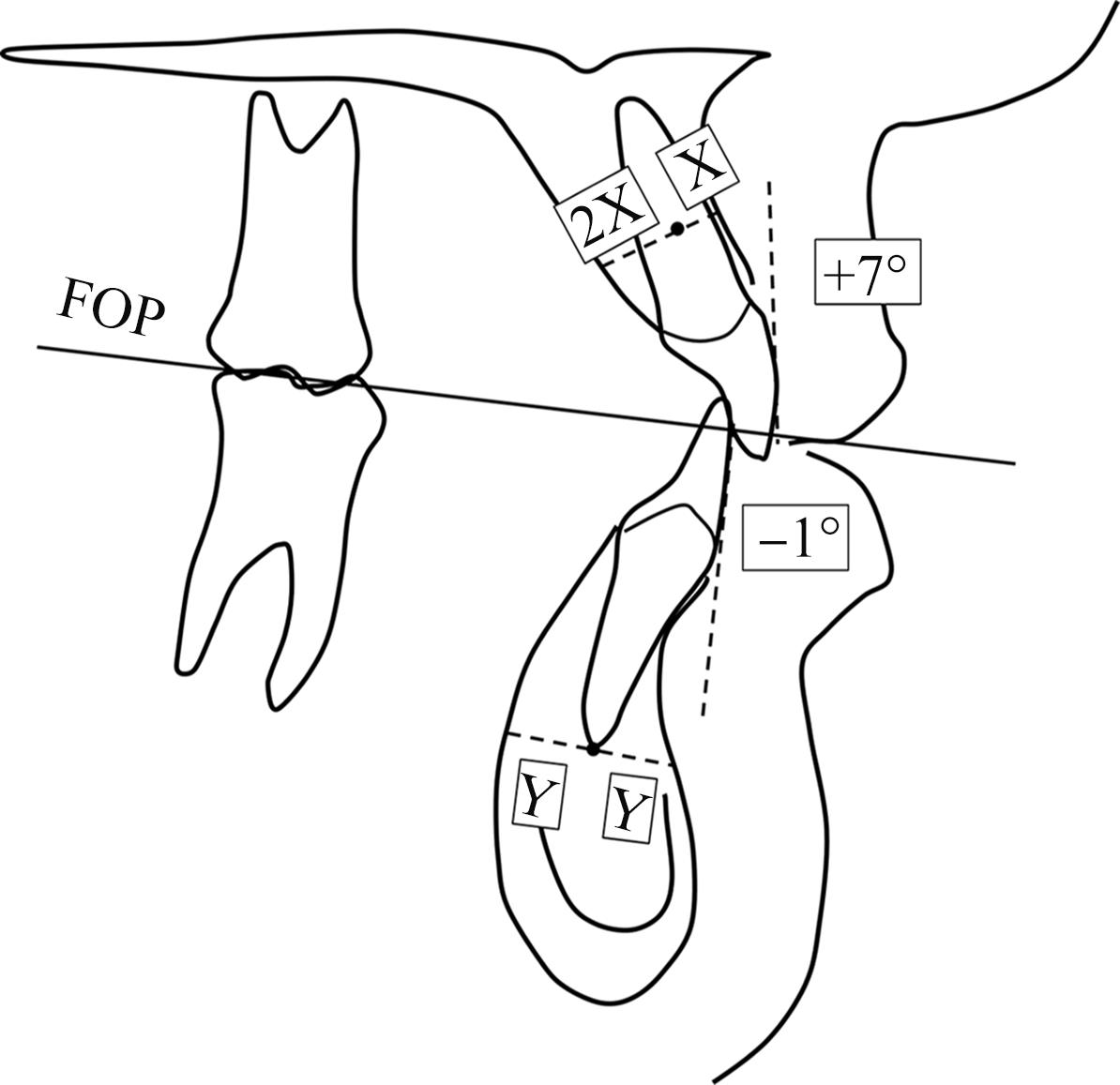
 龈向中点
 龈向中点
 平面(functional occlusal plane,FOP)