国际口腔医学杂志 ›› 2024, Vol. 51 ›› Issue (6): 713-721.doi: 10.7518/gjkq.2024083
• 论著 • 上一篇
刘翼1( ),邱江珊2,申道南1,关鑫3,丁一1()
Yi Liu1(),Jiangshan Qiu2,Daonan Shen1,Xin Guan3,Yi Ding1()
摘要:
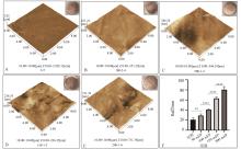
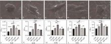
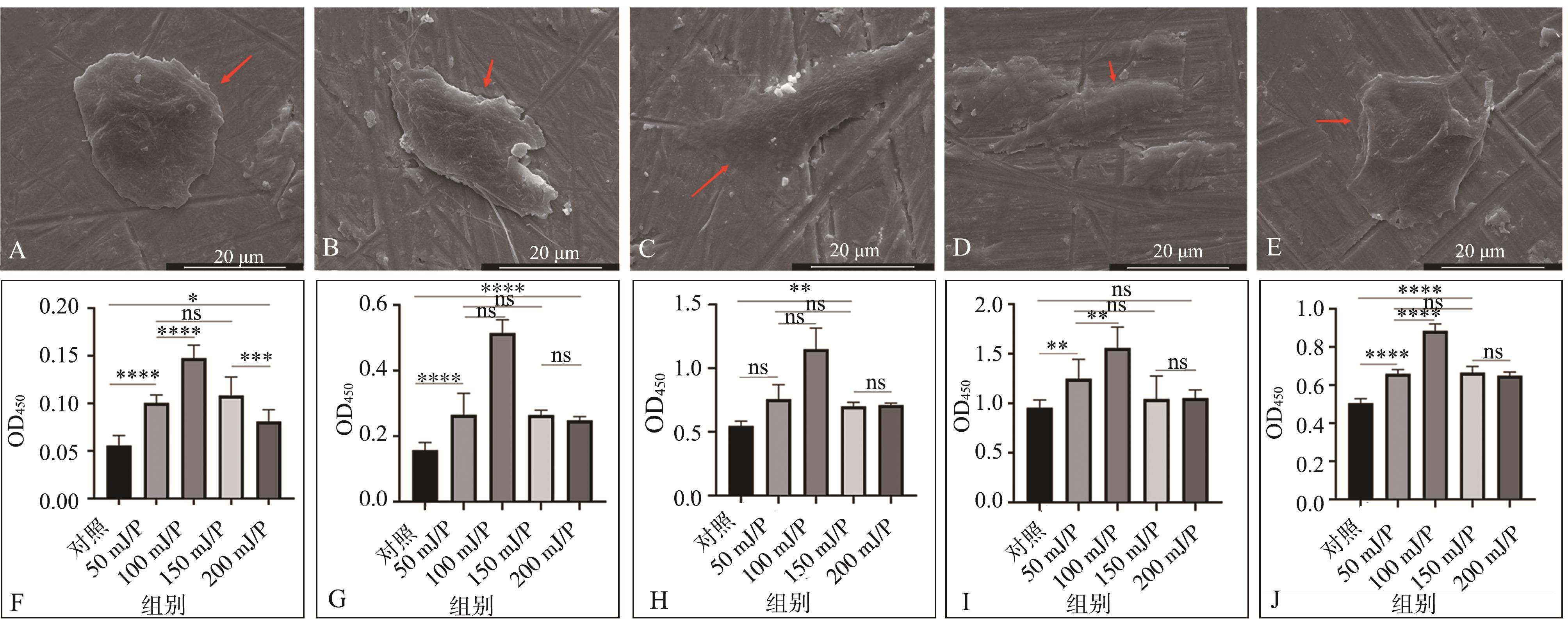
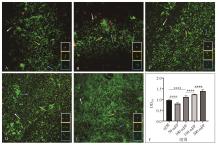
目的 探究不同脉冲能量Er: YAG激光照射后钛盘表面形貌与粗糙度的变化,研究不同钛盘表面形貌对细胞生物学行为以及细菌生物学特性的影响,探索Er: YAG激光改性钛表面的合适参数。 方法 将Ti6Al4V钛盘随机分为5组,设置不同的参数(0、50、100、150、200 mJ/P)进行Er: YAG激光照射,通过原子力显微镜观察钛盘表面形貌和粗糙度的变化,使用扫描电子显微镜、酶联免疫吸附试验检测钛盘表面MG63细胞的黏附、增殖和成骨分化能力,通过结晶紫染色和活/死细菌染色观察钛盘表面生物膜的形成。 结果 激光对钛盘表面形貌的改变程度以及钛盘表面粗糙度与脉冲能量成正比,组间差异具有统计学意义(P<0.05)。细胞实验结果显示:100 mJ/P组钛盘表面MG63细胞的早期黏附形态、增殖和成骨分化能力明显优于其他组,差异有统计学意义(P<0.05)。细菌实验结果显示:50 mJ/P组钛盘表面生物膜形成明显低于未处理组,差异有统计学意义(P<0.05)。 结论 在100 mJ/P处理后钛盘表面MG63细胞表现出良好的黏附、增殖及分化能力,在50 mJ/P处理时细菌生物膜形成最少。50~100 mJ/P可能是Er: YAG激光改性种植体表面的适宜参数区间。
中图分类号:
| 1 | Bosshardt DD, Chappuis V, Buser D. Osseointegration of titanium, titanium alloy and zirconia dental implants: current knowledge and open questions[J]. Periodontol 2000, 2017, 73(1): 22-40. |
| 2 | Herrera D, Berglundh T, Schwarz F, et al. Prevention and treatment of peri-implant diseases-the EFP S3 level clinical practice guideline[J]. J Clin Perio-dontol, 2023, 50(): 4-76. |
| 3 | de Bruyn H, Christiaens V, Doornewaard R, et al. Implant surface roughness and patient factors on long-term peri-implant bone loss[J]. Periodontol 2000, 2017, 73(1): 218-227. |
| 4 | Sinjab K, Sawant S, Ou A, et al. Impact of surface characteristics on the peri-implant microbiome in health and disease[J]. J Periodontol, 2024, 95(3): 244-255. |
| 5 | 杨帮成, 周学东, 于海洋, 等. 钛种植体表面改性方法[J]. 华西口腔医学杂志, 2019, 37(2): 124-129. |
| Yang BC, Zhou XD, Yu HY, et al. Advances in titanium dental implant surface modification[J]. West China J Stomatol, 2019, 37(2): 124-129. | |
| 6 | Sotova C, Yanushevich O, Kriheli N, et al. Dental implants: modern materials and methods of their surface modification[J]. Materials, 2023, 16(23): 7383. |
| 7 | Saran R, Ginjupalli K, George SD, et al. LASER as a tool for surface modification of dental biomate-rials: a review[J]. Heliyon, 2023, 9(6): e17457. |
| 8 | Gupta R, Naveena AH, Vadali M, et al. Enhancement of cell attachment on Ti6Al4V via surface modifications using pulsed laser surface melting[J]. Surf Coat Technol, 2023, 474: 130080. |
| 9 | Bayrak M, Kocak-Oztug NA, Gulati K, et al. Inf-luence of clinical decontamination techniques on the surface characteristics of SLA titanium implant[J]. Nanomaterials, 2022, 12(24): 4481. |
| 10 | Lee JS, Son K, Hwang SM, et al. Effect of electrocautery and laser treatment on the composition and morphology of surface-modified titanium implants[J]. Bioengineering, 2023, 10(11): 1251. |
| 11 | Eghbali N, Naffakh-Moosavy H, Sadeghi Mohammadi S, et al. The influence of laser frequency and groove distance on cell adhesion, cell viability, and antibacterial characteristics of Ti-6Al-4V dental implants treated by modern fiber engraving laser[J]. Dent Mater, 2021, 37(3): 547-558. |
| 12 | Yang D, Xu TY, Fan L, et al. MicroRNA-216b enhances cisplatin-induced apoptosis in osteosarcoma MG63 and SaOS-2 cells by binding to JMJD2C and regulating the HIF1α/HES1 signaling axis[J]. J Exp Clin Cancer Res, 2020, 39(1): 201. |
| 13 | Hassani A, Khoshfetrat AB, Rahbarghazi R, et al. Collagen and nano-hydroxyapatite interactions in alginate-based microcapsule provide an appropriate osteogenic microenvironment for modular bone tissue formation[J]. Carbohydr Polym, 2022, 277: 118807. |
| 14 | Shailaja A, Bruce TF, Gerard P, et al. Comparison of cell viability assessment and visualization of Aspergillus niger biofilm with two fluorescent probe staining methods[J]. Biofilm, 2022, 4: 100090. |
| 15 | Parga A, Manoil D, Brundin M, et al. Gram-negative quorum sensing signalling enhances biofilm formation and virulence traits in gram-positive pathogen Enterococcus faecalis [J]. J Oral Microbiol, 2023, 15(1): 2208901. |
| 16 | Haney EF, Trimble MJ, Hancock REW. Microtiter plate assays to assess antibiofilm activity against bacteria[J]. Nat Protoc, 2021, 16(5): 2615-2632. |
| 17 | Kligman S, Ren Z, Chung CH, et al. The impact of dental implant surface modifications on osseointegration and biofilm formation[J]. J Clin Med, 2021, 10(8): 1641. |
| 18 | Xiong SB, Lu XG, Zhang SQ, et al. Osteogenic properties of bioactive titanium in inflammatory environment[J]. Dent Mater, 2023, 39(10): 929-937. |
| 19 | Wang CY, Wang X, Lu R, et al. Influence of surface nanotopography and wettability on early phases of peri-implant soft tissue healing: an in-vivo study in dogs[J]. BMC Oral Health, 2023, 23(1): 651. |
| 20 | Jiang PL, Zhang YM, Hu R, et al. Advanced surface engineering of titanium materials for biomedical applications: from static modification to dynamic responsive regulation[J]. Bioact Mater, 2023, 27: 15-57. |
| 21 | Accioni F, Vázquez J, Merinero M, et al. Latest trends in surface modification for dental implantology: innovative developments and analytical applications[J]. Pharmaceutics, 2022, 14(2): 455. |
| 22 | Revathi A, Mitun D, Balla VK, et al. Surface properties and cytocompatibility of Ti-6Al-4V fabricated using laser engineered net shaping[J]. Mater Sci Eng C Mater Biol Appl, 2019, 100: 104-116. |
| 23 | Wehner C, Laky M, Shokoohi-Tabrizi HA, et al. Effects of Er: YAG laser irradiation of different tita-nium surfaces on osteoblast response[J]. J Mater Sci Mater Med, 2021, 32(3): 22. |
| 24 | Shin SI, Min HK, Park BH, et al. The effect of Er: YAG laser irradiation on the scanning electron microscopic structure and surface roughness of various implant surfaces: an in vitro study[J]. Lasers Med Sci, 2011, 26(6): 767-776. |
| 25 | Moeintaghavi A, Bagheri H, Pour MY, et al. Effects of diode, CO2, Er: YAG, and Er and Cr: YSGG on titanium implant surfaces by scanning electron microscopy[J]. Adv Mater Sci Eng, 2021, 2021(1): 3551097. |
| 26 | Shirazi S, Ravindran S, Cooper LF. Topography-mediated immunomodulation in osseointegration; ally or enemy[J]. Biomaterials, 2022, 291: 121903. |
| 27 | Im JS, Choi H, An HW, et al. Effects of surface treatment method forming new nano/micro hierarchical structures on attachment and proliferation of osteoblast-like cells[J]. Materials, 2023, 16(16): 5717. |
| 28 | Ayobian-Markazi N, Fourootan T, Zahmatkesh A. An in vitro evaluation of the responses of human osteoblast-like SaOs-2 cells to SLA titanium surfaces irradiated by erbium: yttrium-aluminum-garnet (Er: YAG) lasers[J]. Lasers Med Sci, 2014, 29(1): 47-53. |
| 29 | Ayobian-Markazi N, Karimi M, Safar-Hajhosseini A. Effects of Er: YAG laser irradiation on wettability, surface roughness, and biocompatibility of SLA titanium surfaces: an in vitro study[J]. Lasers Med Sci, 2015, 30(2): 561-566. |
| 30 | Kotsakis GA, Olmedo DG. Peri-implantitis is not periodontitis: scientific discoveries shed light on microbiome-biomaterial interactions that may determine disease phenotype[J]. Periodontol 2000, 2021, 86(1): 231-240. |
| 31 | Belibasakis GN, Manoil D. Microbial community-driven etiopathogenesis of peri-implantitis[J]. J Dent Res, 2021, 100(1): 21-28. |
| 32 | Sakanaka A, Kuboniwa M, Shimma S, et al. Fusobacterium nucleatum metabolically integrates commensals and pathogens in oral biofilms[J]. mSystems, 2022, 7(4): e0017022. |
| 33 | Choi S, Jo YH, Luke Yeo IS, et al. The effect of surface material, roughness and wettability on the adhesion and proliferation of Streptococcus gordonii, Fusobacterium nucleatum and Porphyromonas gingivalis [J]. J Dent Sci, 2023, 18(2): 517-525. |
| 34 | Assery N, Alomeir N, Zeng Y, et al. The effect of Er: YAG laser treatment on biofilm formation on titanium and zirconia disc surfaces[J]. J Periodontol, 2023, 94(3): 344-353. |
| 35 | Stein JM, Conrads G, Abdelbary MMH, et al. Antimicrobial efficiency and cytocompatibility of diffe-rent decontamination methods on titanium and zirconium surfaces[J]. Clin Oral Implants Res, 2023, 34(1): 20-32. |
| 36 | Birang E, Birang R, Narimani T, et al. Investigation of the antibacterial effect of laser irradiation and chemical agent on human oral biofilms contamina-ted titanium discs[J]. Photodiagnosis Photodyn Ther, 2019, 25: 259-264. |
| 37 | Polak D, Shani-Kdoshim S, Alias M, et al. The in vitro efficacy of biofilm removal from titanium sur-faces using Er: YAG laser: comparison of treatment protocols and ablation parameters[J]. J Periodontol, 2022, 93(1): 100-109. |
| [1] | 孙旭,邓振南,文才,赵颖. Er: YAG激光照射种植体表面微形貌变化的扫描电子显微镜观察[J]. 国际口腔医学杂志, 2023, 50(6): 669-673. |
| [2] | 章善,沈树平,张舫,杨卫东. Er: YAG激光光子激活光声流技术对根管壁牙本质失水状况及牙根抗压强度的影响[J]. 国际口腔医学杂志, 2022, 49(1): 55-59. |
| [3] | 祁星颖,郑国莹,隋磊. 钛种植体表面形貌对成骨的影响[J]. 国际口腔医学杂志, 2018, 45(5): 527-533. |
| [4] | 陶婷婷 李长真 杨恒 丁一. 锥形束CT在牙周影像学检查中的应用研究初探[J]. 国际口腔医学杂志, 2014, 41(4): 412-414. |
|
||