国际口腔医学杂志 ›› 2026, Vol. 53 ›› Issue (5): 667-675.doi: 10.7518/gjkq.2026131
朱晓宇( ),程晓雨,刘梦潇,李冉,王林娜,侯彦()
Xiaoyu Zhu(),Xiaoyu Cheng,Mengxiao Liu,Ran Li,Linna Wang,Yan Hou()
摘要:
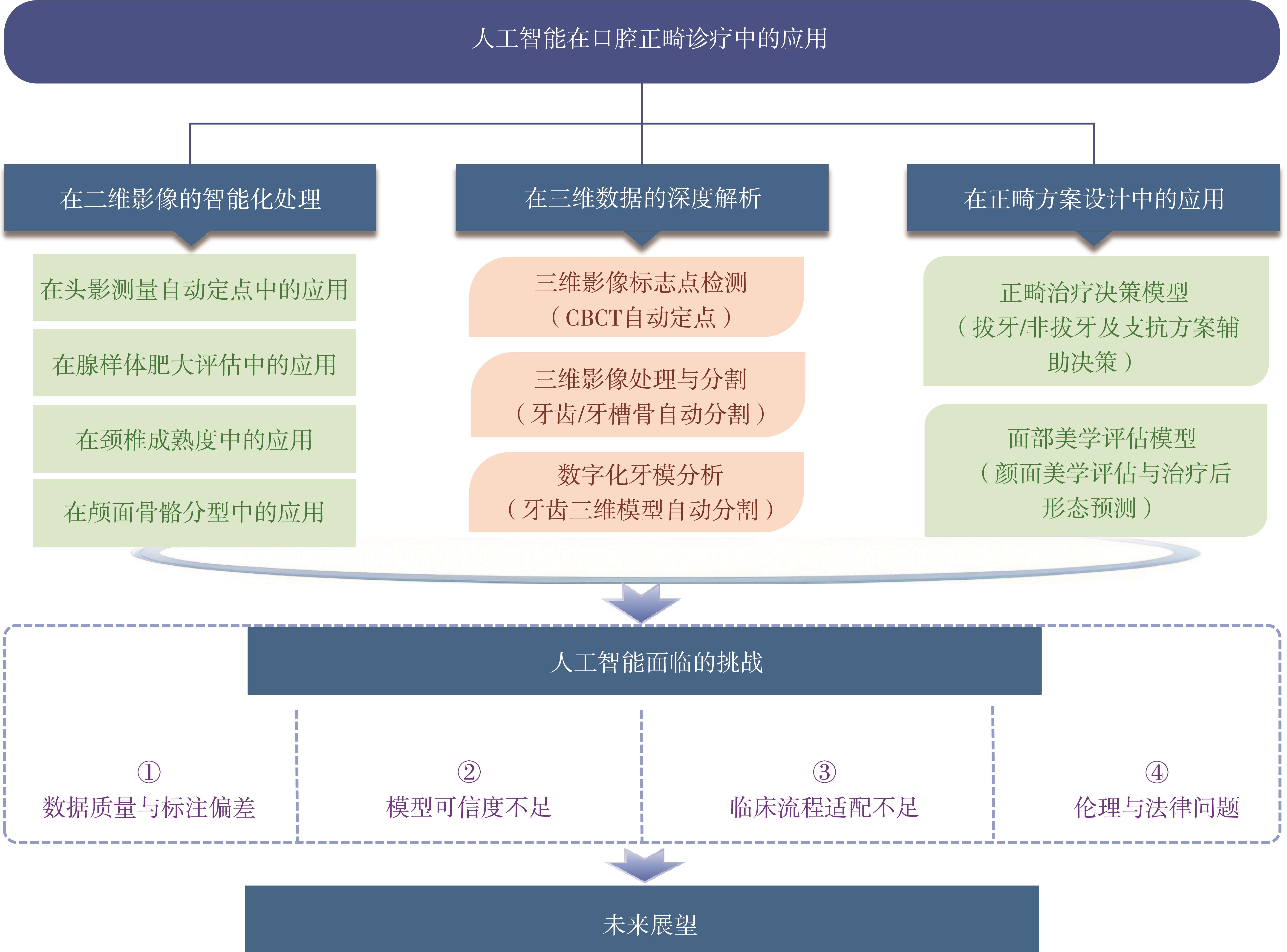
随着机器学习和深度学习技术的发展,人工智能(AI)在口腔正畸诊疗中的应用逐渐增多,主要涉及二维影像自动分析、三维影像处理、数字化牙模分析、治疗方案辅助决策及颜面形态预测等方面。本文围绕AI在口腔正畸诊疗中的主要应用场景进行综述,分析其在提高影像分析效率、辅助诊断评估和支持治疗方案设计中的应用价值,并从数据质量、模型泛化能力、可解释性、临床流程适配及伦理法律风险等方面讨论其临床转化面临的问题,旨在推动口腔正畸诊疗向数智化方向高质量发展。
中图分类号:
| [1] | Shan T, Tay FR, Gu L. Application of artificial intelligence in dentistry[J]. J Dent Res, 2021, 100(3): 232-244. |
| [2] | Sarvamangala DR, Kulkarni RV. Convolutional neural networks in medical image understanding: a survey[J]. Evol Intell, 2022, 15(1): 1-22. |
| [3] | Kim H, Shim E, Park J, et al. Web-based fully automated cephalometric analysis by deep learning[J]. Comput Meth Programs Biomed, 2020, 194: 105513. |
| [4] | Arik SÖ, Ibragimov B, Xing L. Fully automated quantitative cephalometry using convolutional neural networks[J]. J Med Imag, 2017, 4(1): 014501. |
| [5] | Qian JH, Cheng M, Tao YB, et al. CephaNet: an improved faster R-CNN for cephalometric landmark detection[C]//2019 IEEE 16th International Symposium on Biomedical Imaging (ISBI 2019). Venice: IEEE, 2019: 868-871. |
| [6] | Song Y, Qiao X, Iwamoto Y, et al. Automatic cephalometric landmark detection on X-ray images using a deep-learning method[J]. Appl Sci, 2020, 10(7): 2547. |
| [7] | Moon JH, Hwang HW, Yu Y, et al. How much deep learning is enough for automatic identification to be reliable[J]. Angle Orthod, 2020, 90(6): 823-830. |
| [8] | Jiang FL, Guo YT, Yang C, et al. Artificial intelligence system for automated landmark localization and analysis of cephalometry[J]. Dentomaxillofacial Radiol, 2023, 52(1): 20220081. |
| [9] | 何琴, 卜艳, 林光磊, 等. Uceph头影测量软件在口腔正畸实验教学中的应用研究[J]. 科教导刊, 2022(16): 101-103. |
| He Q, Bu Y, Lin GL, et al. Application of uceph cephalometric software in orthodontic experimental teaching[J]. Guide Sci Educ, 2022(16): 101-103. | |
| [10] | 刘恒朗, 冯琳静, 王莉丽. 智能手机应用程序与电脑软件在头影测量中的一致性评价[J]. 口腔医学研究, 2021, 37(8): 702-707. |
| Liu HL, Feng LJ, Wang LL. Agreement evaluation of cephalometric analysis between smartphone application and computer software[J]. J Oral Sci Res, 2021, 37(8): 702-707. | |
| [11] | Shen Y, Li XH, Liang X, et al. A deep-learning-based approach for adenoid hypertrophy diagnosis[J]. Med Phys, 2020, 47(5): 2171-2181. |
| [12] | Liu JL, Li SH, Cai YM, et al. Automated radiographic evaluation of adenoid hypertrophy based on VGG-lite[J]. J Dent Res, 2021, 100(12): 1337-1343. |
| [13] | Zhao TT, Zhou JW, Yan JR, et al. Automated adenoid hypertrophy assessment with lateral cephalo-metry in children based on artificial intelligence[J]. Diagnostics, 2021, 11(8): 1386. |
| [14] | Makaremi M, Lacaule C, Mohammad-Djafari A. Deep learning and artificial intelligence for the determination of the cervical vertebra maturation degree from lateral radiography[J]. Entropy, 2019, 21(12): 1222. |
| [15] | Kim EG, Oh IS, So JE, et al. Estimating cervical vertebral maturation with a lateral cephalogram u-sing the convolutional neural network[J]. J Clin Med, 2021, 10(22): 5400. |
| [16] | Zhou J, Zhou H, Pu LL, et al. Development of an artificial intelligence system for the automatic evaluation of cervical vertebral maturation status[J]. Diagnostics, 2021, 11(12): 2200. |
| [17] | Seo H, Hwang J, Jeong T, et al. Comparison of deep learning models for cervical vertebral maturation stage classification on lateral cephalometric radiographs[J]. J Clin Med, 2021, 10(16): 3591. |
| [18] | Nan L, Tang M, Liang BH, et al. Automated sagittal skeletal classification of children based on deep learning[J]. Diagnostics, 2023, 13(10): 1719. |
| [19] | Kim HJ, Kim KD, Kim DH. Deep convolutional neural network-based skeletal classification of ce-phalometric image compared with automated-tra-cing software[J]. Sci Rep, 2022, 12: 11659. |
| [20] | Chen XY, Lian CF, Deng HH, et al. Fast and accurate craniomaxillofacial landmark detection via 3D faster R-CNN[J]. IEEE Trans Med Imaging, 2021, 40(12): 3867-3878. |
| [21] | Dot G, Schouman T, Chang S, et al. Automatic 3-dimensional cephalometric landmarking via deep learning[J]. J Dent Res, 2022, 101(11): 1380-1387. |
| [22] | Cui ZM, Fang Y, Mei L, et al. A fully automatic AI system for tooth and alveolar bone segmentation from cone-beam CT images[J]. Nat Commun, 2022, 13: 2096. |
| [23] | Jang TJ, Kim KC, Cho HC, et al. A fully automated method for 3D individual tooth identification and segmentation in dental CBCT[J]. IEEE Trans Pattern Anal Mach Intell, 2022, 44(10): 6562-6568. |
| [24] | Tian SK, Dai N, Zhang B, et al. Automatic classification and segmentation of teeth on 3D dental mo-del using hierarchical deep learning networks[J]. IEEE Access, 2019, 7: 84817-84828. |
| [25] | Zhang JD, Li CP, Song Q, et al. Automatic 3D tooth segmentation using convolutional neural networks in harmonic parameter space[J]. Graph Models, 2020, 109: 101071. |
| [26] | Xie XQ, Wang L, Wang AM. Artificial neural network modeling for deciding if extractions are necessary prior to orthodontic treatment[J]. Angle Orthod, 2010, 80(2): 262-266. |
| [27] | Jung SK, Kim TW. New approach for the diagnosis of extractions with neural network machine learning[J]. Am J Orthod Dentofacial Orthop, 2016, 149(1): 127-133. |
| [28] | El-Dawlatly MM, Abdelmaksoud AR, Amer OM, et al. Evaluation of the efficiency of computerized algorithms to formulate a decision support system for deepbite treatment planning[J]. Am J Orthod Dentofacial Orthop, 2021, 159(4): 512-521. |
| [29] | Li PL, Kong DY, Tang T, et al. Orthodontic treatment planning based on artificial neural networks[J]. Sci Rep, 2019, 9: 2037. |
| [30] | Knoops PGM, Papaioannou A, Borghi A, et al. A machine learning framework for automated diagnosis and computer-assisted planning in plastic and reconstructive surgery[J]. Sci Rep, 2019, 9: 13597. |
| [31] | Tanikawa C, Yamashiro T. Development of novel artificial intelligence systems to predict facial morphology after orthognathic surgery and orthodontic treatment in Japanese patients[J]. Sci Rep, 2021, 11(1): 15853. |
| [32] | Park YS, Choi JH, Kim Y, et al. Deep learning-based prediction of the 3D postorthodontic facial changes[J]. J Dent Res, 2022, 101(11): 1372-1379. |
| [33] | McKinnon MR, Simpson EK, Henneberg M. Grow-th patterns and individual variation in mid-sagittal facial soft tissue depth from childhood to adulthood[J]. J Forensic Sci, 2018, 63(6): 1641-1651. |
| [34] | Liu ZY, Chen G, Dai FF, et al. Analysis of correlation of 3-dimensional lip vermilion morphology and dentoskeletal forms in young Chinese adults on the basis of sex and skeletal patterns[J]. Am J Orthod Dentofacial Orthop, 2021, 159(5): e423-e437. |
| [35] | Ramos AL, Sakima MT, Pinto Ados S, et al. Upper lip changes correlated to maxillary incisor retrac-tion—a metallic implant study[J]. Angle Orthod, 2005, 75(4): 499-505. |
| [36] | Hodges A, Rossouw PE, Campbell PM, et al. Prediction of lip response to four first premolar extractions in white female adolescents and adults[J]. Angle Orthod, 2009, 79(3): 413-421. |
| [37] | Scott Conley R, Jernigan C. Soft tissue changes after upper premolar extraction in Class Ⅱ camouflage therapy[J]. Angle Orthod, 2006, 76(1): 59-65. |
| [38] | Cui ZM, Li CJ, Wang WP. ToothNet: automatic tooth instance segmentation and identification from cone beam CT images[C]//2019 IEEE/CVF Confe-rence on Computer Vision and Pattern Recognition (CVPR). Los Angeles: IEEE, 2019: 6361-6370. |
| [39] | 张丽鑫. 人工智能医疗器械临床准入评估标准数据集构建路径研究[D]. 北京: 北京协和医学院, 2022. |
| Zhang LX. Study on the construction path of dataset for clinical access assessment standards of artificial intelligence medical devices[D]. Beijing: Peking Union Medical College, 2022. | |
| [40] | Schwendicke F, Samek W, Krois J. Artificial intelligence in dentistry: chances and challenges[J]. J Dent Res, 2020, 99(7): 769-774. |
| [41] | Vasey B, Nagendran M, Campbell B, et al. Repor-ting guideline for the early-stage clinical evaluation of decision support systems driven by artificial intelligence: DECIDE-AI[J]. BMJ, 2022, 377: e070904. |
| [42] | Collins GS, Moons KGM, Dhiman P, et al. TRIPOD+AI statement: updated guidance for reporting clinical prediction models that use regression or machine learning methods[J]. BMJ, 2024, 385: e078378. |
| [43] | Ⅱ Price WN. Regulating black-box medicine[J]. Mich Law Rev, 2017, 116(3): 421-474. |
| [44] | 李润生. 论医疗人工智能“黑箱”难题的应对策略和规制进路[J]. 东南大学学报(哲学社会科学版), 2021, 23(6): 83-92, 146. |
| Li RS. On the countermeasures and regulatory approaches to the black box of medical artificial intelligence[J]. J Southeast Univ (Philos Soc Sci), 2021, 23(6): 83-92, 146. | |
| [45] | 谢贤聚, 白玉兴. 医学人工智能的特点及在口腔正畸中的规范化开发[J]. 实用口腔医学杂志, 2022, 38(6): 702-706. |
| Xie XJ, Bai YX. Characteristics of medical artificial intelligence and its standardized development in orthodontics[J]. J Pract Stomatol, 2022, 38(6): 702-706. | |
| [46] | Tang L, Li JX, Fantus S. Medical artificial intelligence ethics: a systematic review of empirical stu-dies[J]. Digit Health, 2023, 9: 20552076231186064. |
| [47] | Maliha G, Gerke S, Cohen IG, et al. Artificial intelligence and liability in medicine[J]. Milbank Quart, 2021, 99(3): 629-647. |
| [1] | 刘璐玮,严斌. 三维颅颌面软硬组织智能头影测量分析的研究进展[J]. 国际口腔医学杂志, 2026, 53(5): 615-623. |
| [2] | 蔡宛伶,郑庆华,张岚,黄定明. 人工智能在牙体牙髓疾病诊断中的应用[J]. 国际口腔医学杂志, 2026, 53(5): 631-638. |
| [3] | 常俐俐,万冕,徐欣. 人工智能在牙体间接修复中的应用[J]. 国际口腔医学杂志, 2026, 53(5): 639-646. |
| [4] | 王若普,郭陟永,花语菲,马中凯,朱桂全,李春洁. 人工智能赋能机器人外科的现状与挑战[J]. 国际口腔医学杂志, 2026, 53(5): 647-656. |
| [5] | 周文媛,范娟,熊再道,朱林,余泽正,王璐,金龙,张盼盼,顾永春. 卷积神经网络ResNet-101和Inception-v3对口腔曲面体层片中下颌第一磨牙远舌根变异的诊断[J]. 国际口腔医学杂志, 2026, 53(3): 328-334. |
| [6] | 方伟,王炳蔚,张军,刘景. 机器学习在龋病预测模型中的应用[J]. 国际口腔医学杂志, 2026, 53(3): 335-343. |
| [7] | 张琳涵,汤亚玲. 拉曼光谱技术在口腔鳞状细胞癌和口腔潜在恶性疾病诊断和治疗中的应用进展[J]. 国际口腔医学杂志, 2026, 53(1): 107-115. |
| [8] | 黄超,石佳,潘珮玥,于乐,唐甜. 人工智能时代下口腔医学教师的角色重构[J]. 国际口腔医学杂志, 2026, 53(1): 134-142. |
| [9] | 张俭, 白雪, 何小倩, 葛振林. 多模态数据融合技术在口腔正畸领域的应用及研究进展[J]. 国际口腔医学杂志, 2025, 52(6): 764-770. |
| [10] | 林超英,张岚,黄定明. 人工智能在根管治疗中的研究进展[J]. 国际口腔医学杂志, 2025, 52(5): 572-578. |
| [11] | 周小洁,侯本祥. 基于深度学习技术诊断龋病方法的研究进展[J]. 国际口腔医学杂志, 2025, 52(5): 579-585. |
| [12] | 孙睿哲,倪前伟,高瞻. 数字化技术在颌面部恶性肿瘤近距离照射治疗中的应用进展[J]. 国际口腔医学杂志, 2025, 52(1): 18-24. |
| [13] | 汪云毅,朱珠,张峰. 人工智能在头影测量自动定点算法上的研究进展[J]. 国际口腔医学杂志, 2024, 51(5): 630-641. |
| [14] | 陈新月,潘晓予,杨燕,贾媛媛,陈亮. 卷积神经网络在牙体牙髓病学中的应用进展[J]. 国际口腔医学杂志, 2024, 51(4): 483-488. |
| [15] | 王楠楠,贺红,花放. 正畸相关釉质脱矿危险因素的研究进展[J]. 国际口腔医学杂志, 2024, 51(1): 91-98. |
|
||