国际口腔医学杂志 ›› 2021, Vol. 48 ›› Issue (5): 549-555.doi: 10.7518/gjkq.2021084
许琳( ),王如意,勾薪瑞,王晓莉,李宇()
Xu Lin(),Wang Ruyi,Gou Xinrui,Wang Xiaoli,Li Yu()
摘要:
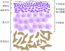
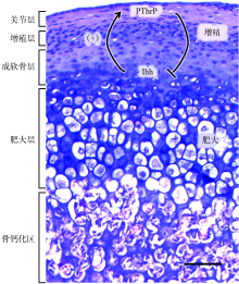
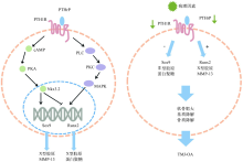
下颌髁突软骨(MCC)不同于生长板和身体其他部位的关节软骨,属于继发性纤维软骨,是颞下颌关节的重要组成部分。由于下颌骨附有牙齿行使咀嚼功能的特殊性,MCC在一生中受机械刺激影响不断改建,随年龄增长可能发生退行性变。甲状旁腺激素相关蛋白(PTHrP)最早在恶性体液性高钙血症中发现,在许多组织中以旁分泌或者自分泌的方式发挥调节骨代谢的作用,在MCC的发育、改建和退行性变中具有重要意义。本文就PTHrP在MCC中的作用进行阐述,为PTHrP在正畸功能矫形和颞下颌关节修复再生提供思路和研究方向。
中图分类号:
| [1] |
Lanske B, Karaplis AC, Lee K, et al. PTH/PTHrP receptor in early development and Indian hedgehog-re-gulated bone growth[J]. Science, 1996, 273(5275):663-666.
pmid: 8662561 |
| [2] |
Kronenberg HM. PTHrP and skeletal development[J]. Ann N Y Acad Sci, 2006, 1068:1-13.
doi: 10.1196/annals.1346.002 |
| [3] |
Stocum DL, Roberts WE. PartⅠ: development and physiology of the temporomandibular joint[J]. Curr Osteoporos Rep, 2018, 16(4):360-368.
doi: 10.1007/s11914-018-0447-7 pmid: 29948821 |
| [4] |
Nickel JC, Iwasaki LR, Gonzalez YM, et al. Mechanobehavior and ontogenesis of the temporomandibular joint[J]. J Dent Res, 2018, 97(11):1185-1192.
doi: 10.1177/0022034518786469 pmid: 30004817 |
| [5] |
Hirouchi H, Kitamura K, Yamamoto M, et al. Developmental characteristics of secondary cartilage in the mandibular condyle and sphenoid bone in mice[J]. Arch Oral Biol, 2018, 89:84-92.
doi: 10.1016/j.archoralbio.2017.12.027 |
| [6] |
Tamimi D, Kocasarac HD, Mardini S. Imaging of the temporomandibular joint[J]. Semin Roentgenol, 2019, 54(3):282-301.
doi: S0037-198X(19)30017-3 pmid: 31376868 |
| [7] |
Hinton RJ, Jing Y, Jing J, et al. Roles of chondrocytes in endochondral bone formation and fracture repair[J]. J Dent Res, 2017, 96(1):23-30.
doi: 10.1177/0022034516668321 pmid: 27664203 |
| [8] |
Allas L, Boumédiene K, Baugé C. Epigenetic dynamic during endochondral ossification and articular cartilage development[J]. Bone, 2019, 120:523-532.
doi: 10.1016/j.bone.2018.10.004 |
| [9] | Bechtold TE, Kurio N, Nah HD, et al. The roles of Indian hedgehog signaling in TMJ formation[J]. Int J Mol Sci, 2019, 20(24):E6300. |
| [10] | Yamazaki K, Suda N, Kuroda T. Distribution of parathyroid hormone-related protein (PTHrP) and typeⅠparathyroid hormone (PTH) PTHrP receptor in developing mouse mandibular condylar cartilage[J]. Ar-ch Oral Biol, 1999, 44(10):853-860. |
| [11] |
Chung UI, Lanske B, Lee K, et al. The parathyroid hormone/parathyroid hormone-related peptide receptor coordinates endochondral bone development by directly controlling chondrocyte differentiation[J]. Proc Natl Acad Sci U S A, 1998, 95(22):13030-13035.
pmid: 9789035 |
| [12] |
Jobert AS, Zhang P, Couvineau A, et al. Absence of functional receptors for parathyroid hormone and parathyroid hormone-related peptide in Blomstrand chondrodysplasia[J]. J Clin Invest, 1998, 102(1):34-40.
pmid: 9649554 |
| [13] |
Karaplis AC, Luz A, Glowacki J, et al. Lethal skeletal dysplasia from targeted disruption of the parathyroid hormone-related peptide gene[J]. Genes Dev, 1994, 8(3):277-289.
doi: 10.1101/gad.8.3.277 |
| [14] |
Tsutsui TW, Riminucci M, Holmbeck K, et al. Deve-lopment of craniofacial structures in transgenic mice with constitutively active PTH/PTHrP receptor[J]. Bone, 2008, 42(2):321-331.
pmid: 18063434 |
| [15] |
Weir EC, Philbrick WM, Amling M, et al. Targeted overexpression of parathyroid hormone-related peptide in chondrocytes causes chondrodysplasia and delayed endochondral bone formation[J]. Proc Natl Acad Sci U S A, 1996, 93(19):10240-10245.
pmid: 8816783 |
| [16] | 张杰, 戴娟, 王美青, 等. 印第安刺猬蛋白信号通路与颞下颌关节改建间的关系[J]. 国际口腔医学杂志, 2013, 40(1):47-50. |
| Zhang J, Dai J, Wang MQ, et al. Indian hedgehog signaling pathway and remodeling of temporoman-dibular joint[J]. Int J Stomatol, 2013, 40(1):47-50. | |
| [17] |
Martin TJ. Parathyroid hormone-related protein, its regulation of cartilage and bone development, and role in treating bone diseases[J]. Physiol Rev, 2016, 96(3):831-871.
doi: 10.1152/physrev.00031.2015 pmid: 27142453 |
| [18] |
Wang L, Shao YY, Ballock RT. Thyroid hormone-mediated growth and differentiation of growth plate chondrocytes involves IGF-1 modulation of beta-ca-tenin signaling[J]. J Bone Miner Res, 2010, 25(5):1138-1146.
doi: 10.1002/jbmr.5 pmid: 20200966 |
| [19] |
Kurio N, Saunders C, Bechtold TE, et al. Roles of Ihh signaling in chondroprogenitor function in postnatal condylar cartilage[J]. Matrix Biol, 2018, 67:15-31.
doi: 10.1016/j.matbio.2018.02.011 |
| [20] | Singh PNP, Shea CA, Sonker SK, et al. Precise spatial restriction of BMP signaling in developing joints is perturbed upon loss of embryo movement[J]. Development, 2018, 145(5): dev153460. |
| [21] |
Chandrasekaran P, Doyran B, Li Q, et al. Biomechanical properties of murine TMJ articular disc and condyle cartilage via AFM-nanoindentation[J]. J Biomech, 2017, 60:134-141.
doi: S0021-9290(17)30331-7 pmid: 28688538 |
| [22] |
Chijimatsu R, Saito T. Mechanisms of synovial joint and articular cartilage development[J]. Cell Mol Life Sci, 2019, 76(20):3939-3952.
doi: 10.1007/s00018-019-03191-5 pmid: 31201464 |
| [23] |
Camirand A, Goltzman D, Gupta A, et al. The role of parathyroid hormone-related protein (PTHrP) in osteoblast response to microgravity: mechanistic implications for osteoporosis development[J]. PLoS One, 2016, 11(7):e0160034.
doi: 10.1371/journal.pone.0160034 |
| [24] |
Huang LJ, Cai XY, Li H, et al. The effects of static pressure on chondrogenic and osteogenic differentiation in condylar chondrocytes from temporomandibular joint[J]. Arch Oral Biol, 2015, 60(4):622-630.
doi: 10.1016/j.archoralbio.2015.01.003 |
| [25] |
Li H, Huang LJ, Xie QY, et al. Study on the effects of gradient mechanical pressures on the proliferation, apoptosis, chondrogenesis and hypertrophy of mandibular condylar chondrocytes in vitro[J]. Arch Oral Biol, 2017, 73:186-192.
doi: 10.1016/j.archoralbio.2016.10.017 |
| [26] |
Jahan E, Matsumoto A, Rafiq AM, et al. Fetal jaw movement affects Ihh signaling in mandibular condylar cartilage development: the possible role of Ihh as mechanotransduction mediator[J]. Arch Oral Biol, 2014, 59(10):1108-1118.
doi: 10.1016/j.archoralbio.2014.06.009 |
| [27] |
Kaul R, O’Brien MH, Dutra E, et al. The effect of altered loading on mandibular condylar cartilage[J]. PLoS One, 2016, 11(7):e0160121.
doi: 10.1371/journal.pone.0160121 |
| [28] |
Li N, Hu P, Wang Y, et al. Tissue interactions are indispensable for cavity formation and disc separation in the temporomandibular joint[J]. Connect Tissue Res, 2021, 62(4):351-358.
doi: 10.1080/03008207.2019.1709452 |
| [29] | Shi Z, Lv J, Xiaoyu L, et al. Condylar degradation from decreased occlusal loading following masticatory muscle atrophy[J]. Biomed Res Int, 2018, 2018:6947612. |
| [30] |
Zhao ZH. A review of the effectiveness and long-term stability of the functional appliance[J]. Chin J Stomatol, 2018, 53(9):590-593.
doi: 10.3760/cma.j.issn.1002-0098.2018.09.004 pmid: 30196617 |
| [31] |
Rabie AB, Tang GH, Xiong H, et al. PTHrP regulates chondrocyte maturation in condylar cartilage[J]. J Dent Res, 2003, 82(8):627-631.
pmid: 12885848 |
| [32] |
Ng AF, Yang YO, Wong RW, et al. Factors regulating condylar cartilage growth under repeated load application[J]. Front Biosci, 2006, 11:949-954.
doi: 10.2741/1851 |
| [33] |
Chen Y, Zhao BJ, Zhu Y, et al. HIF-1-VEGF-Notch mediates angiogenesis in temporomandibular joint osteoarthritis[J]. Am J Transl Res, 2019, 11(5):2969-2982.
pmid: 31217867 |
| [34] |
Cui SJ, Zhang T, Fu Y, et al. DPSCs attenuate experimental progressive TMJ arthritis by inhibiting the STAT1 pathway[J]. J Dent Res, 2020, 99(4):446-455.
doi: 10.1177/0022034520901710 pmid: 31977264 |
| [35] |
Long HQ, Tian PF, Guan YX, et al. Expression of Ihh signaling pathway in condylar cartilage after bite-raising in adult rats[J]. J Mol Histol, 2019, 50(5):459-470.
doi: 10.1007/s10735-019-09840-0 |
| [36] |
Zhou YC, Shu B, Xie R, et al. Deletion of Axin1 in condylar chondrocytes leads to osteoarthritis-like phenotype in temporomandibular joint via activation of β-catenin and FGF signaling[J]. J Cell Physiol, 2019, 234(2):1720-1729.
doi: 10.1002/jcp.v234.2 |
| [37] |
Yuan XL, Liu HQ, Li LF, et al. The roles of endoplasmic reticulum stress in the pathophysiological development of cartilage and chondrocytes[J]. Curr Pharm Des, 2017, 23(11):1693-1704.
doi: 10.2174/1381612822666161025152423 |
| [38] |
Deng ZN, Liu Y, Wang CP, et al. Involvement of PI3K/Akt pathway in rat condylar chondrocytes regulated by PTHrP treatment[J]. Arch Oral Biol, 2014, 59(10):1032-1041.
doi: 10.1016/j.archoralbio.2014.04.012 |
| [39] |
Roberts WE, Stocum DL. Part Ⅱ: temporomandibular joint (TMJ)-regeneration, degeneration, and adaptation[J]. Curr Osteoporos Rep, 2018, 16(4):369-379.
doi: 10.1007/s11914-018-0462-8 pmid: 29943316 |
| [40] | Yang HX, Zhang M, Liu Q, et al. Inhibition of ihh reverses temporomandibular joint osteoarthritis via a PTH1R signaling dependent mechanism[J]. Int J Mol Sci, 2019, 20(15):E3797. |
| [41] |
Sobue T, Yeh WC, Chhibber A, et al. Murine TMJ loading causes increased proliferation and chondrocyte maturation[J]. J Dent Res, 2011, 90(4):512-516.
doi: 10.1177/0022034510390810 pmid: 21248355 |
| [42] |
Wang S, Deng L, Li Y, et al. Differential gene expression in the condylar cartilage of growing rabbits with temporomandibular joint anterior disk displacement-a transcriptomic study[J]. Arch Oral Biol, 2017, 74:92-100.
doi: 10.1016/j.archoralbio.2016.11.006 |
| [43] |
Chen J, Gupta T, Barasz JA, et al. Analysis of microarchitectural changes in a mouse temporomandibular joint osteoarthritis model[J]. Arch Oral Biol, 2009, 54(12):1091-1098.
doi: 10.1016/j.archoralbio.2009.10.001 pmid: 19896116 |
| [44] | 郭敏, 张婧, 鹿蕾, 等. 实验性单侧前牙反𬌗修复体对大鼠髁突软骨中甲状旁腺激素相关蛋白和PTH/PTHrP受体-1表达的影响[J]. 华西口腔医学杂志, 2013,31(2):122-126. |
| Guo M, Zhang J, Lu L, et al. Effects of experimen-tally created unilateral anterior crossbite prosthesis on the expression of parathyroid hormone-related peptide and parathyroid hormone receptor-1 in man-dibular condylar cartilage of rat[J]. West China J Stomatol, 2013,31(2):122-126. | |
| [45] | Chen S, Fu PL, Cong RJ, et al. Strategies to minimize hypertrophy in cartilage engineering and regeneration[J]. Genes Dis, 2015, 2(1):76-95. |
| [46] |
Fischer J, Ortel M, Hagmann S, et al. Role of PTHrP(1-34) pulse frequency versus pulse duration to enhance mesenchymal stromal cell chondrogenesis[J]. J Cell Physiol, 2016, 231(12):2673-2681.
doi: 10.1002/jcp.25369 |
| [47] |
Fahy N, Gardner OFW, Alini M, et al. Parathyroid hormone-related protein gradients affect the progression of mesenchymal stem cell chondrogenesis and hypertrophy[J]. Tissue Eng Part A, 2018, 24(9/10):849-859.
doi: 10.1089/ten.tea.2017.0337 |
| [48] |
Fischer J, Aulmann A, Dexheimer V, et al. Intermittent PTHrP(1-34) exposure augments chondrogenesis and reduces hypertrophy of mesenchymal stromal cells[J]. Stem Cells Dev, 2014, 23(20):2513-2523.
doi: 10.1089/scd.2014.0101 |
| [1] | 李静雅,税钰森,郭永文. 循环牵张应力影响人牙周膜细胞成骨分化机制的研究进展[J]. 国际口腔医学杂志, 2020, 47(6): 652-660. |
| [2] | 陈潇 闫宝勇 左艳萍 盛海莹 苑学微. 整合素β1在大鼠髁突软骨力学适应性改建过程中的表达[J]. 国际口腔医学杂志, 2014, 41(4): 383-386. |
| [3] | 黄男安,李娟,赵志河. 机械力刺激对成骨细胞的作用[J]. 国际口腔医学杂志, 2008, 35(S1): -. |
| [4] | 包广洁1综述 康宏2,Kyriacos A. Athanasiou3 审校. 颞下颌关节盘的结构表征与组织工程研究[J]. 国际口腔医学杂志, 2008, 35(3): 307-307~310. |
| [5] | 钟孝欢,黄生高,. 甲状旁腺激素相关蛋白和机械力对成骨细胞增殖分化的影响[J]. 国际口腔医学杂志, 2006, 33(06): 442-444. |
|