国际口腔医学杂志 ›› 2019, Vol. 46 ›› Issue (6): 687-692.doi: 10.7518/gjkq.2019080
王蕊,盖阔,刘梦齐,蒋丽( )
)
Wang Rui,Gai Kuo,Liu Mengqi,Jiang Li()
摘要:
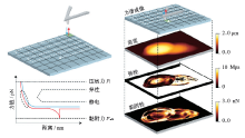
原子力显微镜作为重要的表面成像及力学探测工具,凭借其纳米级(nm)时空分辨率、皮牛级(pN)力灵敏度、液相环境等优势在微生物形貌力学探测中发挥了重要作用。本文简述了基于原子力显微镜的多种成像模式、力谱技术及二者相结合应用的最新进展,并综述了近年来原子力显微镜在细菌与基底、细菌与细菌及细菌与细菌生物膜的黏附力学特性研究方面的应用,为细菌黏附力学的深入研究提供了新思路。
中图分类号:
| [1] | Zhang S, Aslan H, Besenbacher F , et al. Quantitative biomolecular imaging by dynamic nanomechanical mapping[J]. Chem Soc Rev, 2014,43(21):7412-7429. |
| [2] | Garcia R, Herruzo ET . The emergence of multifre-quency force microscopy[J]. Nat Nanotechnol, 2012,7(4):217-226. |
| [3] | Fantner GE, Barbero RJ, Gray DS , et al. Kinetics of antimicrobial peptide activity measured on individual bacterial cells using high-speed atomic force micros-copy[J]. Nat Nanotechnol, 2010,5(4):280-285. |
| [4] | Benning FMC, Sakiyama Y, Mazur A , et al. High-speed atomic force microscopy visualization of the dynamics of the multienzyme fatty acid synthase[J]. ACS Nano, 2017,11(11):10852-10859. |
| [5] | Abu-Lail NI, Camesano TA . Specific and nonspecific interaction forces between Escherichia coli and silicon nitride, determined by poisson statistical analysis[J]. Langmuir, 22(17):7296-7301. |
| [6] | Eskhan AO, Abu-Lail NI . A new approach to de-coupling of bacterial adhesion energies measured by AFM into specific and nonspecific components[J]. Colloid Polym Sci, 2014,292(2):343-353. |
| [7] | El-Kirat-Chatel S, Puymege A, Duong TH , et al. Phenotypic heterogeneity in attachment of marine bacteria toward antifouling copolymers unraveled by AFM[J]. Front Microbiol, 2017,8:1399. |
| [8] | Rosenthal K, Oehling V, Dusny C , et al. Beyond the bulk: disclosing the life of single microbial cells[J]. FEMS Microbiol Rev, 2017,41(6):751-780. |
| [9] | Beaussart A, El-Kirat-Chatel S, Herman P, et al. Sin-gle-cell force spectroscopy of probiotic bacteria[J]. Biophys J, 2013,104(9):1886-1892. |
| [10] | El-Kirat-Chatel S, Beaussart A . Probing bacterial adhesion at the single-molecule and single-cell levels by AFM-based force spectroscopy[J]. Methods Mol Biol, 2018,1814:403-414. |
| [11] | De Keersmaecker H, Frederickx W, Fujita Y , et al. Correlative atomic force and single-molecule fluore-scence microscopy of nucleoprotein complexes[J]. Methods Mol Biol, 2018,1814:339-359. |
| [12] | Dufrêne YF, Martínez-Martín D, Medalsy I , et al. Multiparametric imaging of biological systems by force-distance curve-based AFM[J]. Nat Methods, 2013,10(9):847-854. |
| [13] | Koehler M, Macher G, Rupprecht A , et al. Combined recognition imaging and force spectroscopy: a new mode for mapping and studying interaction sites at low lateral density[J]. Sci Adv Mater, 2017,9(1):128-134. |
| [14] | Mei L, Ren Y, Busscher HJ , et al. Poisson analysis of streptococcal bond-strengthening on saliva-coated enamel[J]. J Dent Res, 2009,88(9):841-845. |
| [15] | Sullan RM, Li JK, Crowley PJ , et al. Binding forces of Streptococcus mutans P1 adhesin[J]. ACS Nano, 2015,9(2):1448-1460. |
| [16] | Heim KP, Sullan RM, Crowley PJ , et al. Identification of a supramolecular functional architecture of Stre-ptococcus mutans adhesin P1 on the bacterial cell surface[J]. J Biol Chem, 2015,290(14):9002-9019. |
| [17] | Cross SE, Kreth J, Zhu L , et al. Nanomechanical properties of glucans and associated cell-surface adhesion of Streptococcus mutans probed by atomic force microscopy under in situ conditions[J]. Micro-biology, 2007,153(Pt 9):3124-3132. |
| [18] | Bank TL, Dosen A, Giese RF , et al. Atomic force spectroscopy evidence of non-specific adhesion of Aggregatibacter actinomycetemcomitans[J]. J Nanosci Nanotechnol, 2011,11(10):8450-8456. |
| [19] | Mei L, Busscher HJ, van der Mei HC, et al. Influence of surface roughness on streptococcal adhesion forces to composite resins[J]. Dent Mater, 2011,27(8):770-778. |
| [20] | Yu P, Wang C, Zhou J , et al. Influence of surface properties on adhesion forces and attachment of Stre-ptococcus mutans to zirconia in vitro[J]. Biomed Res Int, 2016,2016:8901253. |
| [21] | Verran J, Jackson S, Coulthwaite L , et al. The effect of dentifrice abrasion on denture topography and the subsequent retention of microorganisms on abraded surfaces[J]. J Prosthet Dent, 2014,112(6):1513-1522. |
| [22] | Wang C, Zhao Y, Zheng S , et al. Effect of enamel morphology on nanoscale adhesion forces of strepto-coccal bacteria: an AFM study[J]. Scanning, 2015,37(5):313-321. |
| [23] | Mei L, van der Mei HC, Ren Y , et al. Poisson analysis of streptococcal bond strengthening on stainless steel with and without a salivary conditioning film[J]. Langmuir, 2009,25(11):6227-6231. |
| [24] | Fang J, Wang C, Li Y , et al. Comparison of bacterial adhesion to dental materials of polyethylene tereph-thalate (PET) and polymethyl methacrylate (PMMA) using atomic force microscopy and scanning electron microscopy[J]. Scanning, 2016,38(6):665-670. |
| [25] | 蒋丽, 王传勇, 薛晶, 等 . 钛及氧化锆种植材料表面微观形貌对龈下细菌黏附力的影响[C]. 第八届全国口腔材料学术交流会, 上海, 2013-8-17. |
| Jiang L, Wang CY, Xue J , et al. Influence of surface microstucture on subginvial bacterial adhesion force to titanium and zirconia implant materials[C]. The 8th National Symposium on Dental Metarials,Shan-ghai, 2013-8-17. | |
| [26] | Feuillie C, Formosa-Dague C, Hays LM , et al. Mole-cular interactions and inhibition of the staphylococcal biofilm-forming protein SdrC[J]. Proc Natl Acad Sci USA, 2017,114(14):3738-3743. |
| [27] | Postollec F, Norde W, de Vries J, et al. Interactive forces between co-aggregating and non-co-aggre-gating oral bacterial pairs[J]. J Dent Res, 2006,85(3):231-234. |
| [28] | Hwang G, Marsh G, Gao L , et al. Binding force dynamics of Streptococcus mutans-glucosyltrans-ferase B to Candida albicans[J]. J Dent Res, 2015,94(9):1310-1317. |
| [29] | Hwang G, Liu Y, Kim D , et al. Candida albicans mannans mediate Streptococcus mutans exoenzyme GtfB binding to modulate cross-kingdom biofilm development in vivo[J]. PLoS Pathog, 2017,13(6):e1006407. |
| [30] | Prystopiuk V, Feuillie C, Herman-Bausier P , et al. Mechanical forces guiding Staphylococcus aureus cellular invasion[J]. ACS Nano, 2018,12(4):3609-3622. |
| [31] | Becke TD, Ness S, Gürster R , et al. Single molecule force spectroscopy reveals two-domain binding mode of Pilus-1 Tip protein RrgA of Streptococcus pneu-monia to fibronectin[J]. ACS Nano, 2018,12(1):549-558. |
| [32] | Alsteens D, Trabelsi H, Soumillion P , et al. Multipara- metric atomic force microscopy imaging of single bacteriophages extruding from living bacteria[J]. Nat Commun, 2013,4:2926. |
| [33] | Formosa-Dague C, Speziale P, Foster TJ , et al. Zinc-dependent mechanical properties of Staphylococcus aureus biofilm-forming surface protein SasG[J]. Proc Natl Acad Sci USA, 2016,113(2):410-415. |
| [34] | Dufrêne YF, Ando T, Garcia R , et al. Imaging modes of atomic force microscopy for application in mole-cular and cell biology[J]. Nat Nanotechnol, 2017,12(4):295-307. |
| [35] | Liu BH, Yu LC . In-situ, time-lapse study of extracel-lular polymeric substance discharge in Streptococcus mutans biofilm[J]. Colloids Surf B Biointerfaces, 2017,150:98-105. |
| [36] | Dupres V, Menozzi FD, Locht C , et al. Nanoscale mapping and functional analysis of individual ad-hesins on living bacteria[J]. Nat Methods, 2005,2(7):515-520. |
| [37] | El-Kirat-Chatel S, Beaussart A, Boyd CD , et al. Single-cell and single-molecule analysis deciphers the loca-lization, adhesion, and mechanics of the biofilm ad-hesin LapA[J]. ACS Chem Biol, 2014,9(2):485-494. |
| [38] | Beaussart A, Péchoux C, Trieu-Cuot P , et al. Mole-cular mapping of the cell wall polysaccharides of the human pathogen Streptococcus agalactiae[J]. Nano-scale, 2014,6(24):14820-14827. |
| [1] | 冯旭,张祎,李梦红,刘楠,王六一,胡敏. 无托槽隐形矫治对牙周健康影响的研究进展[J]. 国际口腔医学杂志, 2019, 46(2): 166-170. |
| [2] | 刘梦齐,盖阔,蒋丽. 抗菌性口腔种植材料的研究进展[J]. 国际口腔医学杂志, 2018, 45(5): 516-521. |
| [3] | 盖阔, 郝丽英, 蒋丽. 应用原子力显微镜对口腔变异链球菌黏附机制的研究[J]. 国际口腔医学杂志, 2017, 44(3): 320-324. |
| [4] | 郑赛男,蒋丽,李伟. 口腔细菌黏附机制的研究进展[J]. 国际口腔医学杂志, 2016, 43(2): 223-227. |
| [5] | 兰静 王传勇 薛晶 蒋丽 李伟. 含纳米羟磷灰石的过氧化氢漂白剂对釉质表面的影响[J]. 国际口腔医学杂志, 2013, 40(4): 432-435. |
| [6] | 苏伟珠1 王浙君1 撒悦1综述 王贻宁1,2审校. 漂白剂对复合树脂的影响[J]. 国际口腔医学杂志, 2012, 39(4): 537-539. |
| [7] | 余志芬综述 张向宇审校 . 细菌黏附的研究进展[J]. 国际口腔医学杂志, 2009, 36(4): 448-450. |
| [8] | 靖军军综述 郝玉庆审校. 壳聚糖及其衍生物对致龋细菌的作用[J]. 国际口腔医学杂志, 2009, 36(3): 291-293. |
| [9] | 邱瑜蕾,包崇云. 生物材料表面细菌生物膜形成与理化特征[J]. 国际口腔医学杂志, 2008, 35(S1): -. |
| [10] | 何海波,唐休发. 肿瘤细胞生物力学的研究进展[J]. 国际口腔医学杂志, 2004, 31(04): 273-275. |
| [11] | 邱文彦,Richard R.Mu. 原子力显微镜在细菌学研究中的应用[J]. 国际口腔医学杂志, 2002, 29(05): -. |
|