国际口腔医学杂志 ›› 2019, Vol. 46 ›› Issue (2): 149-155.doi: 10.7518/gjkq.2019018
黄婕,林云红( )
)
Jie Huang,Yunhong Lin()
摘要:
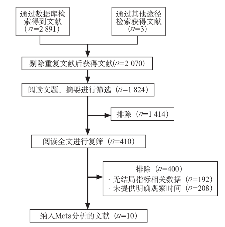
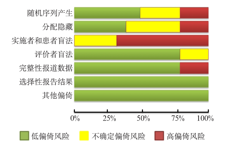
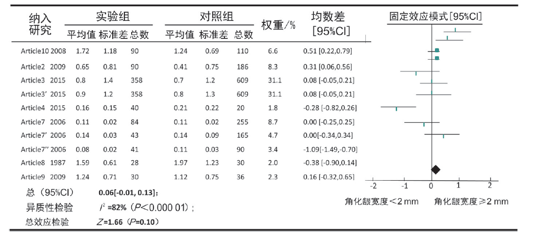
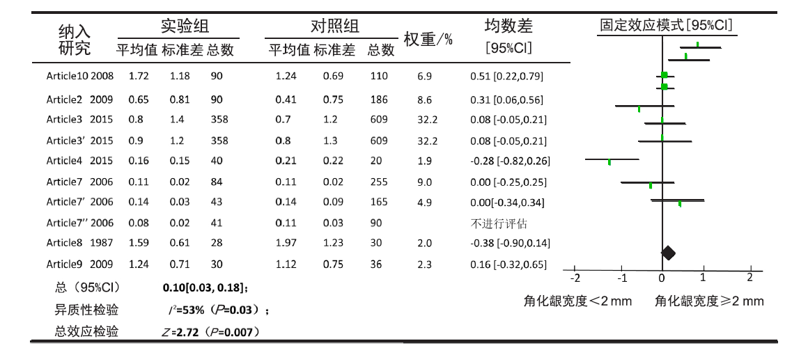
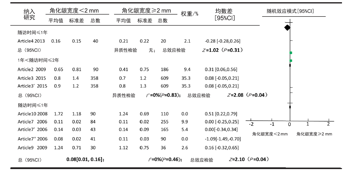
目的 口腔种植体袖口周围角化龈的存在被认为是种植体周围的一个黏膜屏障,角化龈的存在无论从生物学角度还是美学立场都确保了种植体周围组织长期的稳定。本次研究运用了Meta分析的方法,通过对种植修复牙体缺失患者临床对照试验进行总结,评估了口腔种植体周围角化龈宽度与种植体周围骨丧失量的相关性。方法 电子检索EMBASE(Ovid)、Cochrane对照试验中心注册库(CENTRAL)、Medline、Pubmed、中国生物医学文献数据库(CBMdisc)和中国知识基础设施工程(CNKI),手动检索国家医学图书馆(NLM)、坎贝尔图书馆等。检索日期截止至2017年5月,检索要求无发表语言以及发表时间的限制。3位评价者独立使用Cochrane质量评价表评价纳入研究的方法学质量,按照纳入、排除标准筛选相关研究,提取数据并进行风险偏倚评价。采用Revman 5.3软件进行Meta分析。结果 在最终纳入的10项研究中,有8项为回顾性研究,另外2项为前瞻性研究,这10篇文献均为中等偏倚风险。其主要研究内容为口腔种植体周围的角化龈宽度与种植体周围骨丧失量的相关性。以“骨丧失量、骨丧失高度、边缘骨丧失量、随访时间均大于2个月”为结局指标,Meta分析的结果显示:排除有明显异质性研究之后,种植体周围角化龈的宽度与骨吸收无负性相关性(P<0.01),该结果稳定并且证据质量较高;随着角化龈宽度的增加,骨吸收量反而逐渐减少,即角化龈对种植体周围骨组织有保护作用,但是对种植体周围骨高度的影响因素很多,例如:自身免疫力的强弱、饮食习惯、依从性等等,则需要更多高质量的对照试验进一步的研究来证实。结论 种植体周围角化龈的宽度对种植体周围骨丧失程度的影响无负性相关性,因此,在今后的临床工作中,应当尽量保存角化龈,才能更大程度地获得植体周围骨稳定性。
中图分类号:
| [1] | Albrektsson T, Zarb G, Worthington P , et al. The Long-term efficacy of currently used dental implants: a review and proposed criteria of success[J]. Int J Oral Maxillofac Implants, 1986,1(1):11-25. |
| [2] |
Albrektsson T, Buser D, Sennerby L . On crestal/mar-ginal bone loss around dental implants[J]. Int J Oral Maxillofac Implants, 2012,27(4):736-738.
doi: 10.1038/sj.bdj.2012.611 pmid: 23342341 |
| [3] |
Misch CE, Perel ML, Wang HL , et al. Implant success, survival, and failure: the international congress of oral implantologists (ICOI) pisa consensus conference[J]. Implant Dent, 2008,17(1):5-15.
doi: 10.1097/ID.0b013e3181676059 pmid: 18332753 |
| [4] |
Papaspyridakos P, Chen CJ, Singh M , et al. Success criteria in implant dentistry: a systematic review[J]. J Dent Res, 2012,91(3):242-248.
doi: 10.1177/0022034511431252 |
| [5] |
Annibali S, Bignozzi I, La Monaca G , et al. Usefulness of the aesthetic result as a success criterion for im-plant therapy: a review[J]. Clin Implant Dent Relat Res, 2012,14(1):3-40.
doi: 10.1111/j.1708-8208.2009.00234.x pmid: 19673953 |
| [6] |
Fürhauser R, Florescu D, Benesch T , et al. Evaluation of soft tissue around single-tooth implant crowns: the pink esthetic score[J]. Clin Oral Implants Res, 2005,16(6):639-644.
doi: 10.1111/j.1600-0501.2005.01193.x pmid: 16307569 |
| [7] |
Sohrabi K, Mushantat A, Esfandiari S , et al. How successful are small-diameter implants? A literature review[J]. Clin Oral Implants Res, 2012,23(5):515-525.
doi: 10.1111/j.1600-0501.2011.02410.x pmid: 22313216 |
| [8] |
Severino VO, Napimoga MH, de Lima Pereira SA . Expression of IL-6, IL-10, IL-17 and IL-8 in the peri-implant crevicular fluid of patients with peri-implan-titis[J]. Arch Oral Biol, 2011,56(8):823-828.
doi: 10.1016/j.archoralbio.2011.01.006 pmid: 21306703 |
| [9] |
Luo L, Xie P, Gong P , et al. Expression of HMGB1 and HMGN2 in gingival tissues, GCF and PICF of periodontitis patients and peri-implantitis[J]. Arch Oral Biol, 2011,56(10):1106-1111.
doi: 10.1590/S1517-838220110003000047 pmid: 3768754 |
| [10] |
Loe H, Theilade E, Jensen SB . Experimental gingivitis in Man[J]. J Periodontol, 1965,36:177-187.
doi: 10.1902/jop.1965.36.3.177 |
| [11] |
Lang NP, Löe H . The relationship between the width of keratinized gingiva and gingival health[J]. J Perio-dontol, 1972,43(10):623-627.
doi: 10.1902/jop.1972.43.10.623 pmid: 4507712 |
| [12] |
Stetler KJ, Bissada NF . Significance of the width of keratinized gingiva on the periodontal status of teeth with submarginal restorations[J]. J Periodontol, 1987,58(10):696-700.
doi: 10.1902/jop.1987.58.10.696 pmid: 2444693 |
| [13] |
Greenstein G, Cavallaro J . The clinical significance of keratinized gingiva around dental implants[J]. Compend Contin Educ Dent, 2011,32(8):24-32, 34.
doi: 10.1111/j.1444-0938.1976.tb01428.x pmid: 22073807 |
| [14] |
Mehta P, Lim LP . The width of the attached gingiva: much ado about nothing[J]. J Dent, 2010,38(7):517-525.
doi: 10.1016/j.jdent.2010.04.007 pmid: 20403409 |
| [15] |
Passoni BB, Dalago HR, Schuldt Filho G , et al. Does the number of implants have any relation with peri-implant disease[J]. J Appl Oral Sci, 2014,22(5):403-408.
doi: 10.1590/1678-775720140055 pmid: 4245752 |
| [16] |
Kim BS, Kim YK, Yun PY , et al. Evaluation of peri-implant tissue response according to the presence of keratinized mucosa[J]. Oral Surg Oral Med Oral Pathol Oral Radiol Endod, 2009,107(3):e24-e28.
doi: 10.1016/j.tripleo.2008.12.010 pmid: 19217009 |
| [17] |
Ladwein C, Schmelzeisen R, Nelson K , et al. Is the presence of keratinized mucosa associated with periimplant tissue health? A clinical cross-sectional analysis[J]. Int J Implant Dent, 2015,1(1):11.
doi: 10.1186/s40729-015-0009-z pmid: 5005560 |
| [18] |
Buyukozdemir Askin S, Berker E, Akincibay H , et al. Necessity of keratinized tissues for dental implants: a clinical, immunological, and radiographic study[J]. Clin Implant Dent Relat Res, 2015,17(1):1-12.
doi: 10.1111/cid.12079 pmid: 23631746 |
| [19] |
Ferreira CF, Buttendorf AR, de Souza JG, et al. Pre-valence of peri-implant diseases: analyses of associated factors[J]. Eur J Prosthodont Restor Dent, 2015,23(4):199-206.
doi: 10.1992/EJPRD_ pmid: 26767242 |
| [20] |
Poli PP, Beretta M, Grossi GB , et al. Risk indicators related to peri-implant disease: an observational re-trospective cohort study[J]. J Periodont Implant Sci, 2016,46(4):266-276.
doi: 10.5051/jpis.2016.46.4.266 pmid: 27588216 |
| [21] |
Chung DM, Oh TJ, Shotwell JL , et al. Significance of keratinized mucosa in maintenance of dental im-plants with different surfaces[J]. J Periodontol, 2006,77(8):1410-1420.
doi: 10.1902/jop.2006.050393 pmid: 16881810 |
| [22] |
Adibrad M, Shahabuei M, Sahabi M . Significance of the width of keratinized mucosa on the health status of the supporting tissue around implants supporting overdentures[J]. J Oral Implantol, 2009,35(5):232-237.
doi: 10.1563/AAID-JOI-D-09-00035.1 pmid: 19882819 |
| [23] |
Bouri A Jr, Bissada N, Al-Zahrani MS , et al. Width of keratinized gingiva and the health status of the supporting tissues around dental implants[J]. Int J Oral Maxillofac Implants, 2008,23(2):323-326.
doi: 10.1016/j.ijom.2007.09.170 pmid: 18548930 |
| [24] |
Leonhardt A, Renvert S, Dahlén G . Microbial findings at failing implants[J]. Clin Oral Implants Res, 1999,10(5):339-345.
doi: 10.1034/j.1600-0501.1999.100501.x pmid: 10551058 |
| [25] |
Quirynen M, Vogels R, Peeters W , et al. Dynamics of initial subgingival colonization of ‘pristine’ peri-implant pockets[J]. Clin Oral Implants Res, 2006,17(1):25-37.
doi: 10.1111/j.1600-0501.2005.01194.x pmid: 16441782 |
| [26] |
Augthun M, Conrads G . Microbial findings of deep peri-implant bone defects[J]. Int J Oral Maxillofac Implants, 1997,12(1):106-112.
pmid: 9048462 |
| [27] |
Salcetti JM, Moriarty JD, Cooper LF , et al. The clinical, microbial, and host response characteristics of the failing implant[J]. Int J Oral Maxillofac Implants, 1997,12(1):32-42.
doi: 10.1111/j.1365-2591.1997.tb01101.x pmid: 9048452 |
| [28] |
Abrahamsson I, Berglundh T, Wennström J , et al. The peri-implant hard and soft tissues at different implant systems. A comparative study in the dog[J]. Clin Oral Implants Res, 1996,7(3):212-219.
doi: 10.1034/j.1600-0501.1996.070303.x pmid: 9151585 |
| [29] |
Berglundh T, Lindhe J, Marinello C , et al. Soft tissue reaction to de novo plaque formation on implants and teeth. An experimental study in the dog[J]. Clin Oral Implants Res, 1992,3(1):1-8.
doi: 10.1034/j.1600-0501.1992.030101.x |
| [30] |
Ericsson I, Berglundh T, Marinello C , et al. Long-standing plaque and gingivitis at implants and teeth in the dog[J]. Clin Oral Implants Res, 1992,3(3):99-103.
doi: 10.1034/j.1600-0501.1992.030301.x pmid: 1290796 |
| [31] |
Toijanic JA, Ward CB, Gewerth ME , et al. A longitu-dinal clinical comparison of plaque-induced inflam-mation between gingival and peri-implant soft tissues in the maxilla[J]. J Periodontol, 2001,72(9):1139-1145.
doi: 10.1902/jop.2000.72.9.1139 pmid: 11577943 |
| [1] | 龚佳明,赵瑞敏,潘宏伟,郎鑫,余占海,李健学. 动态导航与静态导航对种植体准确性的Meta分析[J]. 国际口腔医学杂志, 2023, 50(5): 538-551. |
| [2] | 李转转,格根塔娜. 牙髓血运重建术和根尖诱导成形术疗效对比的Meta分析[J]. 国际口腔医学杂志, 2023, 50(2): 177-185. |
| [3] | 龚佳明,赵瑞敏,李婉昕,苏琳涵,余占海,李健学. 根盾技术对即刻种植临床效果的影响:基于随机对照研究的Meta分析[J]. 国际口腔医学杂志, 2022, 49(5): 537-547. |
| [4] | 张珊,葛晓磊,李杰,谢新宇,常维维,马文盛. 上颌前方牵引矫治对颌骨生长发育长期影响的Meta分析[J]. 国际口腔医学杂志, 2022, 49(5): 548-555. |
| [5] | 马玉,左玉,张鑫. 光动力疗法辅助治疗牙周炎治疗效果的Meta分析[J]. 国际口腔医学杂志, 2022, 49(3): 305-316. |
| [6] | 周万航,李嫣斐,许日聪,万启军. 牙周非手术治疗对慢性肾脏病危险因素及全身炎症水平影响的Meta分析[J]. 国际口腔医学杂志, 2021, 48(5): 528-535. |
| [7] | 秦小茹,刘梦圆. 牙周病和心肌梗死发生风险相关性队列研究的Meta分析[J]. 国际口腔医学杂志, 2021, 48(2): 165-172. |
| [8] | 刘玲,龚仁国,董秀华,刘入梦. 正畸联合双颌手术治疗前牙区严重骨性开长期稳定性的Meta分析[J]. 国际口腔医学杂志, 2021, 48(2): 173-179. |
| [9] | 汪是琦,常雅琴,陈斌,谭葆春,泥艳红. 植骨术与植骨联用屏障膜在牙周再生治疗中临床疗效对比的系统评价与Meta分析[J]. 国际口腔医学杂志, 2020, 47(6): 644-651. |
| [10] | 侯亚丽,马利. 亚洲人群干扰素调节因子6基因多态性与非综合征型唇腭裂相关性研究的Meta分析[J]. 国际口腔医学杂志, 2020, 47(4): 397-405. |
| [11] | 高洁,马锐,葛振林. 热激活镍钛弓丝矫治效率的系统评价[J]. 国际口腔医学杂志, 2019, 46(4): 393-399. |
| [12] | 潘韦霖,曹钰彬,刘畅,刘济远,李春洁,潘剑,华成舸. 不同翻瓣设计对下颌第三磨牙拔除术后疼痛的影响:系统评价与Meta分析[J]. 国际口腔医学杂志, 2019, 46(2): 142-148. |
| [13] | 黎静, 刘星辰, 李佳园, 李小兵. 稳定咬合板治疗慢性颞下颌关节盘不可复性移位的临床随机对照试验的系统评价[J]. 国际口腔医学杂志, 2017, 44(4): 405-410. |
| [14] | 李唐 李春洁 门乙 杨文宾 韩波 李龙江. Meta分析评价全景片在口腔癌下颌骨侵犯中的诊断效能[J]. 国际口腔医学杂志, 2016, 43(1): 26-. |
| [15] | 杨文宾 李春洁 门乙 韩波 李龙江 潘剑. 保留腮腺咬肌筋膜预防Frey综合征的系统评价与Meta分析[J]. 国际口腔医学杂志, 2013, 40(6): 730-735. |
|