Int J Stomatol ›› 2024, Vol. 51 ›› Issue (6): 713-721.doi: 10.7518/gjkq.2024083
• Original Articles • Previous Articles
Yi Liu1( ),Jiangshan Qiu2,Daonan Shen1,Xin Guan3,Yi Ding1()
CLC Number:
| 1 | Bosshardt DD, Chappuis V, Buser D. Osseointegration of titanium, titanium alloy and zirconia dental implants: current knowledge and open questions[J]. Periodontol 2000, 2017, 73(1): 22-40. |
| 2 | Herrera D, Berglundh T, Schwarz F, et al. Prevention and treatment of peri-implant diseases-the EFP S3 level clinical practice guideline[J]. J Clin Perio-dontol, 2023, 50(): 4-76. |
| 3 | de Bruyn H, Christiaens V, Doornewaard R, et al. Implant surface roughness and patient factors on long-term peri-implant bone loss[J]. Periodontol 2000, 2017, 73(1): 218-227. |
| 4 | Sinjab K, Sawant S, Ou A, et al. Impact of surface characteristics on the peri-implant microbiome in health and disease[J]. J Periodontol, 2024, 95(3): 244-255. |
| 5 | 杨帮成, 周学东, 于海洋, 等. 钛种植体表面改性方法[J]. 华西口腔医学杂志, 2019, 37(2): 124-129. |
| Yang BC, Zhou XD, Yu HY, et al. Advances in titanium dental implant surface modification[J]. West China J Stomatol, 2019, 37(2): 124-129. | |
| 6 | Sotova C, Yanushevich O, Kriheli N, et al. Dental implants: modern materials and methods of their surface modification[J]. Materials, 2023, 16(23): 7383. |
| 7 | Saran R, Ginjupalli K, George SD, et al. LASER as a tool for surface modification of dental biomate-rials: a review[J]. Heliyon, 2023, 9(6): e17457. |
| 8 | Gupta R, Naveena AH, Vadali M, et al. Enhancement of cell attachment on Ti6Al4V via surface modifications using pulsed laser surface melting[J]. Surf Coat Technol, 2023, 474: 130080. |
| 9 | Bayrak M, Kocak-Oztug NA, Gulati K, et al. Inf-luence of clinical decontamination techniques on the surface characteristics of SLA titanium implant[J]. Nanomaterials, 2022, 12(24): 4481. |
| 10 | Lee JS, Son K, Hwang SM, et al. Effect of electrocautery and laser treatment on the composition and morphology of surface-modified titanium implants[J]. Bioengineering, 2023, 10(11): 1251. |
| 11 | Eghbali N, Naffakh-Moosavy H, Sadeghi Mohammadi S, et al. The influence of laser frequency and groove distance on cell adhesion, cell viability, and antibacterial characteristics of Ti-6Al-4V dental implants treated by modern fiber engraving laser[J]. Dent Mater, 2021, 37(3): 547-558. |
| 12 | Yang D, Xu TY, Fan L, et al. MicroRNA-216b enhances cisplatin-induced apoptosis in osteosarcoma MG63 and SaOS-2 cells by binding to JMJD2C and regulating the HIF1α/HES1 signaling axis[J]. J Exp Clin Cancer Res, 2020, 39(1): 201. |
| 13 | Hassani A, Khoshfetrat AB, Rahbarghazi R, et al. Collagen and nano-hydroxyapatite interactions in alginate-based microcapsule provide an appropriate osteogenic microenvironment for modular bone tissue formation[J]. Carbohydr Polym, 2022, 277: 118807. |
| 14 | Shailaja A, Bruce TF, Gerard P, et al. Comparison of cell viability assessment and visualization of Aspergillus niger biofilm with two fluorescent probe staining methods[J]. Biofilm, 2022, 4: 100090. |
| 15 | Parga A, Manoil D, Brundin M, et al. Gram-negative quorum sensing signalling enhances biofilm formation and virulence traits in gram-positive pathogen Enterococcus faecalis [J]. J Oral Microbiol, 2023, 15(1): 2208901. |
| 16 | Haney EF, Trimble MJ, Hancock REW. Microtiter plate assays to assess antibiofilm activity against bacteria[J]. Nat Protoc, 2021, 16(5): 2615-2632. |
| 17 | Kligman S, Ren Z, Chung CH, et al. The impact of dental implant surface modifications on osseointegration and biofilm formation[J]. J Clin Med, 2021, 10(8): 1641. |
| 18 | Xiong SB, Lu XG, Zhang SQ, et al. Osteogenic properties of bioactive titanium in inflammatory environment[J]. Dent Mater, 2023, 39(10): 929-937. |
| 19 | Wang CY, Wang X, Lu R, et al. Influence of surface nanotopography and wettability on early phases of peri-implant soft tissue healing: an in-vivo study in dogs[J]. BMC Oral Health, 2023, 23(1): 651. |
| 20 | Jiang PL, Zhang YM, Hu R, et al. Advanced surface engineering of titanium materials for biomedical applications: from static modification to dynamic responsive regulation[J]. Bioact Mater, 2023, 27: 15-57. |
| 21 | Accioni F, Vázquez J, Merinero M, et al. Latest trends in surface modification for dental implantology: innovative developments and analytical applications[J]. Pharmaceutics, 2022, 14(2): 455. |
| 22 | Revathi A, Mitun D, Balla VK, et al. Surface properties and cytocompatibility of Ti-6Al-4V fabricated using laser engineered net shaping[J]. Mater Sci Eng C Mater Biol Appl, 2019, 100: 104-116. |
| 23 | Wehner C, Laky M, Shokoohi-Tabrizi HA, et al. Effects of Er: YAG laser irradiation of different tita-nium surfaces on osteoblast response[J]. J Mater Sci Mater Med, 2021, 32(3): 22. |
| 24 | Shin SI, Min HK, Park BH, et al. The effect of Er: YAG laser irradiation on the scanning electron microscopic structure and surface roughness of various implant surfaces: an in vitro study[J]. Lasers Med Sci, 2011, 26(6): 767-776. |
| 25 | Moeintaghavi A, Bagheri H, Pour MY, et al. Effects of diode, CO2, Er: YAG, and Er and Cr: YSGG on titanium implant surfaces by scanning electron microscopy[J]. Adv Mater Sci Eng, 2021, 2021(1): 3551097. |
| 26 | Shirazi S, Ravindran S, Cooper LF. Topography-mediated immunomodulation in osseointegration; ally or enemy[J]. Biomaterials, 2022, 291: 121903. |
| 27 | Im JS, Choi H, An HW, et al. Effects of surface treatment method forming new nano/micro hierarchical structures on attachment and proliferation of osteoblast-like cells[J]. Materials, 2023, 16(16): 5717. |
| 28 | Ayobian-Markazi N, Fourootan T, Zahmatkesh A. An in vitro evaluation of the responses of human osteoblast-like SaOs-2 cells to SLA titanium surfaces irradiated by erbium: yttrium-aluminum-garnet (Er: YAG) lasers[J]. Lasers Med Sci, 2014, 29(1): 47-53. |
| 29 | Ayobian-Markazi N, Karimi M, Safar-Hajhosseini A. Effects of Er: YAG laser irradiation on wettability, surface roughness, and biocompatibility of SLA titanium surfaces: an in vitro study[J]. Lasers Med Sci, 2015, 30(2): 561-566. |
| 30 | Kotsakis GA, Olmedo DG. Peri-implantitis is not periodontitis: scientific discoveries shed light on microbiome-biomaterial interactions that may determine disease phenotype[J]. Periodontol 2000, 2021, 86(1): 231-240. |
| 31 | Belibasakis GN, Manoil D. Microbial community-driven etiopathogenesis of peri-implantitis[J]. J Dent Res, 2021, 100(1): 21-28. |
| 32 | Sakanaka A, Kuboniwa M, Shimma S, et al. Fusobacterium nucleatum metabolically integrates commensals and pathogens in oral biofilms[J]. mSystems, 2022, 7(4): e0017022. |
| 33 | Choi S, Jo YH, Luke Yeo IS, et al. The effect of surface material, roughness and wettability on the adhesion and proliferation of Streptococcus gordonii, Fusobacterium nucleatum and Porphyromonas gingivalis [J]. J Dent Sci, 2023, 18(2): 517-525. |
| 34 | Assery N, Alomeir N, Zeng Y, et al. The effect of Er: YAG laser treatment on biofilm formation on titanium and zirconia disc surfaces[J]. J Periodontol, 2023, 94(3): 344-353. |
| 35 | Stein JM, Conrads G, Abdelbary MMH, et al. Antimicrobial efficiency and cytocompatibility of diffe-rent decontamination methods on titanium and zirconium surfaces[J]. Clin Oral Implants Res, 2023, 34(1): 20-32. |
| 36 | Birang E, Birang R, Narimani T, et al. Investigation of the antibacterial effect of laser irradiation and chemical agent on human oral biofilms contamina-ted titanium discs[J]. Photodiagnosis Photodyn Ther, 2019, 25: 259-264. |
| 37 | Polak D, Shani-Kdoshim S, Alias M, et al. The in vitro efficacy of biofilm removal from titanium sur-faces using Er: YAG laser: comparison of treatment protocols and ablation parameters[J]. J Periodontol, 2022, 93(1): 100-109. |
| [1] | Zaimu Yang,Pei Cao,Zhenhua Liu,Qingxian Luan. Correlation study of plasma cell-free extra-mitochondrial mitochondria DNA and periodontitis clinical parameters [J]. Int J Stomatol, 2024, 51(3): 288-295. |
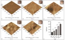
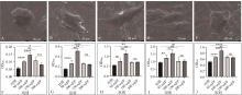
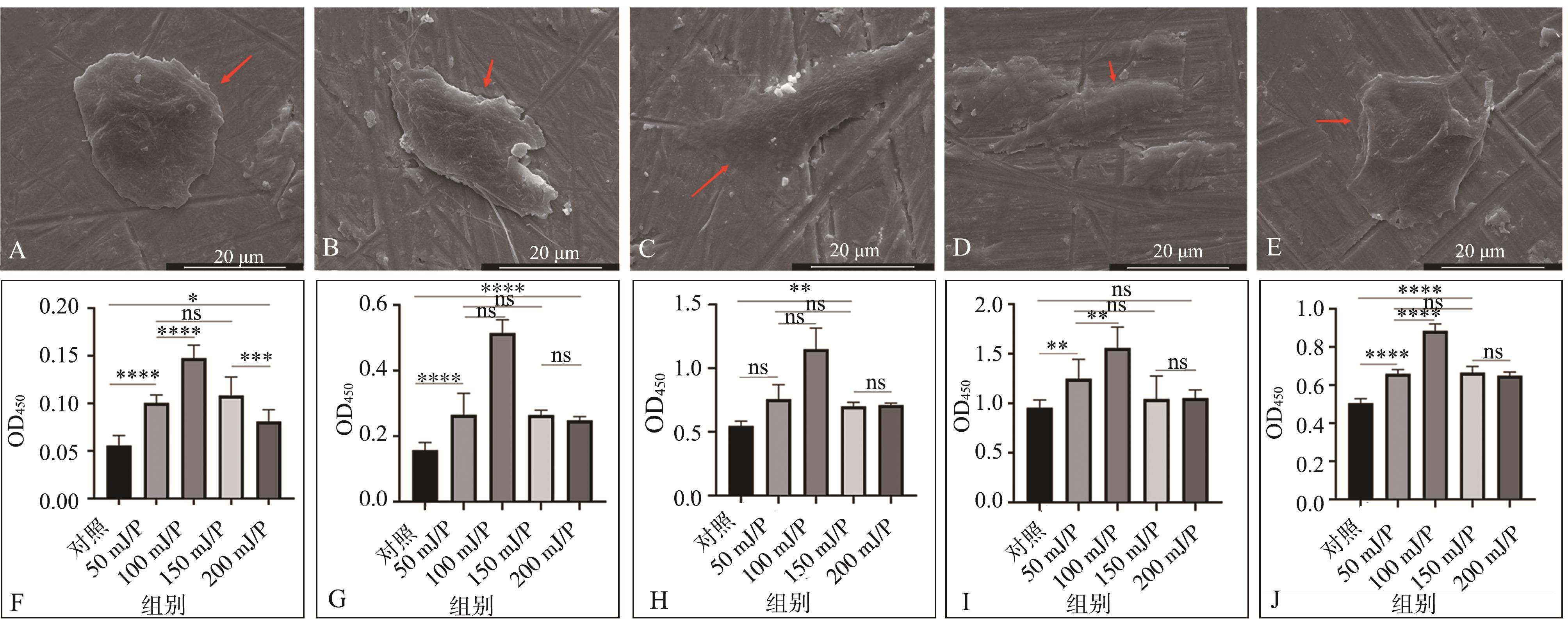
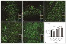
| [2] | Sun Xu,Deng Zhennan,Wen Cai,Zhao Ying. Implant surface micromorphological changes after Er: YAG laser irradiation observed under scanning electron microscope [J]. Int J Stomatol, 2023, 50(6): 669-673. |
| [3] | Zhang Shan,Shen Shuping,Zhang Fang,Yang Weidong. Effect of photon-initiated photoacoustic streaming Er: YAG laser on the water loss of dentin and compressive strength of root [J]. Int J Stomatol, 2022, 49(1): 55-59. |
| [4] | He Rong,Liu Xuejun,Zhou Yukun. Systematic review on the effect of photon-initiated photoacoustic streaming in endodontic irrigation [J]. Int J Stomatol, 2021, 48(6): 644-655. |
| [5] | Tao Tingting, Li Changzhen, Yang Heng, Ding Yi.. The study of cone beam computed tomography applications in periodontal imaging [J]. Inter J Stomatol, 2014, 41(4): 412-414. |
|
||