Int J Stomatol ›› 2022, Vol. 49 ›› Issue (1): 60-65.doi: 10.7518/gjkq.2022011
• Orginal Article • Previous Articles Next Articles
Xia Feifei( ),Qin Wenjuan,Feng Jia,Zhou Xuyang,Sun Ercan,Li Changxue()
| [1] | Fodor D, Pop S, Maniu A, et al. Gray scale and doppler ultrasonography of the benign tumors of parotid gland (pleomorphic adenoma and Warthin’s tumor). Pictorial essay[J]. Med Ultrason, 2010,12(3):238-244. |
| [2] | David E, Cantisani V, De Vincentiis M, et al. Contrast-enhanced ultrasound in the evaluation of paro-tid gland lesions: an update of the literature[J]. Ultrasound, 2016,24(2):104-110. |
| [3] | Comoglu S, Ozturk E, Celik M, et al. Comprehensive analysis of parotid mass: a retrospective study of 369 cases[J]. Auris Nasus Larynx, 2018,45(2):320-327. |
| [4] | Psychogios G. Ultrasonography techniques in the preoperative diagnosis of parotid gland tumors-an updated review of the literature[J]. Med Ultrason, 2021,23(1):122-123. |
| [5] | Kwon MR, Shin JH, Hahn SY, et al. Histogram analysis of greyscale sonograms to differentiate between the subtypes of follicular variant of papillary thyroid cancer[J]. Clin Radiol, 2018, 73(6): 591.e1- 591.e7. |
| [6] | Nam SJ, Yoo J, Lee HS, et al. Quantitative evaluation for differentiating malignant and benign thyroid nodules using histogram analysis of grayscale sonograms[J]. J Ultrasound Med, 2016,35(4):775-782. |
| [7] | Park KW, Shin JH, Hahn SY, et al. The role of histogram analysis of grayscale sonograms to differen-tiate thyroid nodules identified by 18F-FDG PET-CT[J]. Medicine (Baltimore), 2020,99(48):e23252. |
| [8] | Chen J, Liu SX, Tang YD, et al. Performance of diffusion-weighted imaging for the diagnosis of parotid gland malignancies: a Meta-analysis[J]. Eur J Radiol, 2021,134:109444. |
| [9] | Basara Akin I, Ozgul H, Simsek K, et al. Texture a-nalysis of ultrasound images to differentiate simple fibroadenomas from complex fibroadenomas and benign phyllodes tumors[J]. J Ultrasound Med, 2020,39(10):1993-2003. |
| [10] | Raja JV, Khan M, Ramachandra VK, et al. Texture analysis of CT images in the characterization of oral cancers involving buccal mucosa[J]. Dentomaxillofac Radiol, 2012,41(6):475-480. |
| [11] | Zhan KY, Khaja SF, Flack AB, et al. Benign parotid tumors[J]. Otolaryngol Clin North Am, 2016,49(2):327-342. |
| [12] | Larian B. Parotidectomy for benign parotid tumors[J]. Otolaryngol Clin North Am, 2016,49(2):395-413. |
| [13] | Matsuda E, Fukuhara T, Donishi R, et al. Usefulness of a novel ultrasonographic classification based on anechoic area patterns for differentiating warthin tumors from pleomorphic adenomas of the parotid gland[J]. Yonago Acta Med, 2017,60(4):220-226. |
| [14] | Freedman LS, Oberman B, Sadetzki S. Using time-dependent covariate analysis to elucidate the relation of smoking history to Warthin’s tumor risk[J]. Am J Epidemiol, 2009,170(9):1178-1185. |
| [15] | Espinoza S, Felter A, Malinvaud D, et al. Warthin s tumor of parotid gland: surgery or follow-up? Diagnostic value of a decisional algorithm with functio-nal MRI[J]. Diagn Interv Imaging, 2016,97(1):37-43. |
| [16] | Khalife A, Bakhshaee M, Davachi B, et al. The diagnostic value of B-mode sonography in differentiation of malignant and benign tumors of the parotid gland[J]. Iran J Otorhinolaryngol, 2016,28(88):305-312. |
| [17] | Stoia S, Băciuț G, Lenghel M, et al. Ultrasonography techniques in the preoperative diagnosis of parotid gland tumors-an updated review of the literature[J]. Med Ultrason, 2021,23(2):194-202. |
| [18] | Rodriguez Gutierrez D, Awwad A, Meijer L, et al. Metrics and textural features of MRI diffusion to improve classification of pediatric posterior fossa tumors[J]. AJNR Am J Neuroradiol, 2014,35(5):1009-1015. |
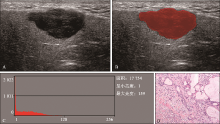
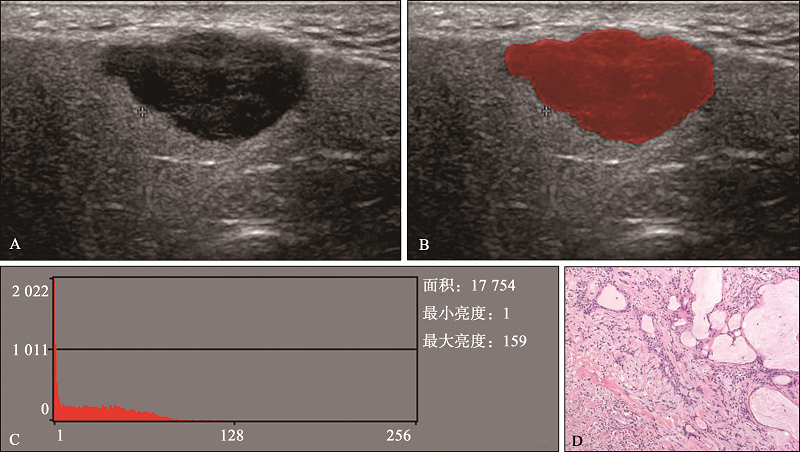
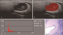
| [19] | 李芳, 徐茂林, 曾书娥, 等. 超声灰度直方图对肿块型肉芽肿性乳腺炎与浸润性导管癌的鉴别诊断[J]. 中国医学影像学杂志, 2020,28(8):602-606. |
| Li F, Xu ML, Zeng SE, et al. Differential diagnosis of massive granulomatous mastitis and invasive ductal carcinoma by histogram analysis of ultrasound gray[J]. Chin J Med Imaging, 2020,28(8):602-606. | |
| [20] | Zhang W, Zhou Y, Xu XQ, et al. A whole-tumor histogram analysis of apparent diffusion coefficient maps for differentiating thymic carcinoma from lym-phoma[J]. Korean J Radiol, 2018,19(2):358-365. |
| [21] | 李毓红, 彭建春, 代月黎. 腮腺多形性腺瘤及腺淋巴瘤的多因素Logistic回归分析[J]. 中国医学影像技术, 2016,32(5):713-716. |
| Li YH, Peng JC, Dai YL. Multi-factor Logistic regression analysis of parotid pleomorphic adenoma and adenolymphoma[J]. Chin J Med Imaging Technol, 2016,32(5):713-716. |
| [1] | Yu Dongyang,Li Shaodong,Han Lei,Shan Ben,Liu Yong,Zhao Zhengyu. Differentiation of pleomorphic adenoma and adenolymphoma of parotid gland by CT morphological features, gender and radiomics [J]. Int J Stomatol, 2023, 50(5): 506-513. |
| [2] | Zhai Xiaojing,Cao Shi,Xin Wenlong,Cao Shan,Zhang Hao. Pleomorphic adenoma with extensive keratin cysts: a case report [J]. Int J Stomatol, 2022, 49(3): 328-331. |
| [3] | Li Tian, Sun Guowen, Tang Enyi.. Research progress on carcinoma ex-pleomorphic adenoma [J]. Inter J Stomatol, 2013, 40(5): 642-644. |
| [4] | HU Yu- hua, LI Jiang.. Advances in the study of mechanisms of malignant tr ansformation of salivary pleomorphic adenoma [J]. Inter J Stomatol, 2007, 34(06): 449-451. |