国际口腔医学杂志 ›› 2023, Vol. 50 ›› Issue (2): 159-165.doi: 10.7518/gjkq.2023037
朱秋艳( ),吴道敏,鲍济波,谢志刚()
Zhu Qiuyan(),Wu Daomin,Bao Jibo,Xie Zhigang.()
摘要:
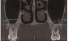
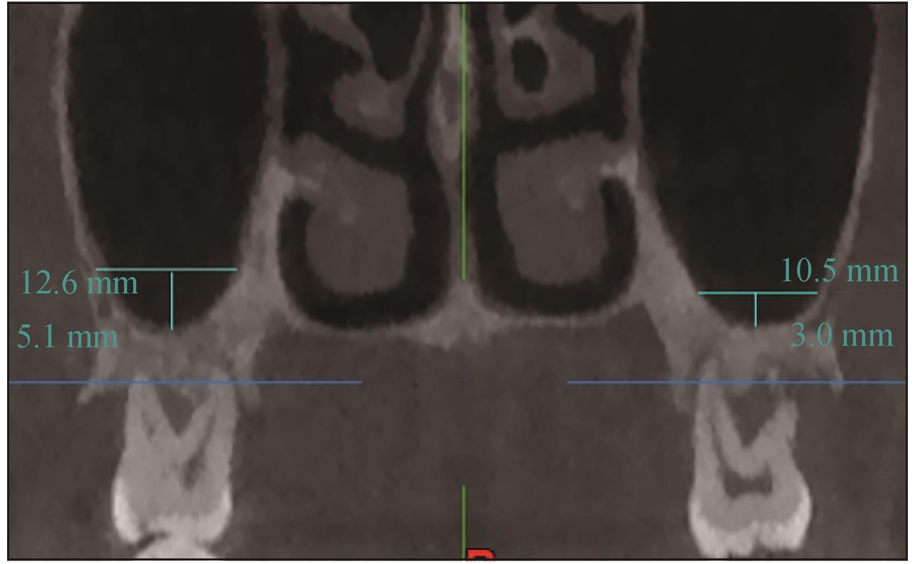
上颌窦底提升术是增加垂直骨高度的可靠的选择之一,移植物稳固需要足够的血管生成,新生血管长入并参与成骨和骨重塑的细胞迁移。这类生物学行为在很大程度上取决于上颌窦腔的三维尺寸,包括剩余骨高度(RBH)、上颌窦膜厚度、上颌窦宽度和上颌窦角度等。不同研究报道了窦腔颊-腭侧宽度对成骨效果的影响,结果不尽相同。上颌窦的三维解剖形态在骨增量手术后的条件性愈合和矿化过程中的作用尚不清楚。本研究就上颌窦窦腔颊-腭侧宽度、上颌窦角度等对窦底提升术后成骨效果的影响做一综述,旨在为临床医生术前进行风险评估和结果预判提供理论参考。
中图分类号:
| 1 | van den Bergh JP, ten Bruggenkate CM, Disch FJ, et al. Anatomical aspects of sinus floor elevations[J]. Clin Oral Implants Res, 2000, 11(3): 256-265. |
| 2 | Pietrokovski J, Massler M. Alveolar ridge resorption following tooth extraction[J]. J Prosthet Dent, 1967, 17(1): 21-27. |
| 3 | Ulm CW, Solar P, Gsellmann B, et al. The edentulous maxillary alveolar process in the region of the maxillary sinus: a study of physical dimension[J]. Int J Oral Maxillofac Surg, 1995, 24(4): 279-282. |
| 4 | Garg AK. Augmentation grafting of the maxillary sinus for placement of dental implants: anatomy, phy-siology, and procedures[J]. Implant Dent, 1999, 8(1): 36-46. |
| 5 | Talo Yildirim T, Güncü GN, Colak M, et al. The relationship between maxillary sinus lateral wall thickness, alveolar bone loss, and demographic variables: a cross-sectional cone-beam computerized tomography study[J]. Med Princ Pract, 2019, 28(2): 109-114. |
| 6 | Boyne PJ, James RA. Grafting of the maxillary sinus floor with autogenous marrow and bone[J]. J Oral Surg, 1980, 38(8): 613-616. |
| 7 | Fugazzotto PA, Vlassis J. Long-term success of sinus augmentation using various surgical approaches and grafting materials[J]. Int J Oral Maxillofac Implants, 1998, 13(1): 52-58. |
| 8 | 郑小菲, 莫安春, 朱娟芳, 等. 上颌窦解剖因素对经牙槽嵴顶上颌窦底提升术成骨效果的影响[J]. 华西口腔医学杂志, 2020, 38(6): 652-656. |
| Zheng XF, Mo AC, Zhu JF, et al. Effect of anatomical parameters of maxillary sinus on the outcomes of transcrestal sinus lift [J]. West China J Stomatol, 2020, 38(6): 652-656. | |
| 9 | Lombardi T, Stacchi C, Berton F, et al. Influence of maxillary sinus width on new bone formation after transcrestal sinus floor elevation: a proof-of-concept prospective cohort study[J]. Implant Dent, 2017, 26(2): 209-216. |
| 10 | Cheng XH, Hu XC, Wan SQ, et al. Influence of la-teral-medial sinus width on no-grafting inlay osteotome sinus augmentation outcomes[J]. J Oral Maxillofac Surg, 2017, 75(8): 1644-1655. |
| 11 | 何添荣, 陈玉英, 许香娜, 等. 上颌窦内外侧宽度对不植骨上颌窦内提升的成骨影响[J]. 口腔生物医学, 2021, 12(4): 257-261. |
| He TR, Chen YY, Xu XN, et al. Influence of lateral-medial sinus width on the outcomes of sinus elevation without graft via transcrestal approach[J]. Oral Biomed, 2021, 12(4): 257-261. | |
| 12 | 范震, 王方, 王佐林. 经牙槽嵴顶上颌窦底提升术: 中华口腔医学会第五届口腔种植专业委员会学术共识[J]. 口腔颌面外科杂志, 2018, 28(1): 1-9. |
| Fan Z, Wang F, Wang ZL. Academic consensus reached in the 5th oral implant committee of Chinese stomatological association-the crestal approach sinus augmentation[J]. J Oral Maxillofac Surg, 2018, 28(1): 1-9. | |
| 13 | 肖剑锐, 李德华, 马威, 等. 上颌窦提升后植骨区垂直高度的变化及分析[J]. 口腔医学研究, 2008, 24(2): 209-211. |
| Xiao JR, Li DH, Ma W, et al. Analysis of the transplanted bone level after maxillary sinus elevation[J]. J Oral Sci Res, 2008, 24(2): 209-211. | |
| 14 | Pizzini A, Basma HS, Li P, et al. The impact of anatomic, patient and surgical factors on membrane perforation during lateral wall sinus floor elevation[J]. Clin Oral Implants Res, 2021, 32(3): 274-284. |
| 15 | 赵士杰, 皮昕. 口腔颌面部解剖学[M]. 北京: 北京大学医学出版社, 2005. |
| Zhao SJ, Pi X. Oral and maxillofacial anatomy[M]. Beijing: Peking University Medical Press, 2005. | |
| 16 | Pikos MA. Maxillary sinus membrane repair: update on technique for large and complete perforations[J]. Implant Dent, 2008, 17(1): 24-31. |
| 17 | Lovasova K, Kachlik D, Rozpravkova M, et al. Three-dimensional CAD/CAM imaging of the ma-xillary sinus in ageing process[J]. Ann Anat, 2018, 218: 69-82. |
| 18 | Whyte A, Boeddinghaus R. Imaging of odontogenic sinusitis[J]. Clin Radiol, 2019, 74(7): 503-516. |
| 19 | Przystańska A, Kulczyk T, Rewekant A, et al. Introducing a simple method of maxillary sinus volume assessment based on linear dimensions[J]. Ann Anat, 2018, 215: 47-51. |
| 20 | Niu LX, Wang J, Yu HJ, et al. New classification of maxillary sinus contours and its relation to sinus floor elevation surgery[J]. Clin Implant Dent Relat Res, 2018, 20(4): 493-500. |
| 21 | 马楠. 不同上颌窦提升术后骨移植材料体积变化的三维重建研究[D]. 昆明: 昆明医科大学, 2019. |
| Ma N. Three-dimensional reconstruction study on the volume change of bone grafts after different ma-xillary sinus lift[D]. Kunming: Kunming Medical University, 2019. | |
| 22 | Spinato S, Bernardello F, Galindo-Moreno P, et al. Maxillary sinus augmentation by crestal access: a retrospective study on cavity size and outcome correlation[J]. Clin Oral Implants Res, 2015, 26(12): 1375-1382. |
| 23 | Stacchi C, Lombardi T, Ottonelli R, et al. New bone formation after transcrestal sinus floor elevation was influenced by sinus cavity dimensions: a prospective histologic and histomorphometric study[J]. Clin Oral Implants Res, 2018, 29(5): 465-479. |
| 24 | Johansson LA, Isaksson S, Lindh C, et al. Maxillary sinus floor augmentation and simultaneous implant placement using locally harvested autogenous bone chips and bone debris: a prospective clinical study[J]. J Oral Maxillofac Surg, 2010, 68(4): 837-844. |
| 25 | Rahpeyma A, Khajehahmadi S, Amini P. Alveolar antral artery: does its diameter correlate with maxillary lateral wall thickness in dentate patients[J]. Iran J Otorhinolaryngol, 2014, 26(76): 163-167. |
| 26 | de Santis E, Lang NP, Ferreira S, et al. Healing at implants installed concurrently to maxillary sinus floor elevation with Bio-Oss® or autologous bone grafts. A histo-morphometric study in rabbits[J]. Clin Oral Implants Res, 2017, 28(5): 503-511. |
| 27 | Busenlechner D, Huber CD, Vasak C, et al. Sinus augmentation analysis revised: the gradient of graft consolidation[J]. Clin Oral Implants Res, 2009, 20(10): 1078-1083. |
| 28 | Johansson LA, Isaksson S, Lindh C, et al. Maxillary sinus floor augmentation and simultaneous implant placement using locally harvested autogenous bone chips and bone debris: a prospective clinical study[J]. J Oral Maxillofac Surg, 2010, 68(4): 837-844. |
| 29 | Zaffe D, D’Avenia F. A novel bone scraper for intraoral harvesting: a device for filling small bone defects[J]. Clin Oral Implants Res, 2007, 18(4): 525-533. |
| 30 | Avila-Ortiz G, Wang HL, Galindo-Moreno P, et al. Influence of lateral window dimensions on vital bone formation following maxillary sinus augmentation[J]. Int J Oral Maxillofac Implants, 2012, 27(5): 1230-1238. |
| 31 | Avila G, Wang HL, Galindo-Moreno P, et al. The influence of the bucco-palatal distance on sinus augmentation outcomes[J]. J Periodontol, 2010, 81(7): 1041-1050. |
| 32 | Soardi CM, Spinato S, Zaffe D, et al. Atrophic ma-xillary floor augmentation by mineralized human bone allograft in sinuses of different size: an histologic and histomorphometric analysis[J]. Clin Oral Implants Res, 2011, 22(5): 560-566. |
| 33 | Kolerman R, Tal H, Moses O. Histomorphometric analysis of newly formed bone after maxillary sinus floor augmentation using ground cortical bone allograft and internal collagen membrane[J]. J Perio-dontol, 2008, 79(11): 2104-2111. |
| 34 | Zhou WJ, Wang F, Magic M, et al. The effect of anatomy on osteogenesis after maxillary sinus floor augmentation: a radiographic and histological analysis[J]. Clin Oral Investig, 2021, 25(9): 5197-5204. |
| 35 | Stacchi C, Rapani A, Lombardi T, et al. Does new bone formation vary in different sites within the same maxillary sinus after lateral augmentation? A prospective histomorphometric study[J]. Clin Oral Implants Res, 2022, 33(3): 322-332. |
| 36 | Scala A, Botticelli D, Rangel IG Jr, et al. Early hea-ling after elevation of the maxillary sinus floor applying a lateral access: a histological study in monkeys[J]. Clin Oral Implants Res, 2010, 21(12): 1320-1326. |
| 37 | Jang HY, Kim HC, Lee SC, et al. Choice of graft material in relation to maxillary sinus width in internal sinus floor augmentation[J]. J Oral Maxillofac Surg, 2010, 68(8): 1859-1868. |
| 38 | Zheng XF, Teng MH, Zhou FJ, et al. Influence of maxillary sinus width on transcrestal sinus augmentation outcomes: radiographic evaluation based on cone beam CT[J]. Clin Implant Dent Relat Res, 2016, 18(2): 292-300. |
| 39 | Jang HY, Kim HC, Lee SC, et al. Choice of graft material in relation to maxillary sinus width in internal sinus floor augmentation[J]. J Oral Maxillofac Surg, 2010, 68(8): 1859-1868. |
| 40 | Lozano-Carrascal N, Salomó-Coll O, Gehrke SA, et al. Radiological evaluation of maxillary sinus anatomy: a cross-sectional study of 300 patients[J]. Ann Anat, 2017, 214: 1-8. |
| 41 | Kang SJ, Shin SI, Herr Y, et al. Anatomical structures in the maxillary sinus related to lateral sinus e-levation: a cone beam computed tomographic analysis[J]. Clin Oral Implants Res, 2013, 24(): 75-81. |
| 42 | Velloso GR, Vidigal GM Jr, de Freitas MM, et al. Tridimensional analysis of maxillary sinus anatomy related to sinus lift procedure[J]. Implant Dent, 2006, 15(2): 192-196. |
| 43 | Cho SC, Wallace SS, Froum SJ, et al. Influence of anatomy on Schneiderian membrane perforations during sinus elevation surgery: three-dimensional analysis[J]. Pract Proced Aesthet Dent, 2001, 13(2): 160-163. |
| 44 | Momeni AK, Roberts CC, Chew FS. Imaging of chronic and exotic sinonasal disease: review[J]. AJR Am J Roentgenol, 2007, 189(6 ): S35-S45. |
| 45 | 王星星, 孙丽娟, 王悦, 等. 冲压法上颌窦底提升术后种植体周围骨改建及影响因素分析[J]. 实用口腔医学杂志, 2021, 37(1): 45-49. |
| Wang XX, Sun LJ, Wang Y, et al. Analysis of bone remodeling around implants and the influencing factors after osteotome sinus floor elevation[J]. J Pract Stomatol, 2021, 37(1): 45-49. |
| [1] | 黄定明, 张岚, 满毅. 牙保存相关上颌窦底提升术的生物学基础[J]. 国际口腔医学杂志, 2023, 50(3): 251-262. |
| [2] | 李洲1 法永红2 王亚锋2 李斌2. 上颌窦内壁分隔的研究进展[J]. 国际口腔医学杂志, 2015, 42(1): 40-43. |
|