国际口腔医学杂志 ›› 2026, Vol. 53 ›› Issue (4): 513-525.doi: 10.7518/gjkq.2026128
• 论著 • 上一篇
舒路红1,2( ),李航基雲1,2,张婷3,4,赵艳3,4,崔古贞3,4,洪伟1,3,廖健1,2()
Luhong Shu1,2(),Hangjiyun Li1,2,Ting Zhang3,4,Yan Zhao3,4,Guzhen Cui3,4,Wei Hong1,3,Jian Liao1,2()
摘要:
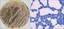
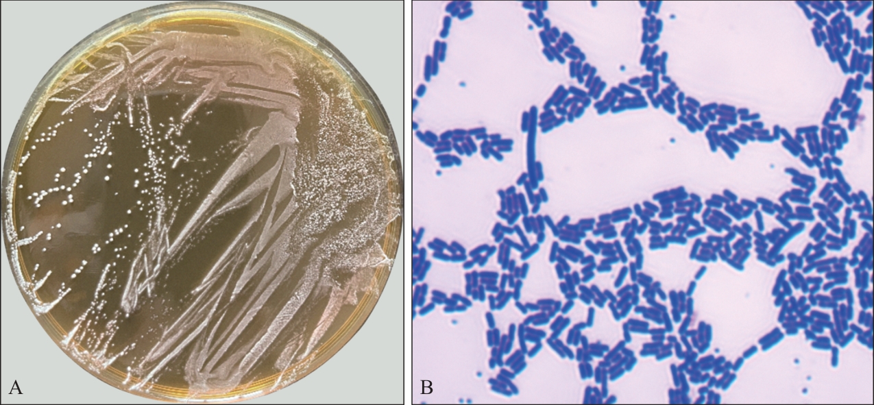
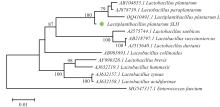
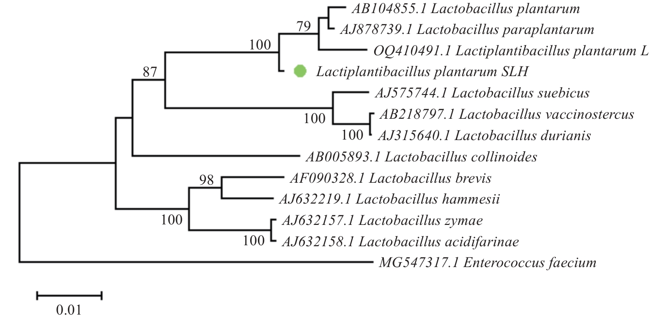
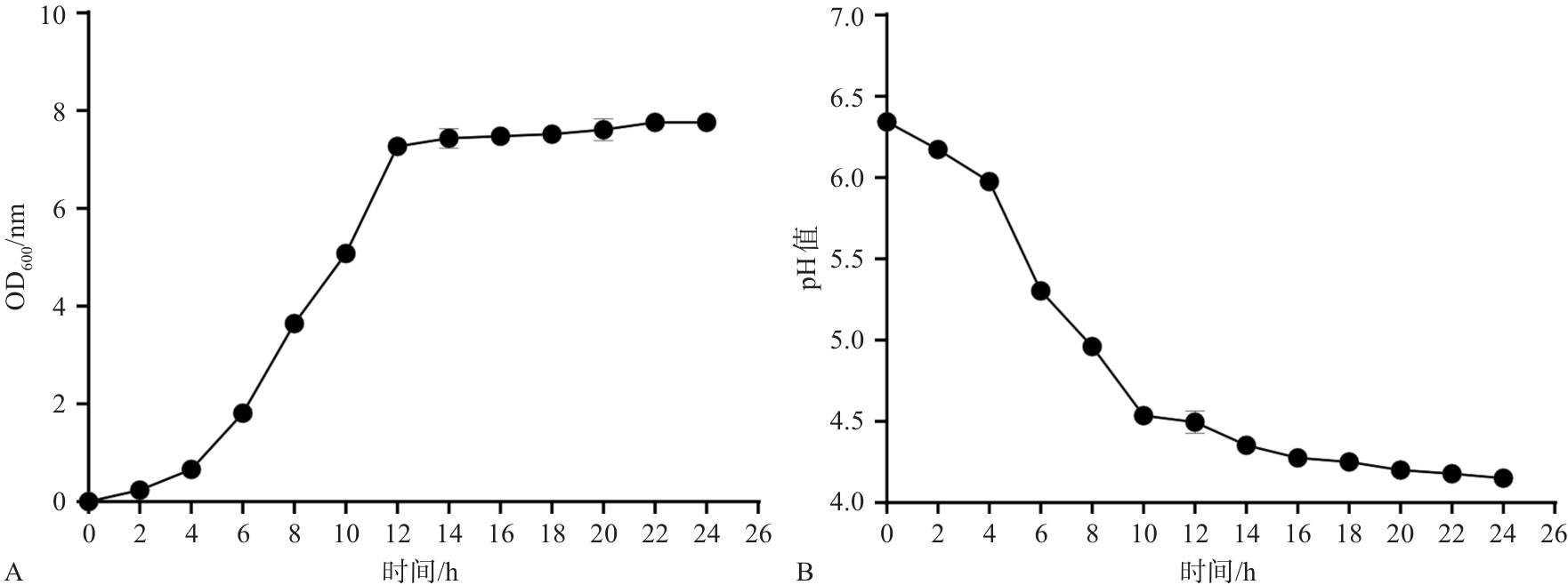
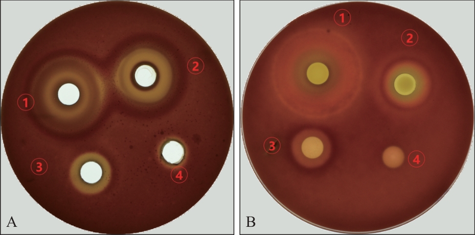
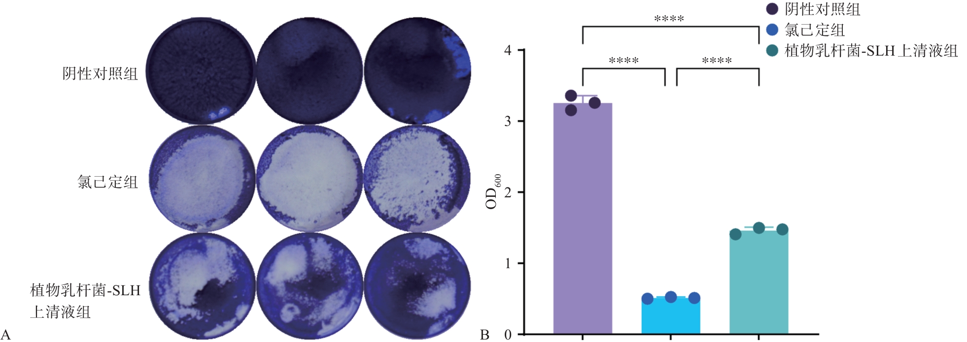
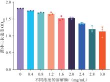
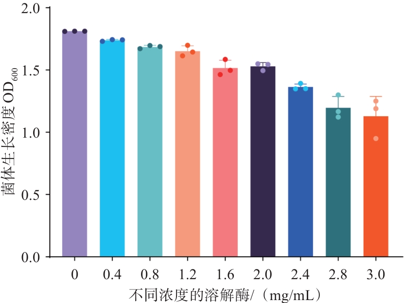
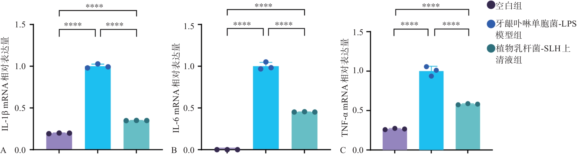
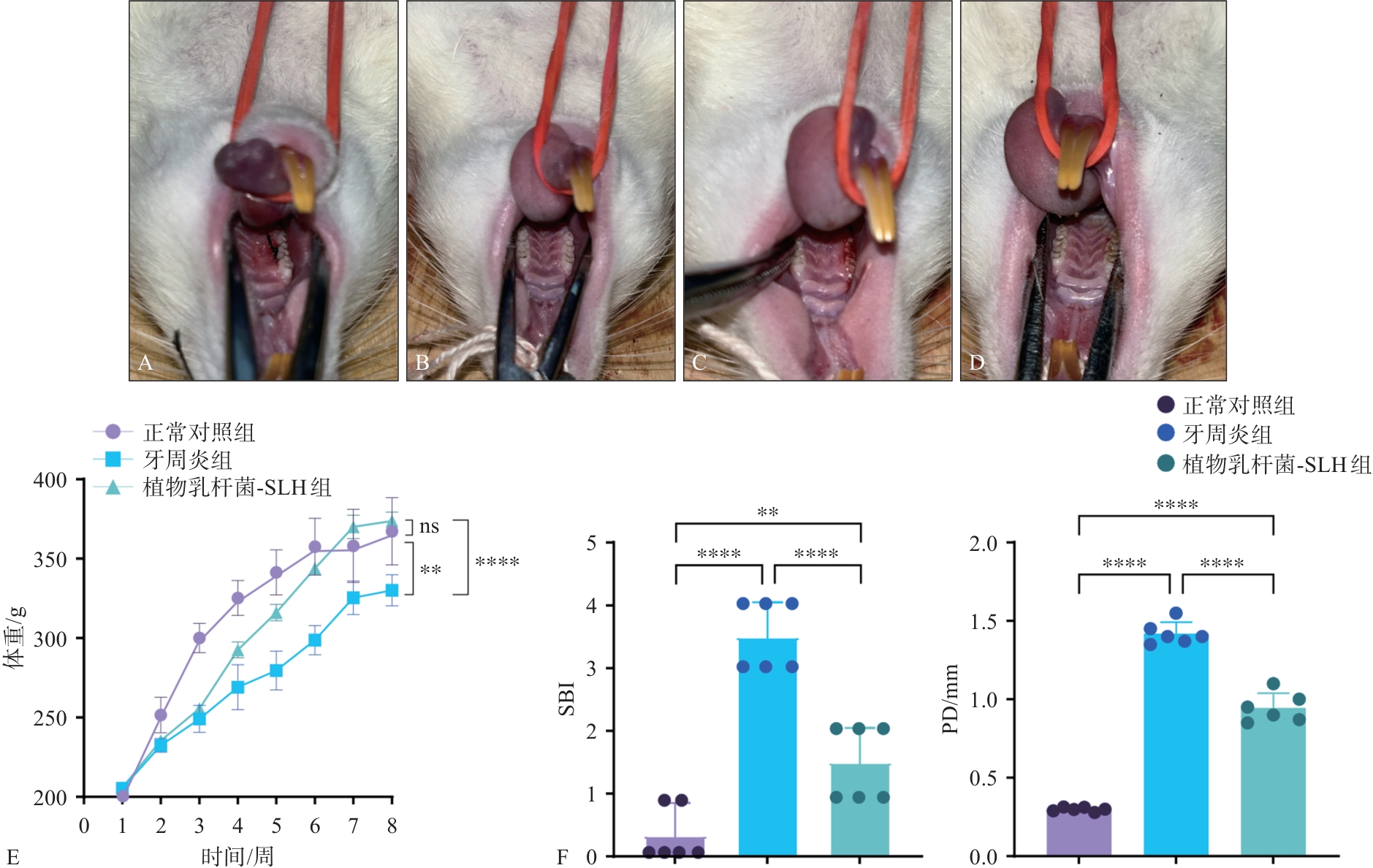
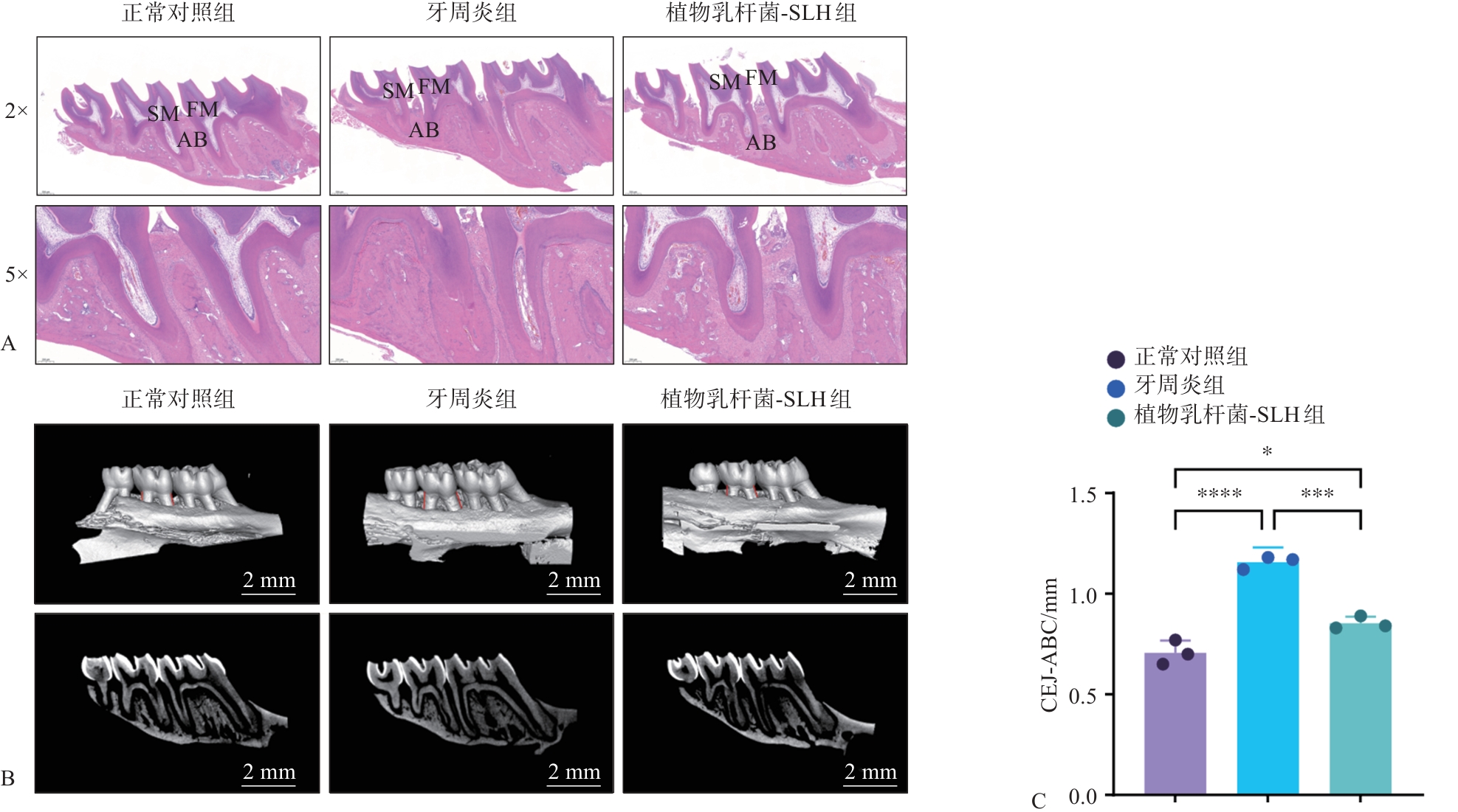
目的 研究临床分离植物乳杆菌-SLH对实验性牙周炎的治疗和缓解作用。 方法 从健康婴儿粪便样品中分离纯化出1株植物乳杆菌-SLH为实验对象,革兰染色后进行形态学观察,绘制其生长曲线和产酸曲线,通过牛津杯琼脂扩散法检测菌体及发酵上清液对具核梭杆菌和牙龈卟啉单胞菌的抑菌活性,结晶紫染色法检测该菌株对牙周生物膜形成的抑制能力,并对菌株的溶菌酶耐受能力、体外抗炎能力进行评估。再用4-0丝线结扎法构建实验性牙周炎大鼠模型,将24只大鼠随机分配为正常对照组、牙周炎组和植物乳杆菌-SLH组,每组8只。用磷酸盐缓冲盐水(PBS)配置 2% 羧甲基纤维素(CMC)溶液作为植物乳杆菌-SLH的重悬溶剂施用于大鼠造模位点的牙周袋内,在正常对照组和牙周炎组分别注射同等量2% CMC的PBS,连续7周给药。给药结束后,对各组大鼠龈沟出血指数及牙周探诊深度进行检测和牙周组织病理学评估。 结果 成功分离纯化得到1株益生菌,经16S rDNA测序鉴定后命名为植物乳杆菌-SLH。抑菌实验及生物膜形成实验表明,该菌株的发酵上清液对牙周致病菌牙龈卟啉单胞菌和具核梭杆菌有抑菌活性(P<0.05)。溶菌酶耐受实验表明,植物乳杆菌-SLH具有耐受较高(3 mg/mL)溶菌酶的能力。另外,植物乳杆菌-SLH可降低炎症细胞模型中白细胞介素(IL)-1β、肿瘤坏死因子-α和IL-6炎症因子的表达(P<0.000 1)。与对照组比较,牙周炎组大鼠龈沟出血指数、牙周探诊深度增加(P<0.000 1)。植物乳杆菌-SLH组与牙周炎组比较,其龈沟出血指数、牙周探诊深度降低(P<0.000 1)。与对照组相比,牙周炎组体重减少(P<0.05),植物乳杆菌-SLH组与牙周炎组比较体重增加(P<0.000 1)。Micro-CT结果显示,牙周炎组骨吸收明显,植物乳杆菌-SLH组比牙周炎组骨吸收减少(P<0.001)。苏木精-伊红染色结果显示,植物乳杆菌-SLH组大鼠牙周组织炎症明显减轻。 结论 植物乳杆菌-SLH对实验性牙周炎具有显著的缓解作用,具有辅助治疗牙周炎的潜力。
中图分类号:
| [1] | Zhang SP, Yu N, Arce RM. Periodontal inflammation: integrating genes and dysbiosis[J]. Periodontol 2000, 2020, 82(1): 129-142. |
| [2] | Jiao J, Jing WD, Si Y, et al. The prevalence and severity of periodontal disease in mainland China: data from the Fourth National Oral Health Survey (2015—2016)[J]. J Clin Periodontol, 2021, 48(2): 168-179. |
| [3] | Ebersole JL, Dawson Ⅲ D, Emecen-Huja P, et al. The periodontal war: microbes and immunity[J]. Pe-riodontol 2000, 2017, 75(1): 52-115. |
| [4] | Lamont RJ, Koo H, Hajishengallis G. The oral microbiota: dynamic communities and host interactions[J]. Nat Rev Microbiol, 2018, 16(12): 745-759. |
| [5] | Abdulkareem AA, Al-Taweel FB, Al-Sharqi AJB, et al. Current concepts in the pathogenesis of periodontitis: from symbiosis to dysbiosis[J]. J Oral Micro-biol, 2023, 15(1): 2197779. |
| [6] | Tonetti MS, Van Dyke TE, GroupWorking 1 of the Joint EFP/AAP Workshop. Periodontitis and atherosclerotic cardiovascular disease: consensus report of the Joint EFP/AAP Workshop on Periodontitis and Systemic Diseases[J]. J Clin Periodontol, 2013, 40(s14): S24-S29. |
| [7] | Baeza M, Morales A, Cisterna C, et al. Effect of periodontal treatment in patients with periodontitis and diabetes: systematic review and meta-analysis[J]. J Appl Oral Sci, 2020, 28: e20190248. |
| [8] | Hajishengallis G, Chavakis T. Local and systemic mechanisms linking periodontal disease and inflammatory comorbidities[J]. Nat Rev Immunol, 2021, 21(7): 426-440. |
| [9] | Bui FQ, Almeida-da-Silva CLC, Huynh B, et al. Association between periodontal pathogens and syste-mic disease[J]. Biomed J, 2019, 42(1): 27-35. |
| [10] | Slots J. Concise evaluation and therapeutic guidelines for severe periodontitis: a public health perspective[J]. Periodontol 2000, 2022, 90(1): 262-265. |
| [11] | Wei Y, Deng YX, Ma ST, et al. Local drug delivery systems as therapeutic strategies against periodontitis: a systematic review[J]. J Control Release, 2021, 333: 269-282. |
| [12] | Gibson GR, Hutkins R, Sanders ME, et al. Expert consensus document: the International Scientific Association for Probiotics and Prebiotics (ISAPP) consensus statement on the definition and scope of prebiotics[J]. Nat Rev Gastroenterol Hepatol, 2017, 14(8): 491-502. |
| [13] | Król-Górniak A, Pomastowski P, Railean-Plugaru V, et al. The study of the molecular mechanism of Lactobacillus paracasei clumping via divalent metal ions by electrophoretic separation[J]. J Chromatogr A, 2021, 1652: 462127. |
| [14] | Khalaf H, Nakka SS, Sandén C, et al. Antibacterial effects of Lactobacillus and bacteriocin PLNC8 αβ on the periodontal pathogen Porphyromonas gingivalis [J]. BMC Microbiol, 2016, 16(1): 188. |
| [15] | Sulijaya B, Yamada-Hara M, Yokoji-Takeuchi M, et al. Antimicrobial function of the polyunsaturated fatty acid KetoC in an experimental model of periodontitis[J]. J Periodontol, 2019, 90(12): 1470-1480. |
| [16] | 张晓旭, 刘欢, 肖苗, 等. 益生菌代谢产物对病原菌的抑菌作用研究进展[J]. 食品与发酵工业, 2023, 49(8): 297-302. |
| Zhang XX, Liu H, Xiao M, et al. Research progress in the antibacterial effect of probiotic metabolites on pathogens[J]. Food Ferment Ind, 2023, 49(8): 297-302. | |
| [17] | Plaza-Diaz J, Ruiz-Ojeda FJ, Gil-Campos M, et al. Mechanisms of action of probiotics[J]. Adv Nutr, 2019, 10: S49-S66. |
| [18] | Abe-Yutori M, Chikazawa T, Shibasaki K, et al. Decreased expression of E-cadherin by Porphyromonas gingivalis-lipopolysaccharide attenuates epithelial barrier function[J]. J Periodontal Res, 2017, 52(1): 42-50. |
| [19] | Yamada M, Takahashi N, Matsuda Y, et al. A bacte-rial metabolite ameliorates periodontal pathogen-induced gingival epithelial barrier disruption via GPR-40 signaling[J]. Sci Rep, 2018, 8(1): 9008. |
| [20] | 翟佳琳, 赵景娜, 王丹丹, 等. 具有抑菌活性植物乳杆菌的筛选及抑菌物质特性的研究[J]. 食品与发酵工业, 2022, 48(13): 84-90. |
| Zhai JL, Zhao JN, Wang DD, et al. Screening of Lactiplantibacillus plantarum with bacteriostatic activity and study on properties of bacteriostatic substances[J]. Food Ferment Ind, 2022, 48(13): 84-90. | |
| [21] | 夏博园, 李艳, 丁旭, 等. 过氧化物酶体增殖物激活受体γ共激活因子-1α在牙周炎诱发大鼠肝损伤中的作用研究[J]. 华西口腔医学杂志, 2021, 39(5): 518-523. |
| Xia BY, Li Y, Ding X, et al. Effect of peroxisome proliferator-activated receptor-γ coactivator-1α on liver injury induced by periodontitis in rats[J]. West China J Stomatol, 2021, 39(5): 518-523. | |
| [22] | 余意, 王超越, 吴正钧, 等. 四株乳杆菌作为口腔益生菌的特性研究[J]. 食品与发酵工业, 2021, 47(15): 77-83. |
| Yu Y, Wang CY, Wu ZJ, et al. Potentials of Lactobacillus strains as oral probiotics candidates[J]. Food Ferment Ind, 2021, 47(15): 77-83. | |
| [23] | Shao Y, Garcia-Mauriño C, Clare S, et al. Primary succession of Bifidobacteria drives pathogen resistance in neonatal microbiota assembly[J]. Nat Microbiol, 2024, 9(10): 2570-2582. |
| [24] | Chen ZH, Leng XY, Zhou F, et al. Screening and identification of probiotic Lactobacilli from the infant gut microbiota to alleviate lead toxicity[J]. Probiotics Antimicrob Proteins, 2023, 15(4): 821-831. |
| [25] | Yu AO, Goldman EA, Brooks JT, et al. Strain diversity of plant-associated Lactiplantibacillus plantarum [J]. Microb Biotechnol, 2021, 14(5): 1990-2008. |
| [26] | Van Holm W, Lauwens K, De Wever P, et al. Probio-tics for oral health: do they deliver what they pro-mise[J]. Front Microbiol, 2023, 14: 1219692. |
| [27] | Guimarães A, Venancio A, Abrunhosa L. Antifungal effect of organic acids from lactic acid bacteria on Penicillium nordicum [J]. Food Addit Contam Part A, 2018, 35(9): 1803-1818. |
| [28] | 杨颖, 陈卫, 张灏, 等. 植物乳杆菌HO-69的口腔益生性质研究[J]. 华西口腔医学杂志, 2008, 26(5): 482-485, 489. |
| Yang Y, Chen W, Zhang H, et al. Probiotic characte-ristics of Lactobacillus plantarum HO-69 applied in oral cavity[J]. West China J Stomatol, 2008, 26(5): 482-485, 489. | |
| [29] | Yoon JH, Oh MS, Lee SY. Effectiveness of organic acids for inactivating pathogenic bacteria inoculated in laboratory media and foods: an updated minireview[J]. Food Sci Biotechnol, 2024, 33(12): 2715-2728. |
| [30] | 张军, 田子罡, 王建华, 等. 有机酸抑菌分子机理研究进展[J]. 畜牧兽医学报, 2011, 42(3): 323-328. |
| Zhang J, Tian ZG, Wang JH, et al. Advances in antimicrobial molecular mechanism of organic acids[J]. Acta Vet Zootechnica Sin, 2011, 42(3): 323-328. | |
| [31] | Wang SY, Nie ZY, Zhu L, et al. Probiotic characte-ristics and the anti-inflammatory effects of Lactiplantibacillus plantarum Z22 isolated from naturally fermented vegetables[J]. Microorganisms, 2024, 12(11): 2159. |
| [32] | de Almeida Brandão D, Spolidorio LC, Johnson F, et al. Dose-response assessment of chemically modified curcumin in experimental periodontitis[J]. J Pe-riodontol, 2019, 90(5): 535-545. |
| [33] | Pudgar P, Povšič K, Čuk K, et al. Probiotic strains of Lactobacillus brevis and Lactobacillus plantarum as adjunct to non-surgical periodontal therapy: 3-month results of a randomized controlled clinical trial[J]. Clin Oral Investig, 2021, 25(3): 1411-1422. |
| [34] | Lam RS, O’Brien-Simpson NM, Lenzo JC, et al. Macrophage depletion abates Porphyromonas gingivalis-induced alveolar bone resorption in mice[J]. J Immunol, 2014, 193(5): 2349-2362. |
| [1] | 刘熠,潘亚萍. 牙槽骨角形吸收的危险因素研究进展[J]. 国际口腔医学杂志, 2026, 53(3): 362-369. |
| [2] | 吴子湘,谭碧云,湛晶蕾,李霞,葛颂. 细胞焦亡参与牙龈卟啉单胞菌促进动脉粥样硬化发生发展的研究进展[J]. 国际口腔医学杂志, 2026, 53(3): 370-380. |
| [3] | 郭怡婧,杜思雨,陈亚冰,王雷. 辛伐他汀前药在牙周炎治疗中的研究进展[J]. 国际口腔医学杂志, 2026, 53(2): 239-246. |
| [4] | 陈可儿,包佳琦,孙伟莲. 颊壁形态影响牙周炎位点牙槽嵴保存的应用进展[J]. 国际口腔医学杂志, 2026, 53(1): 84-90. |
| [5] | 石琦企,秦旭,朱光勋. 铜死亡在牙周炎中的研究进展[J]. 国际口腔医学杂志, 2026, 53(1): 91-97. |
| [6] | 王诗雅,袁国华,邹静. 异位釉质的形成机制及临床诊疗策略[J]. 国际口腔医学杂志, 2025, 52(6): 713-721. |
| [7] | 陈禹黄,梁星,李然. 牙周炎患者缺失牙修复的临床考量及预后评估[J]. 国际口腔医学杂志, 2025, 52(6): 823-831. |
| [8] | 朱然,严静,孙卫斌,吴文蕾,刘玉. 服用抗血栓药物患者牙周基础治疗期间的出血风险管理[J]. 国际口腔医学杂志, 2025, 52(5): 670-676. |
| [9] | 李欢,原韶钟. 牙周炎与非酒精性脂肪性肝病相关性的研究进展[J]. 国际口腔医学杂志, 2025, 52(5): 677-683. |
| [10] | 祝舒钰,周静,谢志刚. 辅助性T细胞17与调节性T细胞之间的制衡效应调控口腔颌面部骨损伤修复的研究进展[J]. 国际口腔医学杂志, 2025, 52(4): 514-525. |
| [11] | 于寰,康健. 牙周内镜在口腔临床诊疗中的应用进展[J]. 国际口腔医学杂志, 2025, 52(4): 544-551. |
| [12] | 李天元, 朱彤欣, 柳庆, 董迎春, 陈斌. 间充质干细胞用于牙周再生临床疗效的系统评价与Meta分析[J]. 国际口腔医学杂志, 2025, 52(3): 296-307. |
| [13] | 别梦瑶,周婕妤,吴亚菲,赵蕾. 牙龈卟啉单胞菌影响血管平滑肌细胞调节性细胞死亡及表型转换的研究进展[J]. 国际口腔医学杂志, 2025, 52(3): 308-316. |
| [14] | 范兴丽,潘乐,赵家园,项秋猛,陈启林. 竞争性内源性RNA在牙周炎中的作用及机制的研究进展[J]. 国际口腔医学杂志, 2025, 52(3): 317-322. |
| [15] | 赵美林,赵依琼,黄姣. Ⅲ期C级牙周炎正畸患者上前牙龈乳头缺陷伴牙龈退缩1例[J]. 国际口腔医学杂志, 2025, 52(3): 333-340. |
|
||