国际口腔医学杂志 ›› 2026, Vol. 53 ›› Issue (2): 166-175.doi: 10.7518/gjkq.2026014
宋东哲1( ),满毅2,黄定明1()
Dongzhe Song1(),Yi Man2,Dingming Huang1()
摘要:
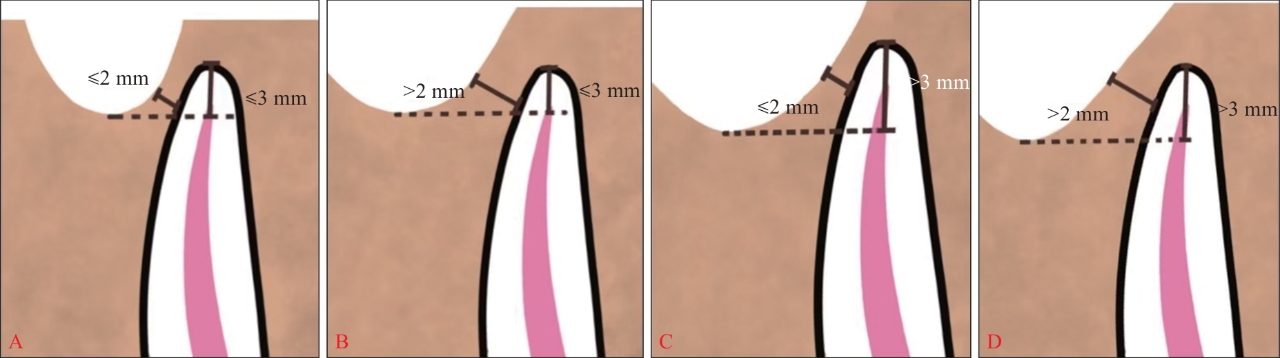
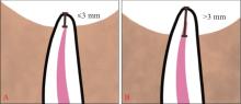
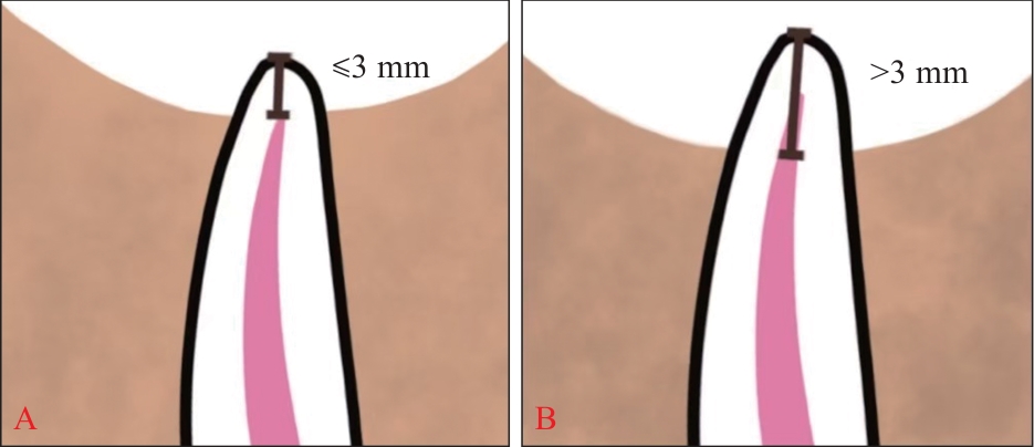
牙保存相关上颌窦底提升术是在控制根管感染、治愈病变的基础上,以最大程度保存天然牙为目标的重要治疗技术。该术式以上颌牙-牙槽骨-上颌窦复合体为解剖基础,其成功实施依赖于对局部多重风险因素的系统评估。本文围绕患牙状态(如牙根位置及长度)、牙槽骨条件(如骨高度、腭大神经血管束)、上颌窦解剖特征(如窦底形态、黏膜厚度)以及牙根与上颌窦的位置关系等方面,系统分析可能影响手术安全性与疗效的局部风险因素,旨在为临床术前评估与方案制定提供依据,从而提升治疗的可预测性与长期成功率。
中图分类号:
| [1] | 黄定明, 张岚, 满毅. 牙保存相关上颌窦底提升术的生物学基础[J]. 国际口腔医学杂志, 2023, 50(3): 251-262. |
| Huang DM, Zhang L, Man Y. Biologic bases of nature tooth-related maxillary sinus floor elevation[J]. Int J Stomatol, 2023, 50(3): 251-262. | |
| [2] | 谭学莲, 满毅, 黄定明. 牙保存相关上颌窦底提升术的临床应用[J]. 国际口腔医学杂志, 2024, 51(4): 381-391. |
| Tan XL, Man Y, Huang DM. Clinical applications of natural tooth-related maxillary sinus floor elevation[J]. Int J Stomatol, 2024, 51(4): 381-391. | |
| [3] | 周学东, 李继遥. 牙体牙髓科诊疗与操作常规[M]. 北京: 人民卫生出版社, 2018: 204-206. |
| Zhou XD, Li JY. Diagnosis, treatment, and opera-ting procedures of cariology and endodontics[M]. Beijing: People’s Medical Publishing House, 2018: 204-206. | |
| [4] | Setzer FC, Kratchman SI. Present status and future directions: surgical endodontics[J]. Int Endod J, 2022, 55(): 1020-1058. |
| [5] | Huang XX, Xu J, Hou BX, et al. Proximity of maxillary molar palatal roots to adjacent structures for en-dodontic microsurgery: a cone-beam computed tomography study[J]. BMC Oral Health, 2025, 25(1): 21. |
| [6] | Wang S, Wang XY, Jiang JH, et al. Relationship between the surgical access line of maxillary poste-rior teeth and the maxillary sinus floor[J]. J Endod, 2022, 48(4): 509-515. |
| [7] | Chaniotis A, Ordinola-Zapata R. Present status and future directions: management of curved and calcified root canals[J]. Int Endod J, 2022, 55(): 656-684. |
| [8] | Wang HG, Xu X, Bian Z, et al. Expert consensus on apical microsurgery[J]. Int J Oral Sci, 2025, 17(1): 2. |
| [9] | Kim S, Kratchman S. Modern endodontic surgery concepts and practice: a review[J]. J Endod, 2006, 32(7): 601-623. |
| [10] | 王捍国. 显微根尖外科手术难度评估系统的初步建立[J]. 实用口腔医学杂志, 2022, 38(5): 557-564. |
| Wang HG. Primary establishment of case difficulty assessment of apical microsurgery[J]. J Pract Stomatol, 2022, 38(5): 557-564. | |
| [11] | 陈益燕, Pradan SP, 杨锦波. 302例显微根尖手术的回顾性研究[J]. 华西口腔医学杂志, 2021, 39(4): 458-463. |
| Chen YY, Pradan SP, Yang JB. A retrospective study of endodontic microsurgery about 302 patients[J]. West China J Stomatol, 2021, 39(4): 458-463. | |
| [12] | Zhang MM, Fang GF, Wang ZH, et al. Clinical outcome and predictors of endodontic microsurgery using cone-beam computed tomography: a retrospective cohort study[J]. J Endod, 2023, 49(11): 1464-1471. |
| [13] | 黄定明, 周学东. 根管治疗难度分析的要点[J]. 中华口腔医学杂志, 2006, 41(9): 532-534. |
| Huang DM, Zhou XD. Difficulty assessment of root canal treatment[J]. Chin J Stomatol, 2026, 41(9): 532-534. | |
| [14] | 梁芷瑩, 赵苑汐, 朱嘉妮, 等. 288例前牙显微根尖手术临床资料的回顾性分析[J]. 国际口腔医学杂志, 2023, 50(2): 166-171. |
| Liang ZY, Zhao YX, Zhu JN, et al. Retrospective analysis of clinical data of 288 cases of endodontic microsurgery on anterior teeth[J]. Int J Stomatol, 2023, 50(2): 166-171. | |
| [15] | Shahabinejad H, Ghassemi A, Pishbin L, et al. Success of ultrasonic technique in removing fractured rotary nickel-titanium endodontic instruments from root canals and its effect on the required force for root fracture[J]. J Endod, 2013, 39(6): 824-828. |
| [16] | 周学东. 成人根管系统形态与根管治疗难度评估[J]. 中国实用口腔科杂志, 2008, 1(1): 5-9. |
| Zhou XD. Assessment of adult root canal system morphology and root canal treatment difficulty[J]. Chin J Pract Stomatol, 2008, 1(1): 5-9. | |
| [17] | Gao S, Jiang Y, Yao YX, et al. Minimally invasive techniques for lateral maxillary sinus floor elevation: small lateral window and one-stage surgery-a 2-5-year retrospective study[J]. Int J Oral Sci, 2023, 15(1): 28. |
| [18] | Stacchi C, Bernardello F, Spinato S, et al. Intraope-rative complications and early implant failure after transcrestal sinus floor elevation with residual bone height ≤5 mm: a retrospective multicenter study[J]. Clin Oral Implants Res, 2022, 33(8): 783-791. |
| [19] | Lundgren S, Cricchio G, Hallman M, et al. Sinus floor elevation procedures to enable implant placement and integration: techniques, biological aspects and clinical outcomes[J]. Periodontol 2000, 2017, 73(1): 103-120. |
| [20] | Lv HX, Sun XL, Wang J, et al. Flapless osteotome-mediated sinus floor elevation using platelet-rich fibrin versus lateral approach using deproteinised bovine bone mineral for residual bone height of 2-6 mm: a randomised trial[J]. Clin Oral Implants Res, 2022, 33(7): 700-712. |
| [21] | Smith BG, Pratt AM, Anderson JA, et al. Targeted endodontic microsurgery: implications of the grea-ter palatine artery[J]. J Endod, 2021, 47(1): 19-27. |
| [22] | Sabeti M, Ihsan MS, Kharat P, et al. The effect of hard tissue defects on the clinical outcome of endo-dontic microsurgery: a systematic review and meta-analysis[J]. Clin Oral Investig, 2023, 27(12): 7079-7089. |
| [23] | Dhamija R, Tewari S, Sangwan P, et al. Impact of platelet-rich plasma in the healing of through-and-through periapical lesions using 2-dimensional and 3-dimensional evaluation: a randomized controlled trial[J]. J Endod, 2020, 46(9):1167-1184. |
| [24] | Dhamija R, Tewari S, Gupta A. Two- and three-dimensional healing assessment after endodontic microsurgery in through-and-through periapical lesions: 5-year follow-up from a randomized control-led trial[J]. Int Endod J, 2024, 57(9): 1180-1199. |
| [25] | Yang XX, Chen X, Zhang YC, et al. Clinical outco-mes of endodontic microsurgery in complicated ca-ses with large or through-and-through lesions: a re-trospective longitudinal study[J]. Clin Oral Investig, 2024, 28(3): 172. |
| [26] | Ricucci D, Rôças IN, Hernández S, et al. “True” versus “bay” apical cysts: clinical, radiographic, histopathologic, and histobacteriologic features[J]. J Endod, 2020, 46(9): 1217-1227. |
| [27] | Lin LM, Huang GTJ, Rosenberg PA. Proliferation of epithelial cell rests, formation of apical cysts, and regression of apical cysts after periapical wound healing[J]. J Endod, 2007, 33(8): 908-916. |
| [28] | Bernardi L, Visioli F, Nör C, et al. Radicular cyst: an update of the biological factors related to lining epithelium[J]. J Endod, 2015, 41(12): 1951-1961. |
| [29] | Li N, Zhang R, Qiao WW, et al. Conservative en-dodontic microsurgery to protect critical anatomical structures-selective curettage: a case series[J]. BMC Oral Health, 2023, 23(1): 615. |
| [30] | Nesari R, Kratchman S, Saad M, et al. Selective curettage: a conservative microsurgical approach to treating large and complicated lesions[J]. J Endod, 2020, 46(11): 1782-1790. |
| [31] | 付琢惠, 谭学莲, 黄定明. 牙源性上颌窦炎的诊疗策略[J]. 国际口腔医学杂志, 2021, 48(3): 367-372. |
| Fu ZH, Tan XL, Huang DM. Diagnosis and treatment of odontogenic maxillary sinusitis[J]. Int J Stomatol, 2021, 48(3): 367-372. | |
| [32] | Lyu MY, Xu DY, Zhang XH, et al. Maxillary sinus floor augmentation: a review of current evidence on anatomical factors and a decision tree[J]. Int J Oral Sci, 2023, 15(1): 41. |
| [33] | Niu LX, Wang J, Yu HJ, et al. New classification of maxillary sinus contours and its relation to sinus floor elevation surgery[J]. Clin Implant Dent Relat Res, 2018, 20(4): 493-500. |
| [34] | Pizzini A, Basma HS, Li P, et al. The impact of anatomic, patient and surgical factors on membrane perforation during lateral wall sinus floor elevation[J]. Clin Oral Implants Res, 2021, 32(3): 274-284. |
| [35] | Monje A, Catena A, Monje F, et al. Maxillary sinus lateral wall thickness and morphologic patterns in the atrophic posterior maxilla[J]. J Periodontol, 2014, 85(5): 676-682. |
| [36] | Kang SJ, Shin SI, Herr Y, et al. Anatomical structures in the maxillary sinus related to lateral sinus elevation: a cone beam computed tomographic ana-lysis[J]. Clin Oral Implants Res, 2013, 24(Suppl A 100): 75-81. |
| [37] | Kwak HH, Park HD, Yoon HR, et al. Topographic anatomy of the inferior wall of the maxillary sinus in Koreans[J]. Int J Oral Maxillofac Surg, 2004, 33(4): 382-388. |
| [38] | Kawakami S, Botticelli D, Nakajima Y, et al. Anatomical analyses for maxillary sinus floor augmentation with a lateral approach: a cone beam computed tomography study[J]. Anat Anz, 2019, 226: 29-34. |
| [39] | Park YB, Jeon HS, Shim JS, et al. Analysis of the anatomy of the maxillary sinus septum using 3-dimensional computed tomography[J]. J Oral Maxillofac Surg, 2011, 69(4): 1070-1078. |
| [40] | Danesh-Sani SA, Movahed A, ElChaar ES, et al. Radiographic evaluation of maxillary sinus lateral wall and posterior superior alveolar artery anatomy: a cone-beam computed tomographic study[J]. Clin Implant Dent Relat Res, 2017, 19(1): 151-160. |
| [41] | 中华口腔医学会口腔种植专业委员会. 上颌窦底提升并发症的专家共识: 出血(第一版)[J]. 中国口腔种植学杂志, 2021, 26(6): 345-348. |
| Chinese Society of Oral Implantology. Expert consensus on complications of sinus floor elevation: bleeding (first edition)[J].Chin J Oral Implantol, 2021, 26(6): 345-348. | |
| [42] | Anamali S, Avila-Ortiz G, Elangovan S, et al. Prevalence of the posterior superior alveolar canal in cone beam computed tomography scans[J]. Clin Oral Implants Res, 2015, 26(1): e8-e12. |
| [43] | 中华口腔医学会口腔种植专业委员会. 上颌窦底提升并发症的专家共识: 上颌窦感染及骨增量材料感染(第一版)[J]. 中国口腔种植学杂志, 2022, 27(2): 71-74. |
| Chinese Society of Oral Implantology. Expert consensus on complications of sinus floor elevation: sinus infection and sinus graft infection (first edition)[J]. Chin J Oral Implantol, 2022, 27(2): 71-74. | |
| [44] | Lin YH, Yang YC, Wen SC, et al. The influence of sinus membrane thickness upon membrane perforation during lateral window sinus augmentation[J]. Clin Oral Implants Res, 2016, 27(5): 612-617. |
| [1] | 谭学莲, 满毅, 黄定明. 牙保存相关上颌窦底提升术的临床应用[J]. 国际口腔医学杂志, 2024, 51(4): 381-391. |
| [2] | 黄定明, 张岚, 满毅. 牙保存相关上颌窦底提升术的生物学基础[J]. 国际口腔医学杂志, 2023, 50(3): 251-262. |
| [3] | 白皓亮,杨禾,赵蕾. 牙周病风险评估及预后判断工具的研究进展[J]. 国际口腔医学杂志, 2021, 48(6): 696-702. |
|