国际口腔医学杂志 ›› 2019, Vol. 46 ›› Issue (1): 20-25.doi: 10.7518/gjkq.2019.01.004
赵文俊,刘媛媛,郝晓琪,王凯利,任家银,郭文豪,郑广宁( )
)
Wenjun Zhao,Yuanyuan Liu,Xiaoqi Hao,Kaili Wang,Jiayin Ren,Wenhao Guo,Guangning Zheng()
摘要:
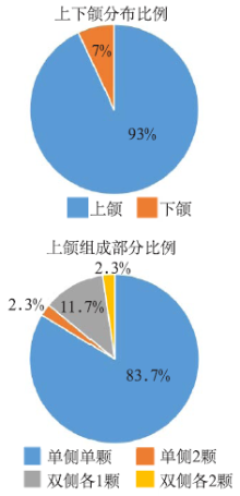
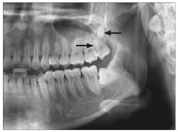
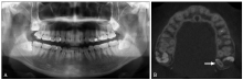
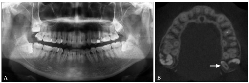
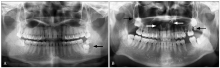
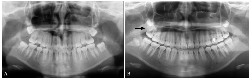
目的 观察分析第三磨牙区多生牙(ST-TMR)的影像资料,分析、总结其影像学特点。方法 收集46例患者的55颗ST-TMR的X线影像资料(曲面体层片或锥形束CT),观察其位置、形态、大小及有无伴发病变等。结果 ST-TMR以上颌单侧单颗多见(36/46),下颌、双侧及同一区域2颗者少见。21颗ST-TMR的锥形束CT显示:埋伏阻生多见(18/21),萌出者少见;多位于第三磨牙远中(9/21),其余分布在第三磨牙的颊(舌)侧、根(冠)方或近中;紧邻上颌窦底壁5颗,进入翼上颌裂1颗;多生牙多体积较小,形态各异,其中呈锥形者多见(13/21),其余如结节状、类似前磨牙者、类似磨牙者少见。可伴发含牙囊肿、前牙区多生牙、邻牙阻生、冠周骨质改变、邻牙根吸收、邻牙龋病、多个牙阻生等病变。结论 ST-TMR的发生女性多于男性,上颌单侧单颗多见;CBCT能够准确观察ST-TMR的位置、形态、大小及有无伴发病变等,为临床医生制定诊疗计划提供依据。
中图分类号:
| [1] |
Reddy GS, Reddy GV, Krishna IV , et al. Nonsyn-dromic bilateral multiple impacted supernumerary mandibular third molars: a rare and unusual case report[J]. Case Rep Dent, 2013,2013:857147.
doi: 10.1155/2013/857147 pmid: 3576790 |
| [2] |
Garvey MT, Barry HJ, Blake M . Supernumerary teeth—an overview of classification, diagnosis and management[J]. J Can Dent Assoc, 1999,65(11):612-616.
pmid: 10658390 |
| [3] |
Koo S, Salvador PS, Ciuffi Júnior J , et al. Bilateral maxillary fourth molars and a supernumerary tooth in maxillary canine region—a case report[J]. SADJ, 2002,57(10):404-406.
pmid: 12523305 |
| [4] | Clementini M, Ottria L, Pandolfi C , et al. Four im-pacted fourth molars in a young patient: a case report[J]. Oral Implantol (Rome), 2013,5(4):100-103. |
| [5] |
Menardía-Pejuan V, Berini-Aytés L, Gay-Escoda C . Supernumerary molars. A review of 53 cases[J]. Bull Group Int Rech Sci Stomatol Odontol, 2000,42(2/3):101-105.
pmid: 11799727 |
| [6] |
Mitsea A, Vardas E, Papachatzopoulou A , et al. The frequency of non-syndromic distomolar teeth in a Greek population sample[J]. J Clin Exp Dent, 2015,7(5):e589-e594.
doi: 10.4317/jced.52411 pmid: 26644834 |
| [7] |
Ceperuelo D, Lozano M, Duran-Sindreu F , et al. Supernumerary fourth molar and dental pathologies in a Chalcolithic individual from the El Mirador Cave site (Sierra de Atapuerca, Burgos, Spain)[J]. Homo, 2015,66(1):15-26.
doi: 10.1016/j.jchb.2014.05.007 pmid: 25456564 |
| [8] |
Schmitd LB, Assao A, Ramalho-Ferreira G , et al. An uncommon occurrence of three-fourth molars con-comitant to hypodontia in a nonsyndromic patient[J]. J Craniofac Surg, 2017,28(2):482-483.
doi: 10.1097/SCS.0000000000003322 |
| [9] |
Shahzad KM, Roth LE . Prevalence and management of fourth molars: a retrospective study and literature review[J]. J Oral Maxillofac Surg, 2012,70(2):272-275.
doi: 10.1016/j.joms.2011.03.063 pmid: 21802814 |
| [10] |
Kara MI, Aktan AM, Ay S , et al. Characteristics of 351 supernumerary molar teeth in Turkish population[J]. Med Oral Patol Oral Cir Bucal, 2012,17(3):e395-e400.
doi: 10.4317/medoral.17605 pmid: 3476093 |
| [11] |
McCrea S . Adjacent dentigerous cysts with the ecto-pic displacement of a third mandibular molar and supernumerary (forth) molar: a rare occurrence[J]. Oral Surg Oral Med Oral Pathol Oral Radiol Endod, 2009,107(6):e15-e20.
doi: 10.1016/j.tripleo.2009.02.002 pmid: 19464637 |
| [12] |
容明灯, 吴慕廉, 黄羽 , 等. 上颌埋伏第三磨牙与多生牙融合1例[J]. 华西口腔医学杂志, 2011,29(1):100-101.
doi: 10.3969/j.issn.1000-1182.2011.01.024 |
|
Rong MD, Wu ML, Huang Y , et al. The fused tooth of maxillary third molar with supernumerary tooth: a case report[J]. West Chin J Stomatol, 2011,29(1):100-101.
doi: 10.3969/j.issn.1000-1182.2011.01.024 |
|
| [13] |
Prakash AR, Reddy PS, Rajanikanth M . Paradental cyst associated with supernumerary tooth fused with third molar: a rare case report[J]. J Oral Maxillofac Pathol, 2012,16(1):131-133.
doi: 10.4103/0973-029X.92991 pmid: 3303508 |
| [14] |
Menditti D, Laino L, Cicciù M , et al. Kissing molars: report of three cases and new prospective on aetio-pathogenetic theories[J]. Int J Clin Exp Pathol, 2015,8(12):15708-15718.
pmid: 26884840 |
| [15] |
Demiriz L, Hazar Bodrumlu E, Içen M , et al. Evalua-tion of the accuracy of cone beam computed tomo- graphy on measuring impacted supernumerary teeth[J]. Scanning, 2016,38(6):579-584.
doi: 10.1002/sca.21303 pmid: 26780989 |
| [16] |
Ferreira-Junior O, de Avila LD, Sampieri MB , et al. Impacted lower third molar fused with a supernume-rary tooth—diagnosis and treatment planning using cone-beam computed tomography[J]. Int J Oral Sci, 2009,1(4):224-228.
doi: 10.4248/IJOS.09056 pmid: 3470106 |
| [17] | 于世凤 . 口腔组织病理学[M]. 7版. 北京: 人民卫生出版社, 2012: 139- 143, 353-354. |
| Yu SF. Oral histopathology[M]. 7th ed. Beijing: People’s Medical Publishing House, 2012: 139- 143, 353-354. | |
| [18] |
Nayak UA, Mathian VM , Veerakumar. Nonsyndrome associated multiple supernumerary teeth: a report of two cases[J]. J Indian Soc Pedod Prev Dent, 2006,24(Suppl 1):S11-S14.
doi: 10.4103/0972-2327.25987 pmid: 16891742 |
| [19] |
Cavalcanti AL, de Alencar CR, de Carvalho Neto LG . Bilateral maxillary and mandibular fourth molars: a case report and literature review[J]. J Investig Clin Dent, 2011,2(4):296-299.
doi: 10.1111/j.2041-1626.2011.00075.x pmid: 25426903 |
| [1] | 孟抒怀,罗锋,裴锡波,万乾炳. 先天性梅毒牙的研究现状[J]. 国际口腔医学杂志, 2021, 48(4): 439-443. |
| [2] | 唐蓓,王扬,王虎,游梦. Stafne骨腔的临床和影像学研究进展[J]. 国际口腔医学杂志, 2021, 48(2): 238-242. |
| [3] | 李继遥, 郑广宁. 原发性牙根纵裂的诊治策略[J]. 国际口腔医学杂志, 2020, 47(4): 373-382. |
| [4] | 赵文俊, 王凯利, 刘莉, 郭文豪, 郑广宁. 2例下颌髁突动脉瘤样骨囊肿[J]. 国际口腔医学杂志, 2018, 45(3): 307-312. |
| [5] | 李蔚 蒋文. 头颈部肌内黏液瘤[J]. 国际口腔医学杂志, 2013, 40(6): 747-749. |
| [6] | 张媛媛. 慢性口腔溃疡的病因和临床鉴别诊断[J]. 国际口腔医学杂志, 2004, 31(S1): -. |
|