国际口腔医学杂志 ›› 2022, Vol. 49 ›› Issue (3): 328-331.doi: 10.7518/gjkq.2022046
翟晓静1( ),曹石2,辛文龙3,曹珊1,张皓3()
Zhai Xiaojing1(),Cao Shi2,Xin Wenlong3,Cao Shan1,Zhang Hao3()
摘要:
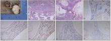
多形性腺瘤是唾液腺中最常见的良性肿瘤,其组织学形态是多样性的。约25%的多形性腺瘤可伴有鳞化,而伴有广泛鳞化及角化囊肿的病例极其罕见,需要与其他囊性病变、鳞状细胞癌、黏液表皮样癌等进行鉴别。本文报道了1例伴有广泛角化囊肿形成的罕见病例,鳞化及角化囊肿成分约占多形性腺瘤的40%。最终,通过文献复习,笔者总结了伴有广泛角化囊肿的多形性腺瘤的临床病理特征、影像学检查、鉴别诊断、治疗及预后。
中图分类号:
| 1 | 王健, 窦红漫, 程平, 等. 腮腺多形性腺瘤伴鳞化及黏液化生1例[J]. 临床与实验病理学杂志, 2019, 35(12): 1492-1493. |
| Wang J, Dou HM, Cheng P, et al. Pleomorphic adenoma of parotid gland with squamous and mucinous metaplasia: a case report [J]. Chin J Clin Exp Pathol, 2019, 35(12): 1492-1493. | |
| 2 | Lim S, Cho I, Park JH, et al. Pleomorphic adenoma with exuberant squamous metaplasia and keratin cysts mimicking squamous cell carcinoma in minor salivary gland[J]. Open J Pathol, 2013, 3(3): 113-116. |
| 3 | Sharma S, Mehendiratta M, Chaudhary N, et al. Squamous metaplasia in pleomorphic adenoma: a dia-gnostic and prognostic enigma[J]. J Pathol Transl Med, 2018, 52(6): 411-415. |
| 4 | Brachtel EF, Pilch BZ, Khettry U, et al. Fine-needle aspiration biopsy of a cystic pleomorphic adenoma with extensive adnexa-like differentiation: differential diagnostic pitfall with mucoepidermoid carcinoma[J]. Diagn Cytopathol, 2003, 28(2): 100-103. |
| 5 | Dardick I, Jeans MT, Sinnott NM, et al. Salivary gland components involved in the formation of squamous metaplasia[J]. Am J Pathol, 1985, 119(1): 33-43. |
| 6 | Lam KY, Ng IO, Chan GS. Palatal pleomorphic adenoma with florid squamous metaplasia: a potential diagnostic pitfall[J]. J Oral Pathol Med, 1998, 27(8): 407-410. |
| 7 | Batrani M, Kaushal M, Sen AK, et al. Pleomorphic adenoma with squamous and appendageal metaplasia mimicking mucoepidermoid carcinoma on cyto- logy[J]. Cytojournal, 2008, 6: 5. |
| 8 | Ramraje SN, Sisodia SM, Goel A. Fine-needle aspiration cytology of pleomorphic adenoma: cytologic variations and diagnostic pitfalls: a report of two ca-ses[J]. Natl J Integr Res Med, 2014, 5(3): 133-137. |
| 9 | Siddaraju N, Murugan P, Basu D, et al. Preoperative cytodiagnosis of cystic pleomorphic adenoma with squamous metaplasia and cholesterol crystals: a case report[J]. Acta Cytol, 2009, 53(1): 101-104. |
| 10 | Hamdan K, Maly B, Elashar R, et al. Mucinous and squamous metaplasia in benign tumors of the paro-tid gland: a potential pitfall in the diagnosis[J]. Otolaryngol Head Neck Surg, 2005, 133(6): 987-988. |
| 11 | Hegde PN, Prasad HLK, Kumar YS, et al. A rare case of an epidermoid cyst in the parotid gland-which was diagnosed by fine needle aspiration cytology[J]. J Clin Diagn Res, 2013, 7(3): 550-552. |
| 12 | AlKindi M, Ramalingam S, Hakeem LA, et al. Giant parotid pleomorphic adenoma with atypical histological presentation and long-term recurrence-free follow-up after surgery: a case report and review of the literature[J]. Case Rep Dent, 2020, 2020: 1-18. |
| 13 | Goulart MC, Freitas-Faria P, Goulart GR, et al. Pleomorphic adenoma with extensive squamous metaplasia and keratin cyst formations in minor salivary gland: a case report[J]. J Appl Oral Sci, 2011, 19(2): 182-188. |
| 14 | Jaishankar HP, Hegde U, Nagpal B. Florid squamous metaplasia and keratin cyst formation in palatal minor salivary gland tumor: a diagnostic challenge[J]. Int J Heal Sci Res, 2016, 6(4): 516-520. |
| 15 | Bajpai M, Pardhe N. Pleomorphic adenoma of submandibular gland with extensive cystic keratinization[J]. J Coll Physicians Surg Pak, 2018, 28(1): 84. |
| 16 | Onodera K, Hu JM, Yamamura Y, et al. A case of pleomorphic adenoma with formation of a large cyst: histomorphometric analysis of tumor cells[J]. Oral Med Pathol, 1998, 3(2): 89-92. |
| 17 | Aker H, Oztürk M, Ozeç I, et al. An unusual buccal adenoma with extensive squamous metaplasia and cyst formation[J]. J Chin Med Assoc, 2003, 66(3): 184-188. |
| [1] | 于冬洋,李绍东,韩雷,单奔,柳勇,赵正宇. CT形态特征、性别联合放射组学鉴别腮腺多形性腺瘤与腺淋巴瘤[J]. 国际口腔医学杂志, 2023, 50(5): 506-513. |
| [2] | 夏飞飞,秦文娟,冯佳,周旭阳,孙二灿,黎昌学. 超声灰度直方图对多形性腺瘤与腺淋巴瘤鉴别诊断效能的初步研究[J]. 国际口腔医学杂志, 2022, 49(1): 60-65. |
| [3] | 李田 孙国文 唐恩溢. 多形性腺瘤的研究进展[J]. 国际口腔医学杂志, 2013, 40(5): 642-644. |
| [4] | 应为民,华成舸. 转移性多形性腺瘤的研究进展[J]. 国际口腔医学杂志, 2008, 35(S1): -. |
| [5] | 胡宇华,李江,. 涎腺多形性腺瘤恶变机制的研究进展[J]. 国际口腔医学杂志, 2007, 34(06): 449-451. |
| [6] | 杨雯珺 张陈平 赵旭东 王铸钢. 多形性腺瘤基因1与唾液腺肿瘤[J]. 国际口腔医学杂志, 2003, 30(05): 359-361. |
|