国际口腔医学杂志 ›› 2021, Vol. 48 ›› Issue (3): 312-317.doi: 10.7518/gjkq.2021036
李明( ),原振英,南欣荣()
Li Ming(),Yuan Zhenying,Nan Xinrong()
摘要:
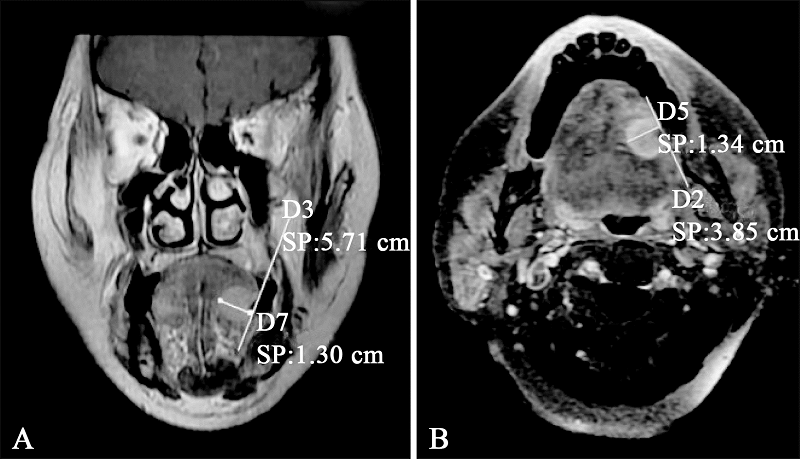
目的 分析磁共振成像(MRI)测量的浸润深度(DOI)与临床颈部淋巴结阴性即cN0期舌鳞状细胞癌患者颈部淋巴结转移的相关性,明确MRI 测量的DOI是否可作为cN0期舌鳞状细胞癌患者颈部淋巴结转移的独立预测因素。方法 经纳入和排除标准筛选后选取61名舌鳞状细胞癌的患者,分析纳入患者中MRI测量的肿瘤DOI及其他临床病理因素(年龄、肿瘤部位、肿瘤T分期、肿瘤分化程度、神经侵犯)与颈部淋巴结转移的关系。结果 61例患者中19例(31%)患者术后病理颈部淋巴结转移阳性(pN+),42例(69%)患者颈部淋巴结转移阴性(pN0)。单因素Logistic回归表明,MRI测量的DOI、T分期及神经侵犯因素与舌鳞状细胞癌患者颈部淋巴结转移有相关性(P<0.05)。多因素Logistic回归表明,仅MRI测量的DOI与颈部淋巴结转移相关(P<0.05)。MRI测量的19例pN+患者DOI均值为(13.2±4.3)mm,42例pN0患者DOI均值为(9.1±4.5)mm,两者差异具有统计学意义(T=3.36,P=0.001)。受试者工作特性曲线(ROC)表明,预测cN0期舌鳞状细胞癌患者颈部淋巴结转移的MRI测量DOI截止值为5.1 mm。结论 MRI测量是预测cN0期舌鳞状细胞癌患者颈部淋巴结转移的独立因素;MRI测量的DOI大于5.1 mm时,cN0期舌鳞状细胞癌患者发生颈部淋巴结转移的风险增加。
中图分类号:
| [1] |
Torre LA, Bray F, Siegel RL, et al. Global cancer statistics, 2012[J]. CA Cancer J Clin, 2015,65(2):87-108.
doi: 10.3322/caac.21262 |
| [2] |
Weiss MH, Harrison LB, Isaacs RS. Use of decision analysis in planning a management strategy for the stage N0 neck[J]. Arch Otolaryngol Head Neck Surg, 1994,120(7):699-702.
doi: 10.1001/archotol.1994.01880310005001 |
| [3] | Atula T, Silvoniemi P, Kurki T, et al. The evaluation and treatment of the neck in carcinoma of the oral cavity[J]. Acta Otolaryngol Suppl, 1997,529:223-225. |
| [4] |
Onercl M, Yilmaz T, Gedikoğlu G. Tumor thickness as a predictor of cervical lymph node metastasis in squamous cell carcinoma of the lower lip[J]. Otolaryngol Head Neck Surg, 2000,122(1):139-142.
doi: 10.1016/S0194-5998(00)70162-8 |
| [5] |
Al-Rajhi N, Khafaga Y, El-Husseiny J, et al. Early stage carcinoma of oral tongue: prognostic factors for local control and survival[J]. Oral Oncol, 2000,36(6):508-514.
pmid: 11036243 |
| [6] |
Pentenero M, Gandolfo S, Carrozzo M. Importance of tumor thickness and depth of invasion in nodal involvement and prognosis of oral squamous cell carcinoma: a review of the literature[J]. Head Neck, 2005,27(12):1080-1091.
doi: 10.1002/(ISSN)1097-0347 |
| [7] |
Yuen AP, Lam KY, Wei WI, et al. A comparison of the prognostic significance of tumor diameter, length, width, thickness, area, volume, and clinicopathological features of oral tongue carcinoma[J]. Am J Surg, 2000,180(2):139-143.
pmid: 11044531 |
| [8] |
Lam P, Au-Yeung KM, Cheng PW, et al. Correlating MRI and histologic tumor thickness in the assessment of oral tongue cancer[J]. AJR Am J Roentgenol, 2004,182(3):803-808.
doi: 10.2214/ajr.182.3.1820803 |
| [9] |
Alsaffar HA, Goldstein DP, King EV, et al. Correlation between clinical and MRI assessment of depth of invasion in oral tongue squamous cell carcinoma[J]. J Otolaryngol Head Neck Surg, 2016,45(1):61.
doi: 10.1186/s40463-016-0172-0 |
| [10] | 尚伟, 郑家伟. 口腔及口咽癌新版TNM分期与NCCN诊治指南部分解读[J]. 中国口腔颌面外科杂志, 2018,16(6):533-546. |
| Shang W, Zheng JW. Interpretation of the new TNM classification and the NCCN guidelines for cancers of the oral cavity and oropharynx[J]. China J Oral Maxillofac Surg, 2018,16(6):533-546. | |
| [11] |
Shintani S, Nakayama B, Matsuura H, et al. Intraoral ultrasonography is useful to evaluate tumor thickness in tongue carcinoma[J]. Am J Surg, 1997,173(4):345-347.
pmid: 9136794 |
| [12] | 冉慕光, 王承光, 陈圣欢. 舌癌影像解剖特点及MRI征象分析[J]. 临床放射学杂志, 2016,35(7):1023-1026. |
| Ran MG, Wang CG, Chen SH. Role of MRI in describing anatomy of tongue carcinoma and evaluating imaging feature[J]. J Clin Radiol, 2016,35(7):1023-1026. | |
| [13] |
Preda L, Chiesa F, Calabrese L, et al. Relationship between histologic thickness of tongue carcinoma and thickness estimated from preoperative MRI[J]. Eur Radiol, 2006,16(10):2242-2248.
doi: 10.1007/s00330-006-0263-9 |
| [14] |
Shah JP, Candela FC, Poddar AK. The patterns of cervical lymph node metastases from squamous carcinoma of the oral cavity[J]. Cancer, 1990,66(1):109-113.
doi: 10.1002/(ISSN)1097-0142 |
| [15] |
Woolgar JA. Histological distribution of cervical lymph node metastases from intraoral/oropharyngeal squamous cell carcinomas[J]. Br J Oral Maxillofac Surg, 1999,37(3):175-180.
doi: 10.1054/bjom.1999.0036 |
| [16] |
Yang X, Tian XR, Wu KL, et al. Prognostic impact of perineural invasion in early stage oral tongue squamous cell carcinoma: Results from a prospective randomized trial[J]. Surg Oncol, 2018,27(2):123-128.
doi: S0960-7404(17)30388-2 pmid: 29937161 |
| [17] |
Mücke T, Mitchell DA, Wagenpfeil S, et al. Incidence and outcome for patients with occult lymph node involvement in T1 and T2 oral squamous cell carcinoma: a prospective study[J]. BMC Cancer, 2014,14:346.
doi: 10.1186/1471-2407-14-346 |
| [18] | Tam S, Amit M, Zafereo M, et al. Depth of invasion as a predictor of nodal disease and survival in patients with oral tongue squamous cell carcinoma[J]. Head Neck, 2019,41(1):177-184. |
| [19] |
Jung J, Cho NH, Kim J, et al. Significant invasion depth of early oral tongue cancer originated from the lateral border to predict regional metastases and prognosis[J]. Int J Oral Maxillofac Surg, 2009,38(6):653-660.
doi: 10.1016/j.ijom.2009.01.004 |
| [20] |
Kwon M, Moon H, Nam SY, et al. Clinical significance of three-dimensional measurement of tumour thickness on magnetic resonance imaging in patients with oral tongue squamous cell carcinoma[J]. Eur Radiol, 2016,26(3):858-865.
doi: 10.1007/s00330-015-3884-z |
| [21] | Goel V, Parihar PS, Parihar A, et al. Accuracy of MRI in prediction of tumour thickness and nodal stage in oral tongue and gingivobuccal cancer with clinical correlation and staging[J]. J Clin Diagn Res, 2016,10(6):TC01-TC05. |
| [22] |
Park JO, Jung SL, Joo YH, et al. Diagnostic accuracy of magnetic resonance imaging (MRI) in the assessment of tumor invasion depth in oral/oropharyngeal cancer[J]. Oral Oncol, 2011,47(5):381-386.
doi: 10.1016/j.oraloncology.2011.03.012 |
| [23] |
Yesuratnam A, Wiesenfeld D, Tsui A, et al. Preoperative evaluation of oral tongue squamous cell carcinoma with intraoral ultrasound and magnetic resonance imaging-comparison with histopathological tumour thickness and accuracy in Guiding patient management[J]. Int J Oral Maxillofac Surg, 2014,43(7):787-794.
doi: 10.1016/j.ijom.2013.12.009 |
| [24] | 刘华, 李龙江, 代晓明. 口腔癌引流区淋巴结反应性增生与微转移关系的研究[J]. 实用口腔医学杂志, 2007,23(3):400-403. |
| Liu H, Li LJ, Dai XM. A study on the relationship between reactive hyperplasia of the draining lymph nodes and oral cancer micrometastasis[J]. J Pract Stomatol, 2007,23(3):400-403. |
| [1] | 李珊,陈林林. 上颌鳞状细胞癌临床颈部淋巴结阴性患者的治疗[J]. 国际口腔医学杂志, 2021, 48(4): 444-449. |
| [2] | 吴南,李斌. 吡咯喹啉醌对舌鳞状细胞癌细胞上皮间质转化的抑制作用研究[J]. 国际口腔医学杂志, 2020, 47(4): 406-412. |
| [3] | 刘彤曦,柯星,杨健. 磁共振成像及其在牙体牙髓专业中的应用[J]. 国际口腔医学杂志, 2019, 46(6): 693-698. |
| [4] | 邓程丹,石冰,李杨. 唇腭裂患者的脑部结构与功能研究进展[J]. 国际口腔医学杂志, 2019, 46(5): 617-620. |
| [5] | 王志强,刘娅丽,马丽娟,杨兰,王若宇,高舒婷. 红景天苷对人舌鳞状细胞癌CAL-27细胞增殖、凋亡、周期及迁移的影响[J]. 国际口腔医学杂志, 2018, 45(6): 678-685. |
| [6] | 王丽萍, 查骏, 葛林虎. 非编码RNA在舌鳞状细胞癌中的研究进展[J]. 国际口腔医学杂志, 2018, 45(4): 420-424. |
| [7] | 秦琨1 张东坡2. 不同材料烤瓷冠与金属冠磁共振成像伪影的比较[J]. 国际口腔医学杂志, 2016, 43(5): 507-510. |
| [8] | 吕春晓 陈嵩. 颞下颌关节病的临床诊断与磁共振成像影像诊断的相关性研究[J]. 国际口腔医学杂志, 2016, 43(1): 47-. |
| [9] | 吴开柳 李思毅 张陈平. 舌鳞状细胞癌颈淋巴结转移的特点和评估处理[J]. 国际口腔医学杂志, 2015, 42(1): 119-122. |
| [10] | 王晓彦1 武云霞2. 尼美舒利对人舌鳞状细胞癌Tca8113细胞ang-2基因表达的影响[J]. 国际口腔医学杂志, 2011, 38(6): 646-648. |
| [11] | 冯正虎, 李春青, 王凌, 韩冰, 聂红兵, 苏雪莲. 血管内皮生长因子-C 在不同浸润方式的舌鳞状细胞癌中的表达[J]. 国际口腔医学杂志, 2011, 38(1): 7-9. |
| [12] | 史琦综述王燕一审校. 口腔内金属对头颅磁共振成像的影响和减少伪影方法的研究进展[J]. 国际口腔医学杂志, 2010, 37(6): 711-713. |
| [13] | 赵芳综述 刘东旭审校. 功能性磁共振在口腔医学中的应用[J]. 国际口腔医学杂志, 2009, 36(5): 586-589. |
| [14] | 霍秋菊综述 李伟忠审校. 环氧化酶-2 及其选择性抑制剂塞来昔布与舌鳞状细胞癌侵袭的研究进展[J]. 国际口腔医学杂志, 2009, 36(3): 344-346,350. |
| [15] | 严少文综述 刘丽审校. 减少金属修复体在磁共振成像中伪影的研究进展[J]. 国际口腔医学杂志, 2008, 35(6): 712-712~714. |
|