国际口腔医学杂志 ›› 2020, Vol. 47 ›› Issue (4): 373-382.doi: 10.7518/gjkq.2020056
• 专家笔谈 • 下一篇
李继遥1( ),郑广宁2
Li Jiyao1(),Zheng Guangning2
摘要:
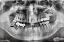
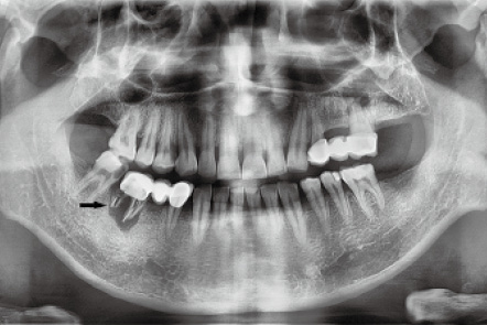
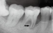
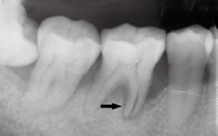
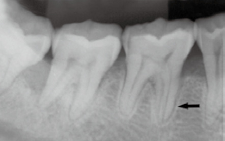
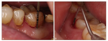
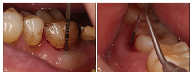
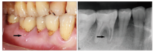
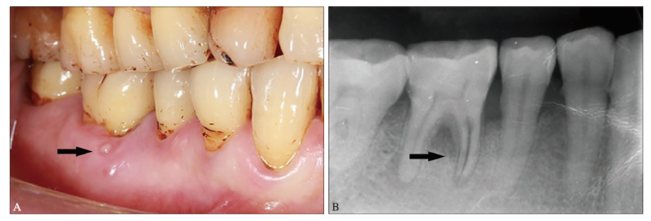
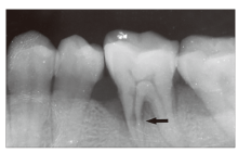
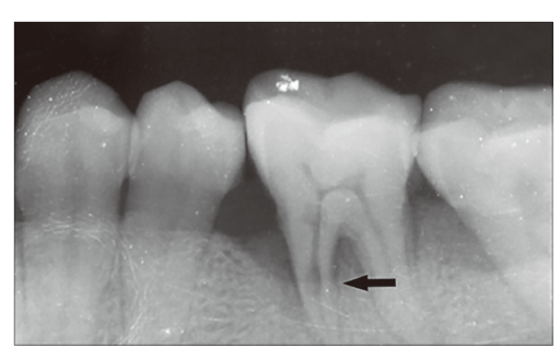
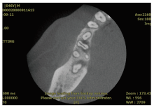
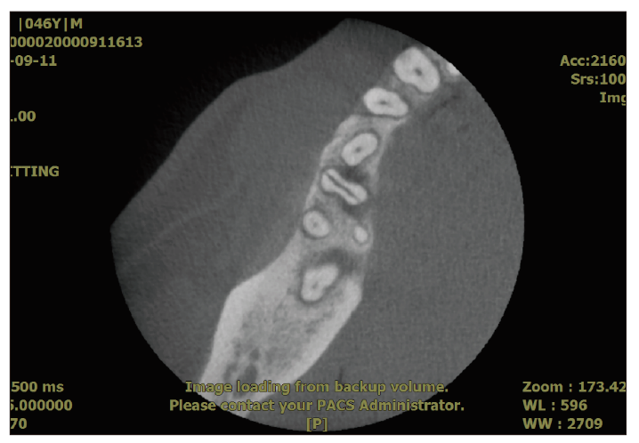
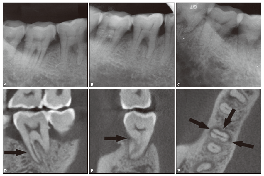
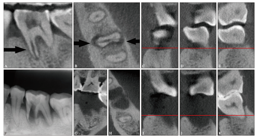
非根管治疗相关的牙根纵裂又被称为原发性牙根纵裂(SVRF),是牙科临床难症。由于临床症状、体征、早期影像学表现缺乏特征性,即便是对于经验丰富的牙医,诊断SVRF也极具挑战性;当SVRF确诊后,又面临截断、修复纵裂牙根或者拔除纵裂患牙的抉择,而拔牙后的修复时机、修复方式也是需要评估的问题。笔者根据文献复习和近3年的临床病例研究结果,对SVRF的早期诊断与鉴别诊断要点、治疗原则与方案进行讨论,以期帮助牙医们提升对SVRF的辨识能力,为临床有效诊断和治疗SVRF提供借鉴。
中图分类号:
| [1] |
Pitts DL, Natkin E. Diagnosis and treatment of verti-cal root fractures[J]. J Endod, 1983,9(8):338-346.
doi: 10.1016/S0099-2399(83)80150-2 pmid: 6579193 |
| [2] |
Yoshino K, Ito K, Kuroda M, et al. Prevalence of vertical root fracture as the reason for tooth extrac-tion in dental clinics[J]. Clin Oral Investig, 2015,19(6):1405-1409.
doi: 10.1007/s00784-014-1357-4 pmid: 25398363 |
| [3] |
Munari LS, Bowles WR, Fok ASL. Relationship be-tween canal enlargement and fracture load of root dentin sections[J]. Dent Mater, 2019,35(5):818-824.
doi: 10.1016/j.dental.2019.02.015 pmid: 30885408 |
| [4] | PradeepKumar AR, Shemesh H, van Loveren C, et al. Impact of apical extent of root canal filling on vertical root fracture: a case-control study[J]. Int En-dod J, 2019,52(9):1283-1289. |
| [5] |
Yoon HG, Oh HK, Lee DY, et al. 3-D finite element analysis of the effects of post location and loading location on stress distribution in root canals of the mandibular 1st molar[J]. J Appl Oral Sci, 2018,26:e20160406.
doi: 10.1590/1678-7757-2016-0406 pmid: 29451648 |
| [6] |
Chai H, Tamse A. Vertical root fracture in buccal roots of bifurcated maxillary premolars from conden-sation of Gutta-percha[J]. J Endod, 2018,44(7):1159-1163.
doi: 10.1016/j.joen.2018.03.017 pmid: 29861061 |
| [7] |
PradeepKumar AR, Shemesh H, Jothilatha S, et al. Diagnosis of vertical root fractures in restored endo-dontically treated teeth: a time-dependent retrospec-tive cohort study[J]. J Endod, 2016,42(8):1175-1180.
doi: 10.1016/j.joen.2016.04.012 pmid: 27339633 |
| [8] | 邹晓英, 岳林. 牙根纵裂的临床诊断[J]. 中国口腔医学继续教育杂志, 2009,12(3):59-64. |
| Zou XY, Yue L. Clinical diagnosis of vertical root fracture[J]. Chin J Stomatol Contin Edu, 2009,12(3):59-64. | |
| [9] | 董艳梅. 牙体牙髓病临床问题解析Ⅸ. 牙根纵裂的病因、危险因素及临床诊断[J]. 中华口腔医学杂志, 2011,46(10):627-630. |
| Dong YM. Analysis of endodontic diseases clinical problems. Ⅸ. The cause, risk fractors and clinical diagnosis of vertical root fracture[J]. Chin J Stomatol, 2011,46(10):627-630. | |
| [10] |
Yang SF, Rivera EM, Walton RE. Vertical root frac-ture in nonendodontically treated teeth[J]. J Endod, 1995,21(6):337-339.
doi: 10.1016/S0099-2399(06)81013-7 pmid: 7673845 |
| [11] |
Chan CP, Tseng SC, Lin CP, et al. Vertical root frac-ture in nonendodontically treated teeth—a clinical report of 64 cases in Chinese patients[J]. J Endod, 1998,24(10):678-681.
doi: 10.1016/s0099-2399(98)80154-4 pmid: 10023252 |
| [12] |
Yoshino K, Ito K, Kuroda M, et al. Duration from initial symptoms to diagnosis of vertical root fracture in dental offices[J]. Bull Tokyo Dent Coll, 2018,59(1):59-61.
doi: 10.2209/tdcpublication.2016-0049 pmid: 29563363 |
| [13] |
See WK, Ho JC, Huang CF, et al. The association between clinical diagnostic factors and the prevalence of vertical root fracture in endodontic surgery[J]. J Formos Med Assoc, 2019,118(3):713-720.
doi: 10.1016/j.jfma.2018.08.022 pmid: 30193836 |
| [14] |
Liao WC, Tsai YL, Wang CY, et al. Clinical and ra-diographic characteristics of vertical root fractures in endodontically and nonendodontically treated teeth[J]. J Endod, 2017,43(5):687-693.
doi: 10.1016/j.joen.2016.12.009 pmid: 28292598 |
| [15] | 李会旭, 刘桂荣, 张艳, 等. 5例原发性牙根纵裂的临床诊断分析及文献回顾[J]. 口腔医学研究, 2018,34(11):1186-1191. |
| Li HX, Liu GR, Zhang Y, et al. Primary vertical root fracture: 5 cases reporet and review of literature[J]. J Oral Sci Res, 2018,34(11):1186-1191. | |
| [16] | Rivera EM, Walton RE. Longitudinal tooth fractures: findings that contribute to complex endodontic dia-gnoses[J]. Endodontic Topics, 2007,16(1):82-111. |
| [17] |
Tamse A, Fuss Z, Lustig J, et al. An evaluation of endodontically treated vertically fractured teeth[J]. J Endod, 1999,25(7):506-508.
doi: 10.1016/S0099-2399(99)80292-1 pmid: 10687518 |
| [18] |
Walton RE, Michelich RJ, Smith GN. The histopa-thogenesis of vertical root fractures[J]. J Endod, 1984,10(2):48-56.
doi: 10.1016/S0099-2399(84)80037-0 pmid: 6586962 |
| [19] |
Walton RE. Vertical root fracture: factors related to identification[J]. J Am Dent Assoc, 2017,148(2):100-105.
doi: 10.1016/j.adaj.2016.11.014 pmid: 28129797 |
| [20] | Tamse A, Tsesis I, Rosen E. Vertical root fractures in dentistry[M]. Berlin: Springer, 2015. |
| [21] |
Gordon MP, Chandler NP. Electronic apex locators[J]. Int Endod J, 2004,37(7):425-437.
doi: 10.1111/j.1365-2591.2004.00835.x pmid: 15189431 |
| [22] | 丁江峰, 江千舟, 陈敏乐. 根尖定位仪定位根管纵折的体外研究[J]. 口腔医学研究, 2013,29(10):939-941. |
| Ding JF, Jiang QZ, Chen ML. Evaluation of apex locators in detection of the vertical root fractures in vitro[J]. J Oral Sci Res, 2013,29(10):939-941. | |
| [23] |
Azabal M, Garcia-Otero D, de la Macorra JC. Ac-curacy of the Justy Ⅱ Apex locator in determining working length in simulated horizontal and vertical fractures[J]. Int Endod J, 2004,37(3):174-177.
doi: 10.1111/j.0143-2885.2004.00776.x pmid: 15009406 |
| [24] | 郑广宁, 李继遥. 牙根折裂的影像诊断[J]. 华西口腔医学杂志, 2016,34(1):1-5. |
| Zheng GN, Li JY. Radiographic diagnosis of vertical root fracture[J]. West China J Stomatol, 2016,34(1):1-5. | |
| [25] |
Special Committee to Revise the Joint AAE/AAOMR Position Statement on use of CBCT in Endodontics. AAE and AAOMR joint position statement: use of cone beam computed tomography in endodontics 2015 update[J]. Oral Surg Oral Med Oral Pathol Oral Radiol, 2015,120(4):508-512.
doi: 10.1016/j.oooo.2015.07.033 pmid: 26346911 |
| [26] |
Leader DM. CBCT is valuable for diagnosis of tooth fracture[J]. Evid Based Dent, 2015,16(1):23-24.
doi: 10.1038/sj.ebd.6401082 pmid: 25909938 |
| [27] |
Long H, Zhou Y, Ye N, et al. Diagnostic accuracy of CBCT for tooth fractures: a meta-analysis[J]. J Dent, 2014,42(3):240-248.
doi: 10.1016/j.jdent.2013.11.024 pmid: 24321294 |
| [28] |
Brady E, Mannocci F, Brown J, et al. A comparison of cone beam tomography and periapical radiography for the detection of vertical root fractures in nonen-dodontically treated teeth[J]. Int Endod J, 2014,47(8):735-746.
doi: 10.1111/iej.12209 |
| [29] |
Guo XL, Li G, Zheng JQ, et al. Accuracy of detec-ting vertical root fractures in non-root filled teeth using cone beam computed tomography: effect of voxel size and fracture width[J]. Int Endod J, 2019,52(6):887-898.
doi: 10.1111/iej.13076 pmid: 30661246 |
| [30] |
Guo XL, Li G, Yin S, et al. Effect of fracture orienta-tion on detection accuracy of vertical root fractures in non-endodontically treated teeth using cone beam computed tomography[J]. Clin Oral Investig, 2019,23(12):4433-4439.
doi: 10.1007/s00784-019-02905-0 pmid: 30982180 |
| [31] |
White SN, Miklus VG, Potter KS, et al. Endodontics and implants, a catalog of therapeutic contrasts[J]. J Evid Based Dent Pract, 2006,6(1):101-109.
doi: 10.1016/j.jebdp.2005.12.013 pmid: 17138408 |
| [32] | Abboud M, Kobren LB, Orentlicher G. Implant complications: biomechanical and esthetic conside-rations—a prosthodontist’s perspective[J]. Compend Contin Educ Dent, 2013,34(Spec No 7):20-24. |
| [33] | De Rouck T, Eghbali R, Collys K, et al. The gingival biotype revisited: transparency of the periodontal probe through the gingival margin as a method to discriminate thin from thick gingiva[J]. J Clin Perio-dontol, 2009,36(5):428-433. |
| [34] |
Chang M, Odman PA, Wennström JL, et al. Esthetic outcome of implant-supported single-tooth replace-ments assessed by the patient and by prosthodontists[J]. Int J Prosthodont, 1999,12(4):335-341.
pmid: 10635203 |
| [35] | 徐玮哲, 宋东哲, 谭学莲, 等. 右侧下颌第一磨牙近中原发性牙根纵裂行活髓保存治疗1例[J]. 华西口腔医学杂志, 2019,37(5):563-567. |
| Xu WZ, Song DZ, Tan XL, et al. Vital pulp preserva-tion treatment in mandibular right first molar with vertical root fractures: a case report[J]. West China J Stomatol, 2019,37(5):563-567. | |
| [36] |
Ku HM, Oh YR, Lee ES, et al. Using autofluorescence to detect bacterial contamination in root fractures[J]. J Dent, 2019,86:27-32.
doi: 10.1016/j.jdent.2019.05.024 pmid: 31121242 |
| [37] |
Floratos SG, Kratchman SI. Surgical management of vertical root fractures for posterior teeth: report of four cases[J]. J Endod, 2012,38(4):550-555.
doi: 10.1016/j.joen.2011.12.030 pmid: 22414848 |
| [38] |
Zhou W, Zheng Q, Tan X, et al. Comparison of mineral trioxide aggregate and iRoot BP plus root repair material as root-end filling materials in endo-dontic microsurgery: a prospective randomized con-trolled study[J]. J Endod, 2017,43(1):1-6.
doi: 10.1016/j.joen.2016.10.010 pmid: 27986096 |
| [39] |
Okaguchi M, Kuo T, Ho YC. Successful treatment of vertical root fracture through intentional replantation and root fragment bonding with 4-META/MMA-TBB resin[J]. J Formos Med Assoc, 2019,118(3):671-678.
doi: 10.1016/j.jfma.2018.08.004 pmid: 30145002 |
| [40] |
Takeuchi S, Sekita T, Kobayashi K. Adhesive approach using internal coping for vertical root fractured teeth with flared root canals[J]. N Y State Dent J, 2015,81(4):29-33.
pmid: 26373031 |
| [41] |
Hadrossek PH, Dammaschke T. New treatment option for an incomplete vertical root fracture—a preliminary case report[J]. Head Face Med, 2014,10:9.
doi: 10.1186/1746-160X-10-9 pmid: 24670232 |
| [42] |
Sugaya T, Tomita M, Motoki Y, et al. Periodontal tissue repair after sealing of the gap in vertical root fracture[J]. Odontology, 2017,105(2):202-207.
doi: 10.1007/s10266-016-0270-5 pmid: 27655624 |
| [43] |
Corbella S, Taschieri S, Samaranayake L, et al. Im-plant treatment choice after extraction of a vertically fractured tooth. Aproposal for a clinical classification of bony defects based on a systematic review of literature[J]. Clin Oral Implants Res, 2014,25(8):946-956.
doi: 10.1111/clr.12164 pmid: 23560723 |
| [1] | 董栩,徐欣. 牙隐裂临床研究进展[J]. 国际口腔医学杂志, 2021, 48(6): 668-674. |
| [2] | 孟抒怀,罗锋,裴锡波,万乾炳. 先天性梅毒牙的研究现状[J]. 国际口腔医学杂志, 2021, 48(4): 439-443. |
| [3] | 蔡潇潇. 美学区数字化种植策略与流程[J]. 国际口腔医学杂志, 2019, 46(6): 621-630. |
| [4] | 赵文俊,刘媛媛,郝晓琪,王凯利,任家银,郭文豪,郑广宁. 46例患者第三磨牙区多生牙的影像学分析[J]. 国际口腔医学杂志, 2019, 46(1): 20-25. |
| [5] | 赵文俊, 王凯利, 刘莉, 郭文豪, 郑广宁. 2例下颌髁突动脉瘤样骨囊肿[J]. 国际口腔医学杂志, 2018, 45(3): 307-312. |
| [6] | 李蔚 蒋文. 头颈部肌内黏液瘤[J]. 国际口腔医学杂志, 2013, 40(6): 747-749. |
| [7] | 李丽综述 吴红崑审校. 隐匿龋的研究进展[J]. 国际口腔医学杂志, 2009, 36(6): 704-707. |
| [8] | 伍俊. 眶颧联合骨折的治疗进展[J]. 国际口腔医学杂志, 2004, 31(S1): -. |
| [9] | 张媛媛. 慢性口腔溃疡的病因和临床鉴别诊断[J]. 国际口腔医学杂志, 2004, 31(S1): -. |
|