国际口腔医学杂志 ›› 2026, Vol. 53 ›› Issue (4): 537-544.doi: 10.7518/gjkq.2026026
• 论著 • 上一篇
李晶晶1( ),邵琳琳2,俞青1()
Jingjing Li1(),Linlin Shao2,Qing Yu1()
摘要:
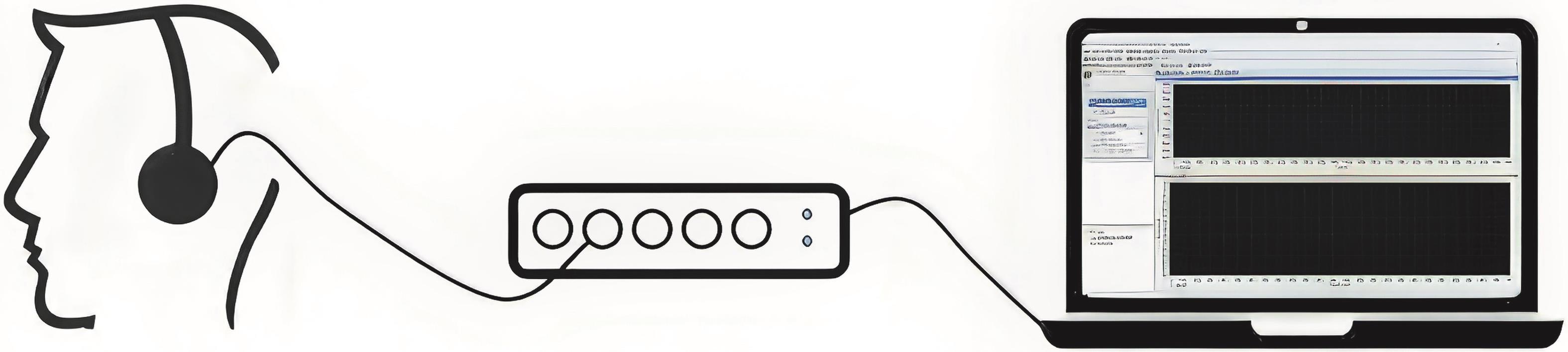
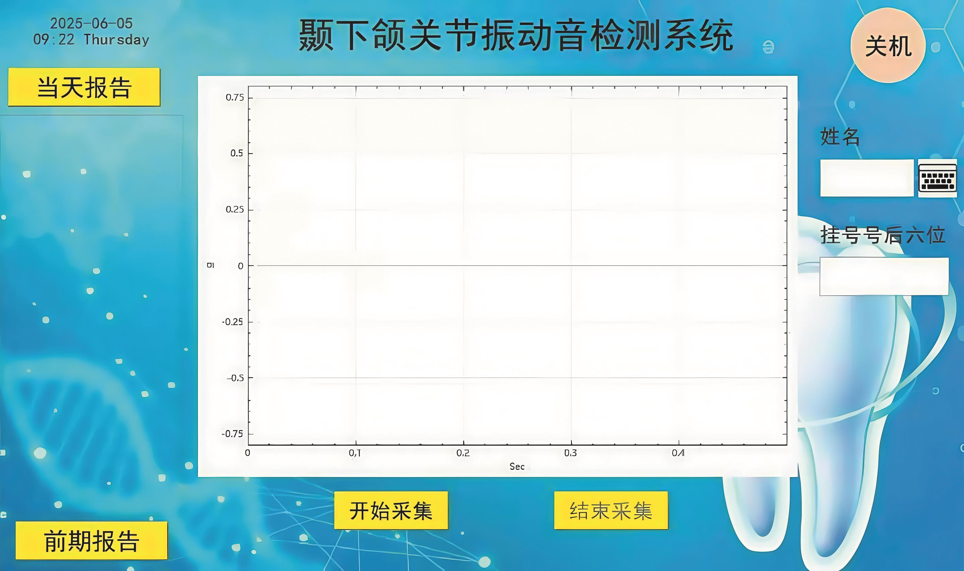
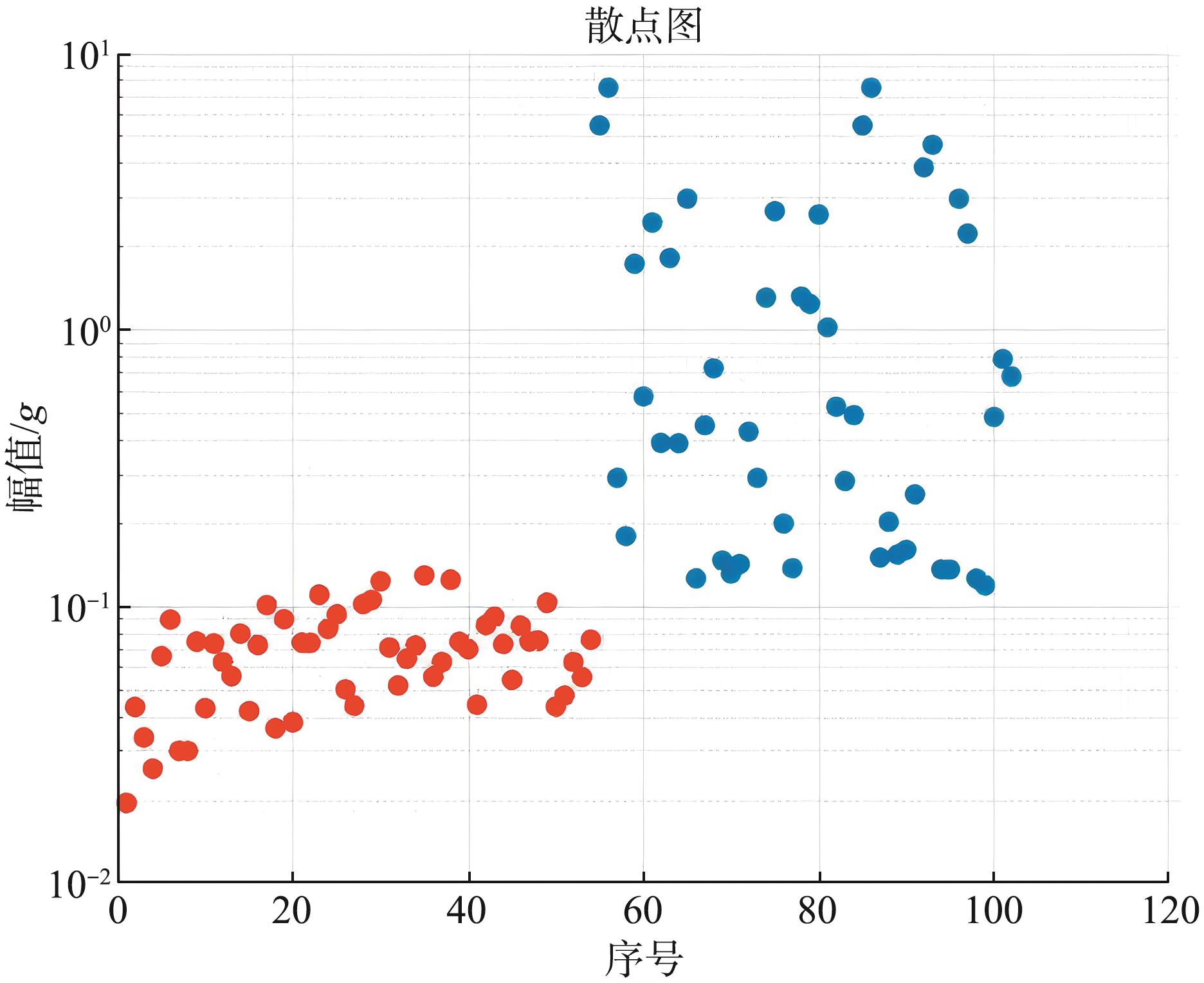
目的 本试验通过搭建颞下颌关节声学检测系统,比较患有可复性关节盘前移位(ADDWR)和健康的颞下颌关节在开闭口运动中关节音振动参数的差异。 方法 选择51个ADDWR关节和51个健康关节为研究对象,通过颞下颌关节声学检测系统检测最大张闭口时TMJ的振动参数,包括峰幅值、振动总能量、不同频段(频率>800 Hz和≤800 Hz)的振动能量,使用Mann-Whitney非参数检验进行组间比较。 结果 ADDWR关节的振动总能量、峰幅值、各频段(>800 Hz和≤800 Hz)的振动能量均明显高于对照组(P<0.01),ADDWR关节振动多发生在开口期。 结论 与健康关节相比,ADDWR关节振动有特征性变化;颞下颌关节声学检测系统可作为一种动态、无创、经济的辅助检查和早期筛查工具,为未来颞下颌关节紊乱病的诊断研究提供新思路。
中图分类号:
| [1] | Durham J, Newton-John TR, Zakrzewska JM. Temporomandibular disorders[J]. BMJ, 2015, 350: h-1154. |
| [2] | Schiffman E, Ohrbach R, Truelove E, et al. Diagnostic criteria for temporomandibular disorders (DC/TMD) for clinical and research applications: Recommendations of the international RDC/TMD consortium network* and orofacial pain special interest group[J]. J Oral Facial Pain Headache, 2014, 28(1): 6-27. |
| [3] | Miernik M, Więckiewicz W. The basic conservative treatment of temporomandibular joint anterior disc displacement without reduction: review[J]. Adv Clin Exp Med, 2015, 24(4): 731-735. |
| [4] | 李榕, 赵青. 基于颞下颌关节思考成人安氏Ⅱ2分类错牙合畸形的治疗[J]. 国际口腔医学杂志, 2024, 51(6): 687-698. |
| Li R, Zhao Q. Treatment of adult class Ⅱ division 2 malocclusion based on temporomandibular joint[J]. Int J Stomatol, 2024, 51(6): 687-698. | |
| [5] | Dworkin SF, LeResche L, DeRouen T, et al. Asse-ssing clinical signs of temporomandibular disorders: reliability of clinical examiners[J]. J Prosthet Dent, 1990, 63(5): 574-579. |
| [6] | Leader JK, Robert Boston J, Rudy TE, et al. Quan-titative description of temporomandibular joint sounds: defining clicking, popping, egg shell crackling and footsteps on gravel[J]. J Oral Rehabil, 2001, 28(5): 466-478. |
| [7] | Deng M, Long X, Dong H, et al. Electrosonogra-phic characteristics of sounds from temporomandi-bular joint disc replacement[J]. Int J Oral Maxillofac Surg, 2006, 35(5): 456-460. |
| [8] | Tanzilli RA, Tallents RH, Katzberg RW, et al. Temporomandibular joint sound evaluation with an electronic device and clinical evaluation[J]. Clin Orthod Res, 2001, 4(2): 72-78. |
| [9] | Kręcisz K, Bączkowicz D, Kawala-Sterniuk A. Using nonlinear vibroartrographic parameters for age-related changes assessment in knee arthrokinematics[J]. Sensors, 2022, 22(15): 5549. |
| [10] | Widmalm SE, Dong Y, Li BX, et al. Unbalanced lateral mandibular deviation associated with TMJ sound as a sign in TMJ disc dysfunction diagnosis[J]. J Oral Rehabil, 2016, 43(12): 911-920. |
| [11] | Bączkowicz D, Majorczyk E. Joint motion quality in chondromalacia progression assessed by vibroa-coustic signal analysis[J]. PM R, 2016, 8(11): 1065-1071. |
| [12] | Łysiak A, Marciniak T, Bączkowicz D. Repeatability of the vibroarthrogram in the temporomandibular joints[J]. Sensors (Basel), 2022, 22(23): 9542. |
| [13] | Vasavi M, Ramaraju AV, Sajjan MCS, et al. An eva-luation of the effect of wearing complete dentures on temporomandibular joint vibrations over time using the joint vibration analyzer[J]. J Indian Pros-thodont Soc, 2023, 23(1): 30-37. |
| [14] | Md WK, Teresa Sierpinska MD P, Mba JR. Can devices for the instrumental analysis of temporomandibular joints be used interchangeably[J]. Cranio, 2021, 39(2): 165-171. |
| [15] | 奚晓菁, 李嘉良, 王琛, 等. 可复性关节盘前移位患者关节振动的特征性研究[J]. 口腔医学, 2017, 37(4): 336-340. |
| Xi XJ, Li JL, Wang C, et al. Characteristics of joint vibration of patients with anterior disc displacement with reduction in TMJ[J]. Stomatology, 2017, 37(4): 336-340. | |
| [16] | 黄超, 许向亮, 孙玉春, 等. 下颌髁突功能面三维运动轨迹的初步推算和模拟[J]. 中华口腔医学杂志, 2018, 53(10): 669-673. |
| Huang C, Xu XL, Sun YC, et al. A preliminary study on the three-dimensional trajectory of condyle[J]. Chin J Stomatol, 2018, 53(10): 669-673. | |
| [17] | Lee YH. Functional anatomy and vibration characteristics of the healthy temporomandibular joint: a narrative review[J]. J Korean Dent Sci, 2024, 17(1): 14-35. |
| [18] | Huang ZS, Lin XF, Li XL. Characteristics of temporomandibular joint vibrations in anterior disk displacement with reduction in adults[J]. Cranio, 2011, 29(4): 276-283. |
| [19] | Manfredini D, Basso D, Salmaso L, et al. Temporomandibular joint click sound and magnetic resonan-ce-depicted disk position: which relationship[J]. J Dent, 2008, 36(4): 256-260. |
| [20] | Young AL. Internal derangements of the temporomandibular joint: a review of the anatomy, diagnosis, and management[J]. J Indian Prosthodont Soc, 2015, 15(1): 2-7. |
| [21] | Oster C, Katzberg RW, Tallents RH, et al. Characteri-zation of temporomandibular joint sounds. A preliminary investigation with arthrographic correlation[J]. Oral Surg Oral Med Oral Pathol, 1984, 58(1): 10-16. |
| [22] | de Oliveira LRLB, Alves IDS, Vieira APF, et al. Temporomandibular joint: from anatomy to internal derangement[J]. Radiol Bras, 2023, 56(2): 102-109. |
| [23] | 夏文棣, 傅开元. 颞下颌关节盘前移位对下颌功能和形态的影响及其生物学机制的研究进展[J]. 中华口腔医学杂志, 2016, 51(3): 182-184. |
| Xia WD, Fu KY. The influences of anterior disc displacement on oral mandibular function and morpho-logy and their biological mechanisms[J]. Chin J Stomatol, 2016, 51(3): 182-184. | |
| [24] | Topaloglu Yasan G, Adiloglu S, Tuz HH, et al. Eva-luation of clinical signs and magnetic resonance imaging findings in patients with temporomandibular disorders[J]. J Craniomaxillofac Surg, 2023, 51(7/8): 441-447. |
| [25] | Xu Y, Zhan J, Zheng Y, et al. Synovial fluid dyna-mics with small disc perforation in temporomandi-bular joint[J]. J Oral Rehabil, 2012, 39(10): 719-726. |
| [26] | Lalue-Sanches M, Gonzaga AR, Guimaraes AS, et al. Disc displacement with reduction of the temporomandibular joint: the real need for treatment[J]. J Pain Relief, 2015, 4(5): 2-5. |
| [27] | Gupta B, Thumati P, Radke J. Temporomandibular joint vibrations from totally asymptomatic subjects[J]. Cranio, 2016, 34(3): 169-175. |
| [28] | Deregibus A, Castroflorio T, De Giorgi I, et al. Could different TMJ disc positions observed in MRI cause different sounds? Analysis on a group of subjects with ADD with reduction: a pilot study[J]. Cranio, 2014, 32(4): 265-274. |
| [29] | Jacewicz E, Alexander JM, Fox RA. Introduction to the special issue on perception and production of sounds in the high-frequency range of human speecha[J]. J Acoust Soc Am, 2023, 154(5): 3168-3172. |
| [30] | Widmalm SE, Bae HEK, Djurdjanovic D, et al. Inau-dible temporomandibular joint vibrations[J]. Cranio, 2006, 24(3): 207-212. |
| [31] | Grochala J, Grochala D, Kajor M, et al. A novel method of temporomandibular joint hypermobility diagnosis based on signal analysis[J]. J Clin Med, 2021, 10(21): 5145. |
| [32] | Sharma S, Crow HC, Kartha K, et al. Reliability and diagnostic validity of a joint vibration analysis device[J]. BMC Oral Health, 2017, 17(1): 56. |
| [33] | Srinivasan R, Gupta S, Chander NG, et al. Clinical validation of the tmd-e tracker for diagnosing temporomandibular disorders: a reliability and accuracy assessment[J]. J Oral Biol Craniofac Res, 2025, 15(4): 799-803. |
| [34] | Paiva G, Paiva PF, de Oliveira ON. Vibrations in the temporomandibular joints in patients examined and treated in a private clinic[J]. Cranio, 1993, 11(3): 202-205. |
| [35] | 王碧娟, 梁甲兴. 食物硬度对口颌系统发育的影响[J]. 国外医学·口腔医学分册, 2003, 30(5): 411-413. |
| Wang BJ, Liang JX. The effect of food hardness on orofacial development[J]. Foreign Med Sci (Stomatol), 2003, 30(5): 411-413. | |
| [36] | Ferreira LA, Grossmann E, Januzzi E, et al. Diag-nosis of temporomandibular joint disorders: indication of imaging exams[J]. Braz J Otorhinolaryngol, 2016, 82(3): 341-352. |
| [37] | Kulesa-Mrowiecka M, Barański R, Kłaczyński M. sEMG and vibration system monitoring for differential diagnosis in temporomandibular joint disorders[J]. Sensors (Basel), 2022, 22(10): 3811. |
| [1] | 李榕,赵青. 基于颞下颌关节思考成人安氏Ⅱ2分类错 畸形的治疗[J]. 国际口腔医学杂志, 2024, 51(6): 687-698. |
| [2] | 张静,钟亦思,郑耘昊,张莉,熊鑫. 不同类型颞下颌关节紊乱病患者焦虑抑郁情绪的差异[J]. 国际口腔医学杂志, 2024, 51(3): 296-302. |
| [3] | 李婧瑜 王艳民综述 白丁审校. 正中关系的研究现状[J]. 国际口腔医学杂志, 2013, 40(4): 508-510. |
| [4] | 杨娴睿1 李春洁2 史宗道3 苏乃川4. 其他疾病误诊为颞下颌关节紊乱病及有关此类文献命名术语的探讨[J]. 国际口腔医学杂志, 2013, 40(1): 28-32. |
| [5] | 黄玉辉 王璟综述 赖文莉审校. Activator 矫治对颞下颌关节的影响及其与颞下颌关节紊乱病的关系[J]. 国际口腔医学杂志, 2011, 38(5): 563-566. |
| [6] | 邢晓伟综述 王东审校. 负荷改变对颞下颌关节软骨和软骨下骨的影响[J]. 国际口腔医学杂志, 2010, 37(5): 586-588. |
| [7] | 王璟,王晟综述 赖文莉审校. 正畸治疗与颞下颌关节紊乱病关系的研究进展[J]. 国际口腔医学杂志, 2009, 36(1): 38-38~40. |
| [8] | 蒋峰, 谷志远, 张雄, 陈建平. 76 例颞下颌关节紊乱病患者许氏位片中髁突位置的分析[J]. 国际口腔医学杂志, 2008, 35(2): 111-113. |
| [9] | 王喜军1综述 易新竹2审校. 雌激素与颞下颌关节紊乱病[J]. 国际口腔医学杂志, 2008, 35(2): 138-140. |
| [10] | 黄卓珊综述 林雪峰审校. 头颈姿势位与颞下颌关节紊乱病的相关性[J]. 国际口腔医学杂志, 2008, 35(1): 74-76. |
| [11] | 尚海燕,陈永进,. 心理因素与颞下颌关节紊乱病的研究进展[J]. 国际口腔医学杂志, 2007, 34(04): 275-277. |
| [12] | 周艺群,吴慧玲,谷志远,顾昕,. 关节盘移位后髁突软骨c-fos基因转录及表达的研究[J]. 国际口腔医学杂志, 2007, 34(04): 239-242. |
| [13] | 焦国华,张志光,. 颞下颌关节紊乱病疼痛的分子机制[J]. 国际口腔医学杂志, 2007, 34(02): 116-118. |
| [14] | 李雪铃综述 林雪峰审校. 磨牙症与颞下颌关节紊乱病关系的研究[J]. 国际口腔医学杂志, 2007, 34(01): 59-59~61. |
| [15] | 陈睿,江婷,阎英. 颞下颌关节紊乱病与颈椎功能紊乱的相互关系[J]. 国际口腔医学杂志, 2005, 32(06): 480-482. |
|
||