国际口腔医学杂志 ›› 2025, Vol. 52 ›› Issue (3): 323-332.doi: 10.7518/gjkq.2025039
周婕妤1( ),赵蕾1,吴亚菲1,李勇2,赵寰1()
Jieyu Zhou1(),Lei Zhao1,Yafei Wu1,Yong Li2,Huan Zhao1()
摘要:
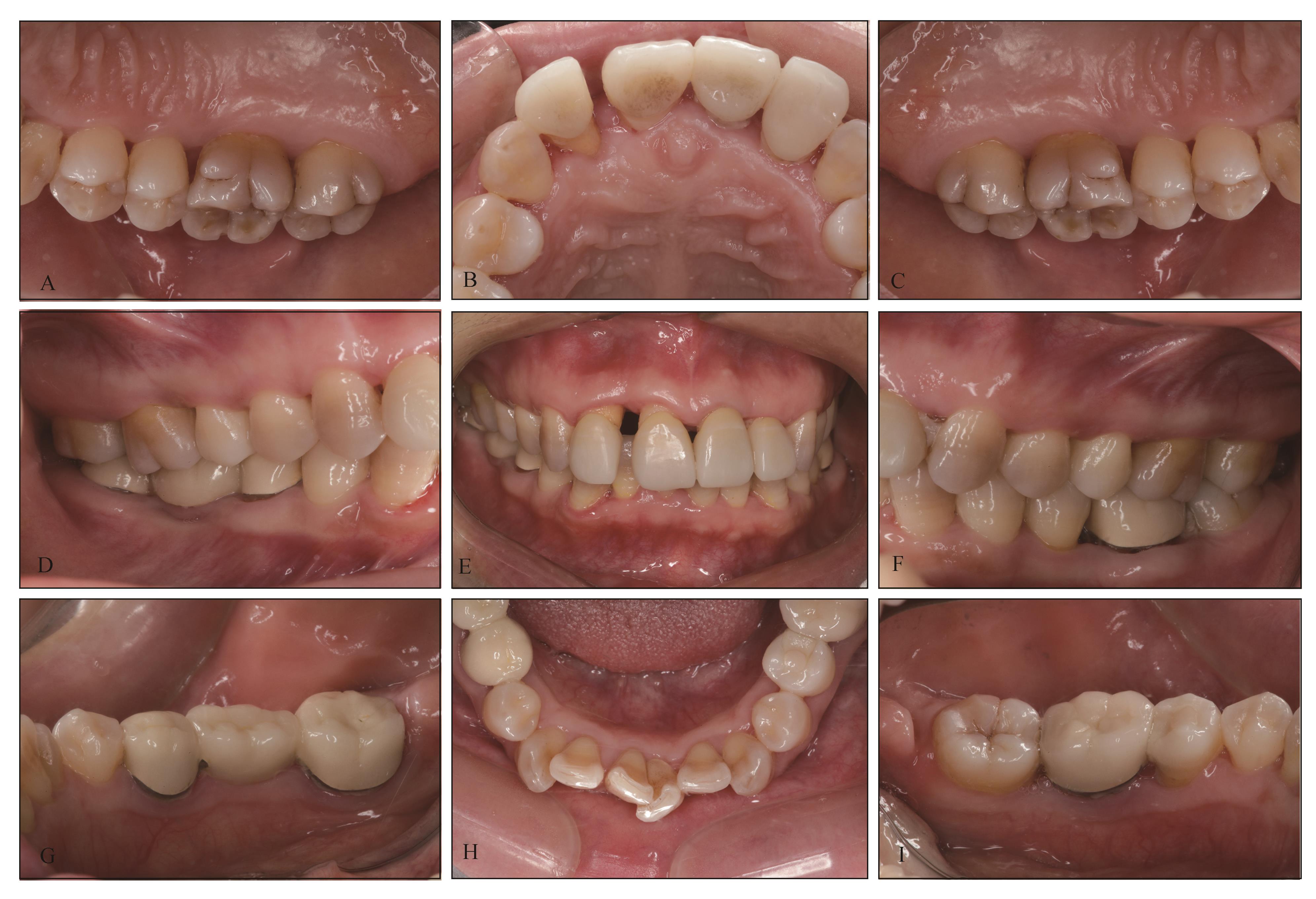
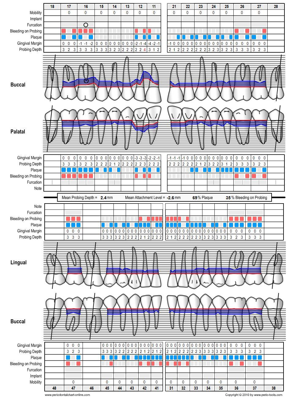
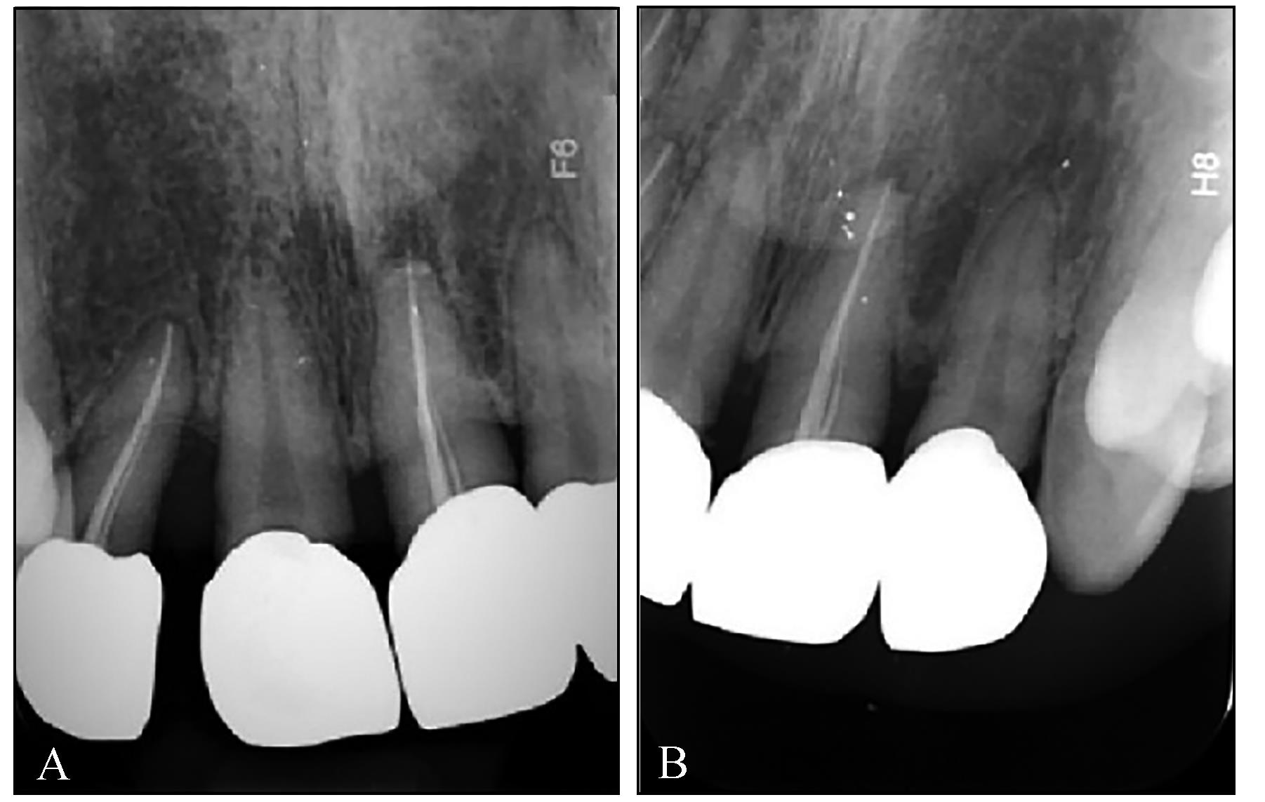
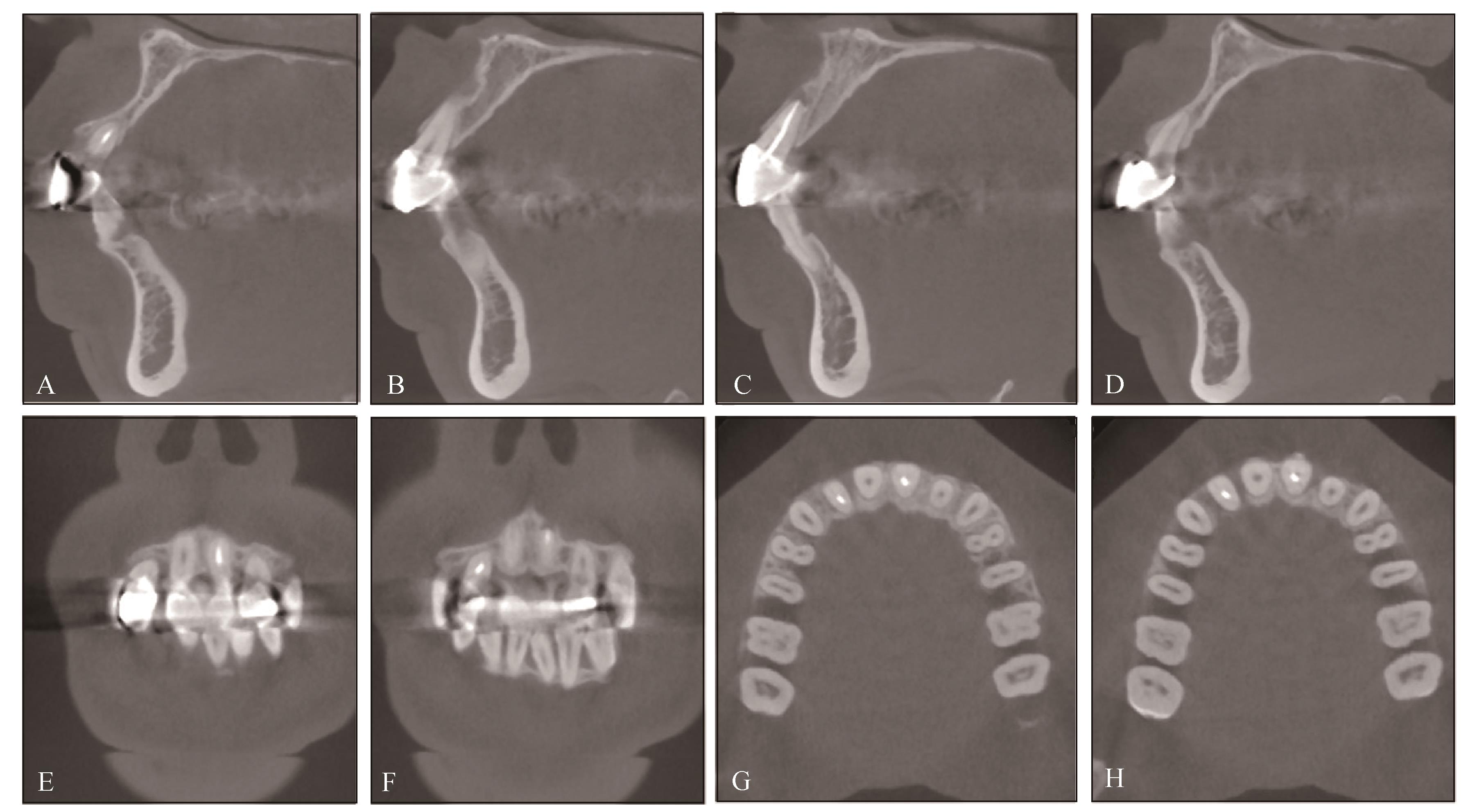
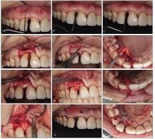
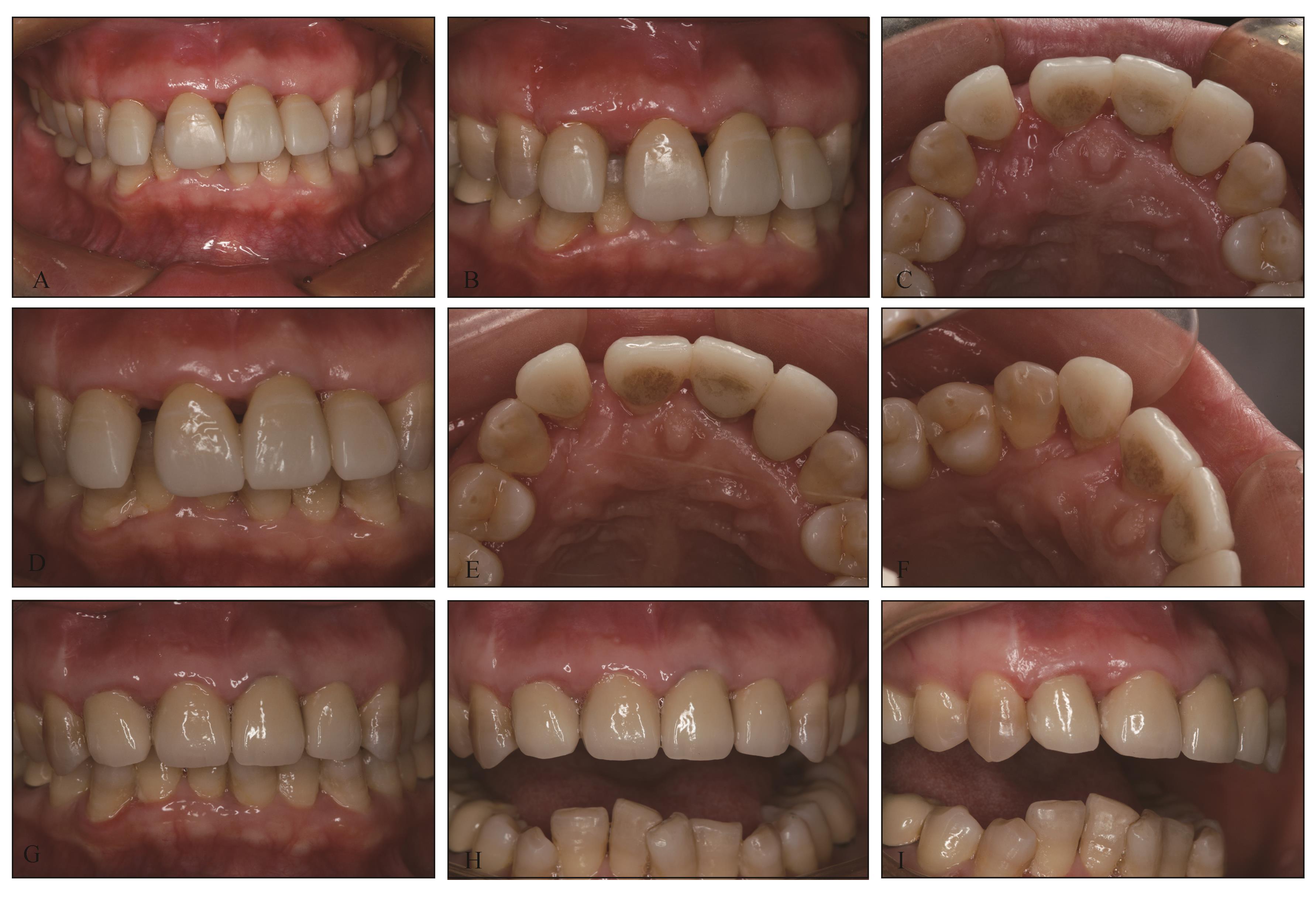
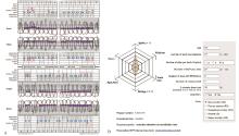
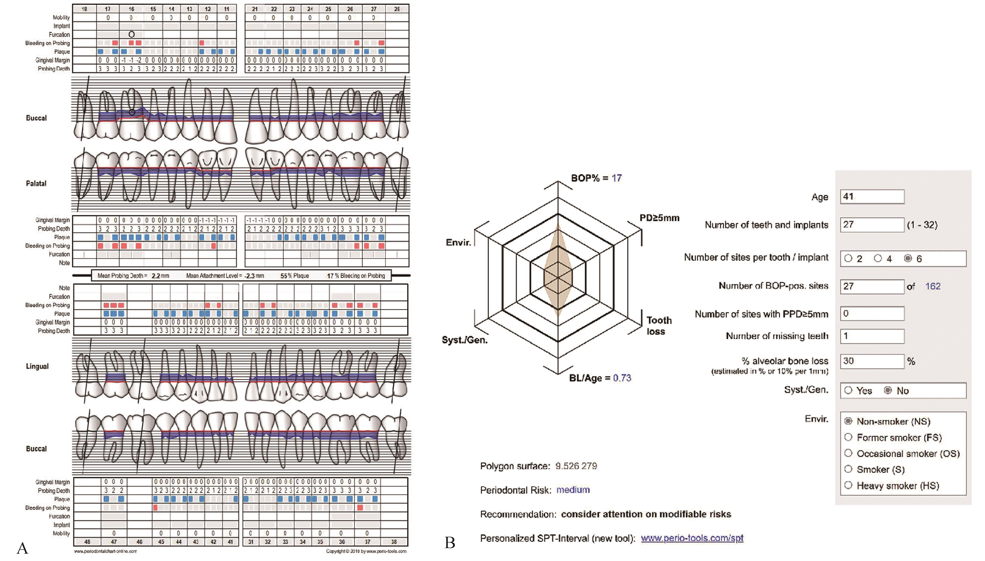
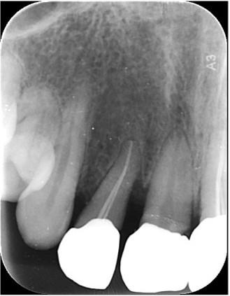
前牙美学区由于牙周炎、局部解剖因素、机械损伤或异常 力等原因易出现牙龈退缩、牙齿移位形成的“黑三角”的现象,是目前口腔临床治疗的难点之一。血管化腭骨膜间结缔组织(VIP-CT)瓣是一种上腭前蒂结缔组织瓣,血供充足,可促进骨移植物成骨,改善邻间隙软组织缺损。本文报道了1例前牙区牙龈退缩伴牙齿移位、龈乳头缺失的病例,经过VIP-CT转瓣移植和骨增量手术后软硬组织状况显著改善,随后联合冠修复恢复邻面触点,最终有效重建了美学区龈乳头,获得了理想的美学结果。
中图分类号:
| 1 | Chen J, Chen J, Chiang C, et al. Esthetic evaluation of natural teeth in anterior maxilla using the pink and white esthetic scores[J]. Clin Implant Dent Relat Res, 2018, 20(5): 770-777. |
| 2 | Singh VP, Uppoor AS, Nayak DG, et al. Black triangle dilemma and its management in esthetic dentis-try[J]. Dent Res J, 2013, 10(3): 296-301. |
| 3 | Ahmad I. Anterior dental aesthetics: gingival perspective[J]. Br Dent J, 2005, 199(4): 195-202. |
| 4 | Tarnow DP, Magner AW, Fletcher P. The effect of the distance from the contact point to the crest of bone on the presence or absence of the interproximal dental papilla[J]. J Periodontol, 1992, 63(12): 995-996. |
| 5 | Cho HS, Jang HS, Kim DK, et al. The effects of interproximal distance between roots on the existence of interdental papillae according to the distance from the contact point to the alveolar crest[J]. J Perio-dontol, 2006, 77(10): 1651-1657. |
| 6 | Joshi K, Baiju CS, Khashu H, et al. Clinical assessment of interdental papilla competency parameters in the esthetic zone[J]. J Esthet Restor Dent, 2017, 29(4): 270-275. |
| 7 | Shanmugam M, Kumar TS, Arun KV, et al. Clinical and histological evaluation of two dressing mate-rials in the healing of palatal wounds[J]. J Indian Soc Periodontol, 2010, 14(4): 241-244. |
| 8 | Agarwal C, Deora S, Abraham D, et al. Vasculari-zed interpositional periosteal connective tissue flap: a modern approach to augment soft tissue[J]. J In-dian Soc Periodontol, 2015, 19(1): 72-77. |
| 9 | Cardaropoli D, Re S, Corrente G. The papilla pre-sence index (PPI): a new system to assess interproximal papillary levels[J]. Int J Periodontics Resto-rative Dent, 2004, 24(5): 488-492. |
| 10 | Belser UC, Grütter L, Vailati F, et al. Outcome eva-luation of early placed maxillary anterior single-tooth implants using objective esthetic criteria: a cross-sectional, retrospective study in 45 patients with a 2- to 4-year follow-up using pink and white esthetic scores[J]. J Periodontol, 2009, 80(1): 140-151. |
| 11 | Papapanou PN, Sanz M, Buduneli N, et al. Perio-dontitis: consensus report of workgroup 2 of the 2017 World Workshop on the Classification of Perio-dontal and Peri-Implant Diseases and Conditions[J]. J Periodontol, 2018, 89(): S173-S182. |
| 12 | Nunn ME, Fan JJ, Su XG, et al. Development of prognostic indicators using classification and regression trees for survival[J]. Periodontol 2000, 2012, 58(1): 134-142. |
| 13 | Lang NP, Tonetti MS. Periodontal risk assessment (PRA) for patients in supportive periodontal therapy (SPT)[J]. Oral Health Prev Dent, 2003, 1(1): 7-16. |
| 14 | Zucchelli G, Mounssif I, Marzadori M, et al. Connective tissue graft wall technique and enamel matrix derivative for the treatment of infrabony defects: case reports[J]. Int J Periodontics Restorative Dent, 2017, 37(5): 673-681. |
| 15 | Jyothi SG, Triveni MG, Mehta DS, et al. Evaluation of single-tooth replacement by an immediate implant covered with connective tissue graft as a biologic barrier[J]. J Indian Soc Periodontol, 2013, 17(3): 354-360. |
| 16 | Rahpeyma A, Khajehahmadi S. Modified VIP-CT flap in late maxillary alveolar cleft surgery[J]. J Craniomaxillofac Surg, 2014, 42(5): 432-437. |
| 17 | Adriaenssens P, Hermans M, Ingber A, et al. Palatal sliding strip flap: soft tissue management to restore maxillary anterior esthetics at stage 2 surgery: a cli-nical report[J]. Int J Oral Maxillofac Implants, 1999, 14(1): 30-36. |
| 18 | Nemcovsky CE, Artzi Z. Split palatal flap. Ⅱ. A surgical approach for maxillary implant uncovering in cases with reduced keratinized tissue: technique and clinical results[J]. Int J Periodontics Restorative Dent, 1999, 19(4): 385-393. |
| 19 | Mathews DP. The pediculated connective tissue graft: a technique for improving unaesthetic implant restorations[J]. Pract Proced Aesthet Dent, 2002, 14(9): 719-724. |
| 20 | 崔传江, 郭吉来, 王欣欣. 血管化骨膜-结缔组织夹层瓣修复上颌美学区大量软组织缺损14例临床分析[J]. 口腔医学, 2016, 36(8): 718-722. |
| Cui CJ, Guo JL, Wang XX. Clinical analysis of soft tissue augmentation by the vascularized interpositional periosteal-connective tissue graft technique in the maxillary aesthetic region[J]. Stomatology, 2016, 36(8): 718-722. | |
| 21 | 宁晔, 吴海珍, 陆钰, 等. VIP-CT瓣在美学区即刻种植即刻修复中的临床应用[J]. 安徽医学, 2016, 37(3): 301-304. |
| Ning Y, Wu HZ, Lu Y, et al. Clinical research of VIP-CT flap in immediate implant and immediate prosthesis in aesthetic zone[J]. Anhui Med J, 2016, 37(3): 301-304. | |
| 22 | Rahpeyma A, Khajehahmadi S. Esthetic management of gingival lesions in anterior maxilla: the role of VIP-CT flap, a technical note[J]. J Surg Tech Case Rep, 2014, 6(1): 12-14. |
| 23 | Muthukumar S, Ajit P, Sundararajan S, et al. Reconstruction of interdental papilla using autogenous bone and connective tissue grafts[J]. J Indian Soc Periodontol, 2016, 20(4): 464-467. |
| [1] | 赵美林,赵依琼,黄姣. Ⅲ期C级牙周炎正畸患者上前牙龈乳头缺陷伴牙龈退缩1例[J]. 国际口腔医学杂志, 2025, 52(3): 333-340. |
| [2] | 高丽,程佳佳,王妮,葛颂. 改良冠向复位隧道技术联合双交叉悬吊缝合技术治疗多牙位牙龈退缩1例[J]. 国际口腔医学杂志, 2025, 52(3): 341-348. |
| [3] | 张婧, 周思颖, 张新铎, 冯玉霞, 李健学. 黏性骨在口腔种植及牙周领域中的研究进展[J]. 国际口腔医学杂志, 2024, 51(4): 433-440. |
| [4] | 吴嘉馨,程兴群,吴红崑. 透明质酸在修复龈乳头退缩中的临床应用进展[J]. 国际口腔医学杂志, 2023, 50(3): 347-352. |
| [5] | 曹正国. 修复治疗相关的牙周问题考量[J]. 国际口腔医学杂志, 2022, 49(1): 1-11. |
| [6] | 郭淑娟, 刘倩, 丁一. 牙周病和植体周病国际新分类简介[J]. 国际口腔医学杂志, 2019, 46(2): 125-134. |
| [7] | 张停停,宗娟娟. 自体结缔组织移植术的研究现状[J]. 国际口腔医学杂志, 2019, 46(1): 89-93. |
| [8] | 郑黎薇, 邹静, 夏斌, 刘英群, 黄洋, 赵今. 儿童乳磨牙金属预成冠的修复治疗[J]. 国际口腔医学杂志, 2017, 44(2): 125-129. |
| [9] | 李雪 彭友俭. 成人前牙区黑三角病因的探讨[J]. 国际口腔医学杂志, 2014, 41(5): 567-570. |
| [10] | 施优灵1 韩光丽2. 正畸治疗中的牙龈退缩[J]. 国际口腔医学杂志, 2014, 41(1): 57-62. |
| [11] | 祝士雯 陈振琦. 唇腭裂近裂隙区牙龈退缩的研究进展[J]. 国际口腔医学杂志, 2013, 40(3): 385-388. |
| [12] | 张晨1 赵绮2综述秦红霞1审校. 冠向复位术治疗MillerⅠ、Ⅱ度牙龈退缩[J]. 国际口腔医学杂志, 2012, 39(5): 639-641. |
| [13] | 支方静综述 莫水学审校. 切牙区黑三角与正畸治疗的研究进展[J]. 国际口腔医学杂志, 2011, 38(5): 611-613. |
| [14] | 林萍, 叶平, 涂慧娟. 评价上颌中切牙龈乳头缺陷的初步报告[J]. 国际口腔医学杂志, 2009, 36(5): 524-526,530. |
| [15] | 张剑,王景云. 无汗型外胚叶发育不全患者的修复治疗[J]. 国际口腔医学杂志, 2004, 31(06): 465-466. |
|