国际口腔医学杂志 ›› 2021, Vol. 48 ›› Issue (4): 405-410.doi: 10.7518/gjkq.2021073
邵冰婷( ),曹丹,严斌()
Shao Bingting(),Cao Dan,Yan Bin()
摘要:
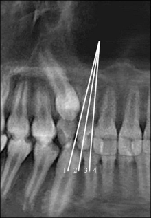
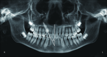
上颌阻生尖牙是临床上常见的难题。早期对可能发生阻生的上颌尖牙进行干预,常常可以减轻其阻生的严重程度,甚至可以使尖牙正常萌出。临床上常通过影像学方法对上颌尖牙阻生进行早期预测。其中影像学检查不仅包括传统的全口曲面体层片等二维影像,也包括近年来发展比较迅速的锥形束CT三维影像。本文通过回顾以往研究来总结目前影像学预测上颌尖牙阻生的研究进展,以指导临床早期预测上颌尖牙阻生并选择合适的干预时机。
中图分类号:
| [1] |
Alqerban A. Impacted maxillary canine in unilateral cleft lip and palate: a literature review[J]. Saudi Dent J, 2019,31(1):84-92.
doi: 10.1016/j.sdentj.2018.11.001 |
| [2] |
Mucedero M, Rozzi M, Milazzo A, et al. Morphometric analysis of the palatal shape and arch dimension in subjects with palatally displaced canine[J]. Eur J Orthod, 2019,41(5):460-467.
doi: 10.1093/ejo/cjy080 pmid: 30602006 |
| [3] |
Becker A, Chaushu S. Etiology of maxillary canine impaction: a review[J]. Am J Orthod Dentofacial Orthop, 2015,148(4):557-567.
doi: 10.1016/j.ajodo.2015.06.013 |
| [4] | Ortiz PM, Tabbaa S, Flores-Mir C, et al. A CBCT investigation of the association between sella-Turcica bridging and maxillary palatal canine impaction[J]. Biomed Res Int, 2018,2018:4329050. |
| [5] |
Sajnani AK. Permanent maxillary canines‒review of eruption pattern and local etiological factors lea-ding to impaction[J]. J Investig Clin Dent, 2015,6(1):1-7.
doi: 10.1111/jicd.12067 |
| [6] |
Alqerban A, Jacobs R, Fieuws S, et al. Radiographic predictors for maxillary canine impaction[J]. Am J Orthod Dentofacial Orthop, 2015,147(3):345-354.
doi: 10.1016/j.ajodo.2014.11.018 pmid: 25726402 |
| [7] |
Naoumova J, Kjellberg H. The use of panoramic radiographs to decide when interceptive extraction is beneficial in children with palatally displaced canines based on a randomized clinical trial[J]. Eur J Orthod, 2018,40(6):565-574.
doi: 10.1093/ejo/cjy002 pmid: 29462471 |
| [8] | 金玲玲, 阮文华. 上颌恒尖牙萌出障碍的研究进展[J]. 口腔医学, 2020,40(2):184-187. |
| Jin LL, Ruan WH. Research progress of the eruption abnormity of permanent maxillary canines[J]. Stoma-tology, 2020,40(2):184-187. | |
| [9] | 安舒, 姜春苗, 詹育香. 上颌尖牙阻生的诊断和早期干预[J]. 国际口腔医学杂志, 2011,38(6):721-724. |
| An S, Jiang CM, Zhan YX. The study on the diagno-sis and prevention of impacted maxillary canines[J]. Inter J Stomatol, 2011,38(6):721-724. | |
| [10] |
Zeno KG, Ghafari JG. Palatally impacted canines: a new 3-dimensional assessment of severity based on treatment objective[J]. Am J Orthod Dentofacial Orthop, 2018,153(3):387-395.
doi: 10.1016/j.ajodo.2017.07.020 |
| [11] |
Kim SH, Son WS, Yamaguchi T, et al. Assessment of the root apex position of impacted maxillary canines on panoramic films[J]. Am J Orthod Dentofacial Orthop, 2017,152(4):489-493.
doi: 10.1016/j.ajodo.2017.01.027 |
| [12] |
Mercuri E, Cassetta M, Cavallini C, et al. Dental anomalies and clinical features in patients with ma-xillary canine impaction[J]. Angle Orthod, 2013,83(1):22-28.
doi: 10.2319/021712-149.1 |
| [13] |
Kanavakis G, Curran KM, Wiseman KC, et al. Eva-luation of crown-root angulation of lateral incisors adjacent to palatally impacted canines[J]. Prog Orthod, 2015,16:4.
doi: 10.1186/s40510-015-0074-0 pmid: 25749110 |
| [14] |
Bertl MH, Foltin A, Lettner S, et al. Association between maxillary lateral incisors’ root volume and pa-latally displaced canines: an instrumental variables approach to the guidance theory[J]. Angle Orthod, 2018,88(6):719-725.
doi: 10.2319/020818-107.1 |
| [15] |
Alhaija ESA, Wazwaz FT. Third molar tooth agenesis and pattern of impaction in patients with palatally displaced canines[J]. Angle Orthod, 2019,89(1):64-70.
doi: 10.2319/031318-203.1 pmid: 30324806 |
| [16] |
Becker A, Chaushu S. Dental age in maxillary canine ectopia[J]. Am J Orthod Dentofacial Orthop, 2000,117(6):657-662.
doi: 10.1016/S0889-5406(00)70174-0 |
| [17] |
Rozylo-Kalinowska I, Kolasa-Raczka A, Kalinowski P. Dental age in patients with impacted maxillary canines related to the position of the impacted teeth[J]. Eur J Orthod, 2011,33(5):492-497.
doi: 10.1093/ejo/cjq123 pmid: 21262933 |
| [18] |
Demirjian A, Goldstein H. New systems for dental maturity based on seven and four teeth[J]. Ann Hum Biol, 1976,3(5):411-421.
pmid: 984727 |
| [19] |
Sajnani A, King N. Dental age of children and adolescents with impacted maxillary canines[J]. J Orofac Orthop, 2012,73(5):359-364.
pmid: 22948209 |
| [20] |
Uribe P, Ransjö M, Westerlund A. Clinical predictors of maxillary canine impaction: a novel approach using multivariate analysis[J]. Eur J Orthod, 2017,39(2):153-160.
doi: 10.1093/ejo/cjw042 |
| [21] |
Ericson S, Kurol J. Early treatment of palatally erup-ting maxillary canines by extraction of the primary canines[J]. Eur J Orthod, 1988,10(4):283-295.
doi: 10.1093/ejo/10.1.283 |
| [22] |
Lindauer SJ, Rubenstein LK, Hang WM, et al. Canine impaction identified early with panoramic radiographs[J]. J Am Dent Assoc, 1992,123(3): 91-92, 95-97.
doi: 10.14219/jada.archive.1992.0307 |
| [23] |
Power SM, Short MB. An investigation into the response of palatally displaced canines to the removal of deciduous canines and an assessment of factors contributing to favourable eruption[J]. Br J Orthod, 1993,20(3):215-223.
pmid: 8399054 |
| [24] | Alejos-Montante K, Martínez-Zumarán A, Torre-Delgadillo G, et al. Early identification of permanent ma-xillary canine impaction: a radiographic comparative study in a Mexican population[J]. J Clin Exp Dent, 2019,11(3):e282-e286. |
| [25] |
Warford JH Jr, Grandhi RK, Tira DE. Prediction of maxillary canine impaction using sectors and angular measurement[J]. Am J Orthod Dentofacial Orthop, 2003,124(6):651-655.
doi: 10.1016/S0889-5406(03)00621-8 |
| [26] |
Sajnani AK, King NM. Early prediction of maxillary canine impaction from panoramic radiographs[J]. Am J Orthod Dentofacial Orthop, 2012,142(1):45-51.
doi: 10.1016/j.ajodo.2012.02.021 pmid: 22748989 |
| [27] |
Alqerban A, Storms AS, Voet M, et al. Early prediction of maxillary canine impaction[J]. Dentomaxillofac Radiol, 2016,45(3):20150232.
doi: 10.1259/dmfr.20150232 |
| [28] | Laurenziello M, Montaruli G, Gallo C, et al. Determinants of maxillary canine impaction: retrospective clinical and radiographic study[J]. J Clin Exp Dent, 2017,9(11):e1304-e1309. |
| [29] | Malik DES, Fida M, Sukhia RH. Correlation between radiographic parameters for the prediction of palatally impacted maxillary canines[J]. J Orthod, 2019,46(1):6-13. |
| [30] |
Cacciatore G, Poletti L, Sforza C. Early diagnosed impacted maxillary canines and the morphology of the maxilla: a three-dimensional study[J]. Prog Orthod, 2018,19(1):20.
doi: 10.1186/s40510-018-0220-6 pmid: 30009340 |
| [31] |
Simões Holz I, Martinelli Carvalho R, Lauris JR, et al. Permanent canine eruption into the alveolar cleft region after secondary alveolar bone grafting: are there prediction factors for impaction[J]. Am J Orthod Dentofacial Orthop, 2018,154(5):657-663.
doi: 10.1016/j.ajodo.2018.01.016 |
| [32] | 慕创创, 李刚. 基于神经网络的深度学习在医学影像中的研究进展[J]. 中华口腔医学杂志, 2019,54(7):492-497. |
| Mu CC, Li G. Research progress in medical imaging based on deep learning of neural network[J]. Chin J Stomatol, 2019,54(7):492-497. | |
| [33] |
Chen S, Wang L, Li G, et al. Machine learning in orthodontics: introducing a 3D auto-segmentation and auto-landmark finder of CBCT images to assess maxillary constriction in unilateral impacted canine patients[J]. Angle Orthod, 2020,90(1):77-84.
doi: 10.2319/012919-59.1 pmid: 31403836 |
| [34] |
Eslami E, Barkhordar H, Abramovitch K, et al. Cone-beam computed tomography vs conventional radio-graphy in visualization of maxillary impacted-canine localization: a systematic review of comparative stu-dies[J]. Am J Orthod Dentofacial Orthop, 2017,151(2):248-258.
doi: S0889-5406(16)30651-5 pmid: 28153153 |
| [35] |
Jawad Z, Carmichael F, Houghton N, et al. A review of cone beam computed tomography for the diagnosis of root resorption associated with impacted canines, introducing an innovative root resorption scale[J]. Oral Surg Oral Med Oral Pathol Oral Radiol, 2016,122(6):765-771.
doi: 10.1016/j.oooo.2016.08.015 |
| [36] |
Cao D, Zhu L, Chen Y, et al. Buccally impacted ma-xillary canines increase the likelihood of root separation in adjacent first premolars[J]. Oral Dis, 2017,23(1):36-41.
doi: 10.1111/odi.12566 pmid: 27500892 |
| [37] | 李铎, 郑博文, 刘奕. 唇腭裂畸形患者蝶鞍鞍桥发生的研究[J]. 中华口腔正畸学杂志, 2018,25(4):196-200. |
| Li D, Zheng BW, Liu Y. Research on prevalence of sella bridge in patients with cleft lip and palate[J]. Chin J Orthod, 2018,25(4):196-200. | |
| [38] |
Ali B, Shaikh A, Fida M. Association between sella turcica bridging and palatal canine impaction[J]. Am J Orthod Dentofacial Orthop, 2014,146(4):437-441.
doi: 10.1016/j.ajodo.2014.06.010 |
| [39] |
Berkhout WE. The ALARA-principle. Backgrounds and enforcement in dental practices[J]. Ned Tijdschr Tandheelkd, 2015,122(5):263-270.
doi: 10.5177/ntvt.2015.5.14227 pmid: 26210218 |
| [40] | 梁宇红, 岳林. 锥形束CT在牙髓根尖周病诊治中的合理应用与思考[J]. 中华口腔医学杂志, 2019,54(9):591-597. |
| Liang YH, Yue L. A discussion on three-dimensional digital imaging technology: application of cone-beam CT in endodontics[J]. Chin J Stomatol, 2019,54(9):591-597. | |
| [41] | Abdelkarim A. Cone-beam computed tomography in orthodontics[J]. Dent J (Basel), 2019,7(3):89. |
| [42] | Grybienė V, Juozėnaitė D, Kubiliūtė K. Diagnostic methods and treatment strategies of impacted maxillary canines: a literature review[J]. Stomatologija, 2019,21(1):3-12. |
| [43] |
Almasoud NN. Extraction of primary canines for interceptive orthodontic treatment of palatally displa-ced permanent canines: a systematic review[J]. Angle Orthod, 2017,87(6):878-885.
doi: 10.2319/021417-105.1 pmid: 28800259 |
| [1] | 柴安,雷荣昌,蒋智升,曲彬彬,郭锦材,李鹤,邵奇. 评估下颌第三磨牙拔除难度分类方法的概述[J]. 国际口腔医学杂志, 2023, 50(6): 718-722. |
| [2] | 田浩楠,林敏,谢丛蔓,任嫒姝. 上颌腭侧阻生尖牙与寰椎后桥相关性的锥形束CT研究[J]. 国际口腔医学杂志, 2021, 48(5): 536-540. |
| [3] | 刘艺,刘奕. 上颌埋伏阻生尖牙与牙弓及腭部形态的相关性[J]. 国际口腔医学杂志, 2021, 48(2): 243-248. |
| [4] | 张旭,李妍熹,李涵识,魏洁雅,鄢鑫语,郑玮,李宇. 正畸软组织侧貌改变预测的研究进展[J]. 国际口腔医学杂志, 2019, 46(1): 105-111. |
| [5] | 陈静, 黄晓峰. 激光在口腔正畸临床应用中的进展[J]. 国际口腔医学杂志, 2017, 44(6): 712-716. |
| [6] | 李小兵,郭维华,黄诗言,徐舒豪,饶南荃,张扬根. 低位乳磨牙伴下颌第一前磨牙水平阻生1例[J]. 国际口腔医学杂志, 2016, 43(4): 377-379. |
| [7] | 刘莉,柯华峰,武传君,田军. 上颌腭侧阻生尖牙非手术与手术助萌的比较研究[J]. 国际口腔医学杂志, 2015, 42(2): 163-165. |
| [8] | 蔡勇涛1,2 罗顺云1 李旭艳2. 45°涡轮钻在第三磨牙拔除术中的改良应用[J]. 国际口腔医学杂志, 2013, 40(5): 577-579. |
| [9] | 姜世同1 刘军2 陈玉长2 王作君3 马德庆1 焦广军1 姜良坤1. 近中水平阻生下颌第三磨牙的矫治探索[J]. 国际口腔医学杂志, 2012, 39(6): 730-732. |
| [10] | 卿萍 高姗姗 朱卓立 樊弘毅 于海洋. 伴靠近下颌神经管埋伏牙的种植修复1 例[J]. 国际口腔医学杂志, 2012, 39(6): 736-738. |
| [11] | 任家银 王虎 李果 赵书平 吴万红 袁珊珊. 成都地区12 岁青少年第三磨牙发育的X 线分析[J]. 国际口腔医学杂志, 2012, 39(3): 297-300. |
| [12] | 赵书平 王虎 李果 任家银 吴万红 袁珊珊. 下颌第三磨牙近中阻生相关因素的头影测量分析[J]. 国际口腔医学杂志, 2012, 39(3): 305-307. |
| [13] | 安舒 姜春苗 詹育香综述 王军审校. 上颌尖牙阻生的诊断和早期干预[J]. 国际口腔医学杂志, 2011, 38(6): 721-725. |
| [14] | 姜世同1 王作君2 安忠军1 焦广军1 姜良坤1 罗圆圆1. 倒位埋伏阻生上中切牙的临床矫治探索[J]. 国际口腔医学杂志, 2011, 38(5): 515-517. |
| [15] | 杨洪1 谢德林2. 微种植支抗正畸矫正下颌磨牙重度阻生1 例[J]. 国际口腔医学杂志, 2011, 38(3): 286-287. |
|