Int J Stomatol ›› 2023, Vol. 50 ›› Issue (1): 82-90.doi: 10.7518/gjkq.2023007
• Methods • Previous Articles Next Articles
Zhu Yeling1( ),Wang Xi2,Sun Tiantong1,Wang Lina1,He Xiaoming1,Li Jiazhi1()
CLC Number:
| 1 | Janson G, Sathler R, Fernandes TM, et al. Correction of Class Ⅱ malocclusion with Class Ⅱ elastics: a systematic review[J]. Am J Orthod Dentofacial Orthop, 2013, 143(3): 383-392. |
| 2 | 胡敏, 相亚宁, 李洪, 等. 4种不同类型颌间牵引对颞下颌关节应力分布影响的三维有限元研究[J]. 华西口腔医学杂志, 2010, 28(2): 145-148. |
| Hu M, Xiang YN, Li H, et al. The stress distribution of the temporomandibular jiont under four types of inter-arch elastic-a three dimentional finite element model analysis[J]. West China J Stomatol, 2010, 28(2): 145-148. | |
| 3 | Wilmes B, Nienkemper M, Nanda R, et al. Palatally anchored maxillary molar mesialization using the mesialslider[J]. J Clin Orthod, 2013, 47(3): 172-179. |
| 4 | Laurentjoye M, Majoufre-Lefebvre C, Siberchicot F, et al. Result of maxillomandibular fixation using intraoral cortical bone screws for condylar fractures of the mandible[J]. J Oral Maxillofac Surg, 2009, 67(4): 767-770. |
| 5 | 傅民魁. 口腔正畸学[M]. 第6版, 北京: 人民卫生出版社, 2013: 233. |
| Fu MK. Orthodontics[M]. 6th ed. Beijing: People’s Medical Publishing House, 2013: 233. | |
| 6 | 付欣, 房玉镇, 叶之慧. 颌间牵引对颞下颌关节区应力分布及位移影响的有限元研究[J]. 中国体视学与图像分析, 2017, 22(4): 477-484. |
| Fu X, Fang YZ, Ye ZH. Finite element analysis of the effect of intermaxillary traction on stress distribution and displacement in temporomandibular joint[J]. Chin J Stereol Image Anal, 2017, 22(4): 477-484. | |
| 7 | 刘进. 正畸治疗中磨牙的拔除和保留(四十五)——牙槽外正畸支抗钉种植的部位及应用[J]. 临床口腔医学杂志, 2009, 25(8): 510-511. |
| Liu J. The location and application of skeletal anchorage[J]. Clin Stomatol, 2009, 25(8): 510-511. | |
| 8 | 邓海艳, 刘海霞. 影响种植支抗稳定性的因素分析[J]. 口腔医学, 2013, 33(7): 496-497. |
| Deng HY, Liu HX. Analysis of factors affecting the stability of planting anchorage[J]. Stomatology, 2013, 33(7): 496-497. | |
| 9 | Jeong GM, Sung SJ, Lee KJ, et al. Finite-element investigation of the center of resistance of the maxillary dentition[J]. Korean J Orthod, 2009, 39(2): 83-94. |
| 10 | Jo AR, Mo SS, Lee KJ, et al. Finite-element analysis of the center of resistance of the mandibular dentition[J]. Korean J Orthod, 2017, 47(1): 21-30. |
| 11 | 徐琳, 段银钟, 谭家莉, 等. 涉及正畸拔除病变第一磨牙矫治牙列拥挤前突的临床研究[J]. 口腔正畸学, 2008, 15(3): 114-117. |
| Xu L, Duan YZ, Tan JL, et al. Clinic study related to the extraction of the unhealthy first molars[J]. Chin J Orthod, 2008, 15(3): 114-117. | |
| 12 | Wise GE, King GJ. Mechanisms of tooth eruption and orthodontic tooth movement[J]. J Dent Res, 2008, 87(5): 414-434. |
| 13 | Andrews LF. Article 2: six element diagnostic record[J]. Andrews J Orthod Orofac Harm, 2001, 2(1): 15-20. |
| 14 | Andrews LF. The 6-elements orthodontic philosophy: treatment goals, classification, and rules for treating[J]. Am J Orthod Dentofacial Orthop, 2015, 148(6): 883-887. |
| 15 | 张珂, 白丁. Andrews口颌面协调六要素在侧貌美学中的应用[J]. 国际口腔医学杂志, 2010, 37(2): 236-239. |
| Zhang K, Bai D. Application of Andrews’ six elements of orofacial harmony in profile esthetics[J]. Int J Stomatol, 2010, 37(2): 236-239. | |
| 16 | Andrews LF. The six elements of orofacial harmony[J]. Andrews J Orthod Orofac Harm, 2000, 1(1): 13-22. |
| 17 | 陈玉梅, 汪海峰, 陈华. 正畸治疗中与颞下颌关节紊乱相关的因素分析[J]. 右江民族医学院学报, 2009, 31(2): 241. |
| Chen YM, Wang HF, Chen H. Analysis of factors related to temporomandibular joint disorder in ortho-dontic treament[J]. J Youjiang Med Col Natl, 2009, 31(2): 241. | |
| 18 | 张超. 微螺钉支抗种植体近中移动下颌第二磨牙的临床研究[J]. 现代预防医学, 2010, 37(17): 3372-3373. |
| Zhang C. Clinical study on gliding orthowire and the mesial movement of mandibular second molar[J]. Modern Prevent Med, 2010, 37(17): 3372-3373. | |
| 19 | Liou EJ, Chen PH, Wang YC, et al. A computed tomographic image study on the thickness of the infrazygomatic crest of the maxilla and its clinical implications for miniscrew insertion[J]. Am J Orthod Dentofacial Orthop, 2007, 131(3): 352-356. |
| 20 | 肖微, 曾妮, 胡帆, 等. 微螺钉支抗考虑的国人正常颌骨皮质厚度CT测量分析[J]. 中华口腔正畸学杂志, 2009, 16(3): 126-129. |
| Xiao W, Zeng N, Hu F, et al. Three dimensional evaluation of the thickness of bone cortex of the upper and lower jaw[J]. Chin J Orthod, 2009, 16(3): 126-129. | |
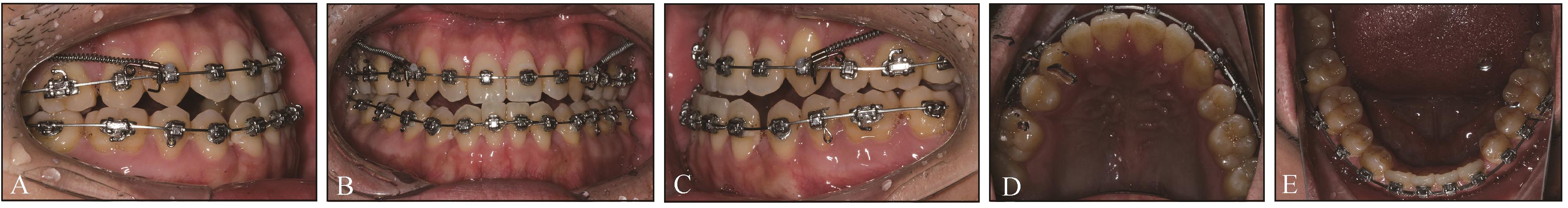
| 21 | 李加志, 刘进, 郭鑫. 种植支抗牵引全牙弓后移的临床初探[J]. 中华口腔正畸学杂志, 2011, 18(2): 76-83. |
| Li JZ, Liu J, Guo X. A pilot study on retraction of the whole dentition with skeletal anchorage[J]. Chin J Orthod, 2011, 18(2): 76-83. | |
| 22 | 曾祥龙. 正畸种植体支抗的发展、类型与应用[J]. 口腔正畸学, 2005, 12(1): 44-48. |
| Zeng XL. The development, pattern and application of orthodontic implant[J]. Chin J Orthod, 2005, 12(1): 44-48. |