国际口腔医学杂志 ›› 2021, Vol. 48 ›› Issue (5): 520-527.doi: 10.7518/gjkq.2021075
刘娟( ),陈斌,闫福华()
Liu Juan(),Chen Bin,Yan Fuhua()
摘要:
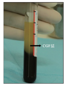
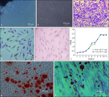
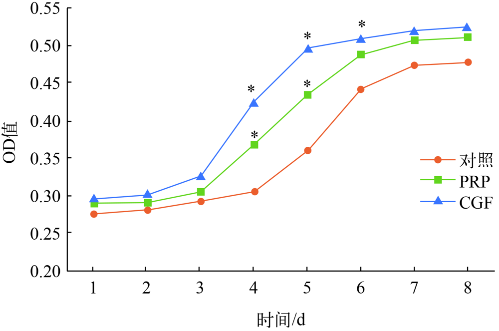
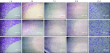
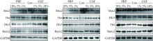
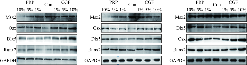
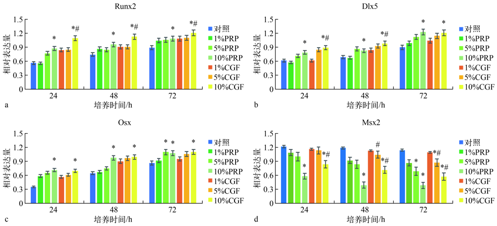
目的 研究富血小板血浆(PRP)和浓缩生长因子(CGF)对人牙周膜细胞(hPDLCs)增殖及成骨分化的影响。方法 分离、培养并鉴定hPDLCs;制备人全血、PRP和CGF,分别进行血小板计数;分别将全血(对照组)、PRP(PRP组)和CGF(CGF组)与hPDLCs共培养,CCK-8法检测细胞增殖能力,细胞划痕实验检测细胞迁移能力;使用1%、5%、10%的PRP和CGF分别对hPDLCs处理24、48、72 h,进行成骨诱导,Western blot法检测成骨相关转录因子Runx2、Osx、Dlx5和Msx2的相对表达量。结果 PRP、CGF可促进hPDLCs的增殖和迁移(P<0.05),提高促进成骨相关因子Runx2、Osx、Dlx5的表达并降低抑制成骨因子Msx2的表达(P<0.05)。结论 PRP、CGF能促进hPDLCs的增殖和迁移,并促进成骨分化相关转录因子的表达。
中图分类号:
| [1] | 闫福华, 李丽丽. 牙周再生治疗研究进展[J]. 口腔医学研究, 2018, 34(3):217-222. |
| Yan FH, Li LL. Development in periodontal regene-rative treatment[J]. J Oral Sci Res, 2018, 34(3):217-222. | |
| [2] |
Seo BM, Miura M, Gronthos S, et al. Investigation of multipotent postnatal stem cells from human pe-riodontal ligament[J]. Lancet, 2004, 364(9429):149-155.
doi: 10.1016/S0140-6736(04)16627-0 |
| [3] | Marx RE, Carlson ER, Eichstaedt RM, et al. Platelet-rich plasma: growth factor enhancement for bone grafts[J]. Oral Surg Oral Med Oral Pathol Oral Ra-diol Endod, 1998, 85(6):638-646. |
| [4] |
Weibrich G, Hansen T, Kleis W, et al. Effect of platelet concentration in platelet-rich plasma on peri-implant bone regeneration[J]. Bone, 2004, 34(4):665-671.
pmid: 15050897 |
| [5] |
Anitua E, Sánchez M, Orive G, et al. The potential impact of the preparation rich in growth factors (PR-GF) in different medical fields[J]. Biomaterials, 2007, 28(31):4551-4560.
doi: 10.1016/j.biomaterials.2007.06.037 |
| [6] | 张宇, 林野, 邱立新, 等. 富血小板血浆促进口腔种植骨再生的临床应用研究[J]. 中华口腔医学杂志, 2004, 39(4):269-272. |
| Zhang Y, Lin Y, Qiu LX, et al. Using platelet-rich plasma (PRP) to improve bone regeneration in im-plant bone defect[J]. Chin J Stomatol, 2004, 39(4):269-272. | |
| [7] |
Choi BH, Zhu SJ, Kim BY, et al. Effect of platelet-rich plasma (PRP) concentration on the viability and proliferation of alveolar bone cells: an in vitro study[J]. Int J Oral Maxillofac Surg, 2005, 34(4):420-424.
doi: 10.1016/j.ijom.2004.10.018 |
| [8] |
Bozkurt Doğan Ş, Öngöz Dede F, Ballı U, et al. Concentrated growth factor in the treatment of adjacent multiple gingival recessions: a split-mouth randomi-zed clinical trial[J]. J Clin Periodontol, 2015, 42(9):868-875.
doi: 10.1111/jcpe.12444 pmid: 26269089 |
| [9] |
Pirpir C, Yilmaz O, Candirli C, et al. Evaluation of effectiveness of concentrated growth factor on osseointegration[J]. Int J Implant Dent, 2017, 3(1):7.
doi: 10.1186/s40729-017-0069-3 |
| [10] |
Honda H, Tamai N, Naka N, et al. Bone tissue engineering with bone marrow-derived stromal cells integrated with concentrated growth factor in Rattus norvegicus calvaria defect model[J]. J Artif Organs, 2013, 16(3):305-315.
doi: 10.1007/s10047-013-0711-7 |
| [11] | 魏中武, 黄谢山, 陈灼庚. 浓缩生长因子在口腔临床中的应用及研究进展[J]. 国际口腔医学杂志, 2020, 47(2):235-243. |
| Wei ZW, Huang XS, Chen ZG. Application and re-search progress on concentrated growth factor in oral clinic[J]. Int J Stomatol, 2020, 47(2):235-243. | |
| [12] |
Loi F, Córdova LA, Pajarinen J, et al. Inflammation, fracture and bone repair[J]. Bone, 2016, 86:119-130.
doi: 10.1016/j.bone.2016.02.020 |
| [13] | Zhang XL, Shi KQ, Jia PT, et al. Effects of platelet-rich plasma on angiogenesis and osteogenesis-associated factors in rabbits with avascular necrosis of the femoral head[J]. Eur Rev Med Pharmacol Sci, 2018, 22(7):2143-2152. |
| [14] |
Yu B, Wang Z. Effect of concentrated growth factors on beagle periodontal ligament stem cells in vitro[J]. Mol Med Rep, 2014, 9(1):235-242.
doi: 10.3892/mmr.2013.1756 |
| [15] |
Fontana S, Olmedo DG, Linares JA, et al. Effect of platelet-rich plasma on the peri-implant bone respon-se: an experimental study[J]. Implant Dent, 2004, 13(1):73-78.
doi: 10.1097/01.ID.0000116455.68968.29 |
| [16] |
Bartold PM, Gronthos S. Standardization of criteria defining periodontal ligament stem cells[J]. J Dent Res, 2017, 96(5):487-490.
doi: 10.1177/0022034517697653 pmid: 28425840 |
| [17] | 刘娟, 赵红宇, 轩东英, 等. 人牙周膜细胞群多向分化潜能的实验研究[J]. 华西口腔医学杂志, 2010, 28(2):185-189. |
| Liu J, Zhao HY, Xuan DY, et al. Differentiation characteristics of human periodontal ligament cell population in vitro[J]. West China J Stomatol, 2010, 28(2):185-189. | |
| [18] | 廖海清, 曹正国. 经典Wnt信号通路在牙周膜细胞成骨分化过程中的调控[J]. 口腔医学研究, 2016, 32(3):224-227. |
| Liao HQ, Cao ZG. Effect of canonical Wnt signalling pathway on the osteogenic differentiation and proli-feration of PDLCs[J]. J Oral Sci Res, 2016, 32(3):224-227. | |
| [19] |
Tamiya H, Ikeda T, Jeong JH, et al. Analysis of the Runx2 promoter in osseous and non-osseous cells and identification of HIF2A as a potent transcription activator[J]. Gene, 2008, 416(1/2):53-60.
doi: 10.1016/j.gene.2008.03.003 |
| [20] |
Nakashima K, Zhou X, Kunkel G, et al. The novel zinc finger-containing transcription factor osterix is required for osteoblast differentiation and bone formation[J]. Cell, 2002, 108(1):17-29.
pmid: 11792318 |
| [21] |
Lee MH, Kim YJ, Kim HJ, et al. BMP-2-induced Runx2 expression is mediated by Dlx5, and TGF-beta 1 opposes the BMP-2-induced osteoblast differentiation by suppression of Dlx5 expression[J]. J Biol Chem, 2003, 278(36):34387-34394.
doi: 10.1074/jbc.M211386200 |
| [22] | 乔静, 欧阳翔英, 曹采方. 富血小板血浆对人牙周膜细胞增殖及分化的影响[J]. 上海口腔医学, 2008, 17(1):60-63. |
| Qiao J, Ouyang XY, Cao CF. The effect of different concentrations of platelet-rich plasma on human pe-riodontal ligament cells in vitro[J]. Shanghai J Sto-matol, 2008, 17(1):60-63. | |
| [23] | 钟声, 闫福华, 卢友光, 等. 富血小板血浆对牙周膜成纤维细胞损伤模型影响的研究[J]. 口腔医学研究, 2005, 21(5):504-506. |
| Zhong S, Yan FH, Lu YG, et al. Effect of platelet-rich plasma (PRP) on periodontal ligament fibroblasts in an in vitro wound healing model[J]. J Oral Sci Res, 2005, 21(5):504-506. | |
| [24] |
Durmuşlar MC, Balli U, Dede FÖ, et al. Histological evaluation of the effect of concentrated growth factor on bone healing[J]. J Craniofac Surg, 2016, 27(6):1494-1497.
doi: 10.1097/SCS.0000000000002873 pmid: 27428921 |
| [1] | 古丽其合热·阿布来提,秦旭,朱光勋. 线粒体自噬在牙周炎发生发展过程中的研究进展[J]. 国际口腔医学杂志, 2024, 51(1): 68-73. |
| [2] | 刘体倩,梁星,刘蔚晴,李晓虹,朱睿. 咬合创伤在牙周炎发生发展中的作用及机制的研究进展[J]. 国际口腔医学杂志, 2023, 50(1): 19-24. |
| [3] | 周灿,曾倩,韦曦. 浓缩生长因子在活髓保存治疗中的应用前景[J]. 国际口腔医学杂志, 2022, 49(6): 684-689. |
| [4] | 洪娅娅,陈学鹏,姒蜜思. 非编码RNA调控牙囊干细胞成骨分化的研究进展[J]. 国际口腔医学杂志, 2022, 49(3): 263-271. |
| [5] | 郭雨婷,吕学超. 药物调控牙髓干细胞成骨分化的研究进展[J]. 国际口腔医学杂志, 2021, 48(6): 737-744. |
| [6] | 李静雅,税钰森,郭永文. 循环牵张应力影响人牙周膜细胞成骨分化机制的研究进展[J]. 国际口腔医学杂志, 2020, 47(6): 652-660. |
| [7] | 魏中武,黄谢山,陈灼庚. 浓缩生长因子在口腔临床中的应用及研究进展[J]. 国际口腔医学杂志, 2020, 47(2): 235-243. |
| [8] | 余晓宏,刘屿,曾莲,杨艳玲,王洲,李卫. 釉基质衍生物对人牙周膜干细胞成骨分化的影响[J]. 国际口腔医学杂志, 2020, 47(1): 24-31. |
| [9] | 周婷茹,李永生. 牙髓干细胞成骨微环境的研究进展[J]. 国际口腔医学杂志, 2019, 46(6): 675-679. |
| [10] | 梅宏翔,张懿丹,张城浩,刘恩言,陈昊,赵志河,廖文. 表没食子儿茶素没食子酸酯在干细胞增殖及成骨分化作用中的研究现状[J]. 国际口腔医学杂志, 2019, 46(4): 431-436. |
| [11] | 胡巍,王译凡,袁一方,李影,郭斌. 节律基因调控成骨和破骨活动机制的研究进展[J]. 国际口腔医学杂志, 2019, 46(3): 302-307. |
| [12] | 杨宇轩,张海霞,王爽. 釉原蛋白在牙周组织再生中的生物学作用[J]. 国际口腔医学杂志, 2019, 46(2): 191-196. |
| [13] | 王瑜,王伟,顾新华. 浓缩生长因子在种植软硬组织增量方面的研究及应用[J]. 国际口腔医学杂志, 2019, 46(2): 218-222. |
| [14] | 李媛媛,程斌,王韵. 长链非编码RNA lnc-p26090对口腔鳞状细胞癌细胞糖酵解及增殖的影响[J]. 国际口腔医学杂志, 2018, 45(6): 628-634. |
| [15] | 李婷婷,张玉峰,王若茜,黄智庆,谢律,薛艺凡,王宇蓝. 石墨烯及其衍生物改性复合材料促成骨机制和应用的研究进展[J]. 国际口腔医学杂志, 2018, 45(6): 673-677. |
|