国际口腔医学杂志 ›› 2021, Vol. 48 ›› Issue (4): 417-425.doi: 10.7518/gjkq.2021070
方苓力( ),谭玺,叶雨丝,黄兰,何瑶()
Fang Lingli(),Tan Xi,Ye Yusi,Huang Lan,He Yao()
摘要:
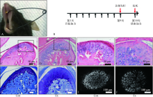
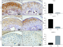
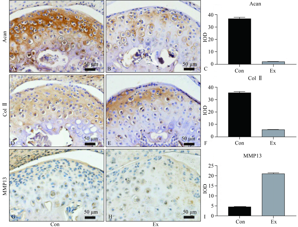
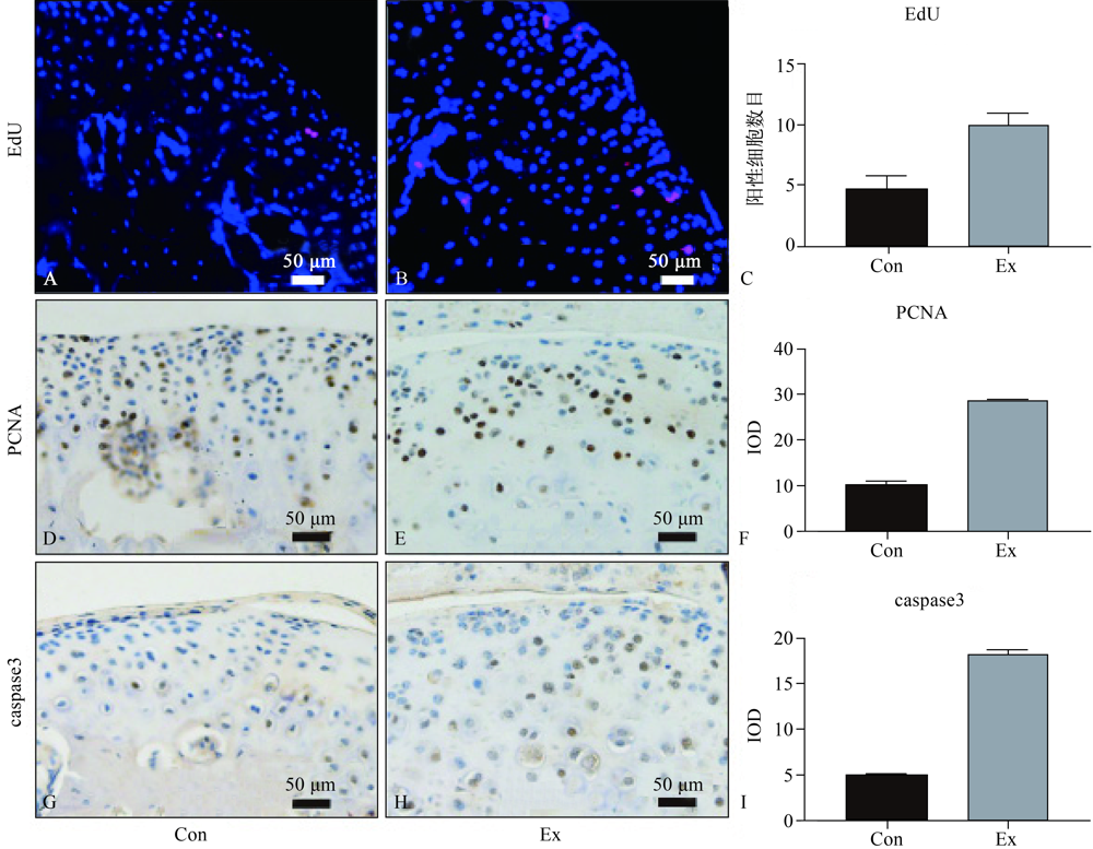
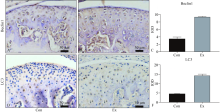
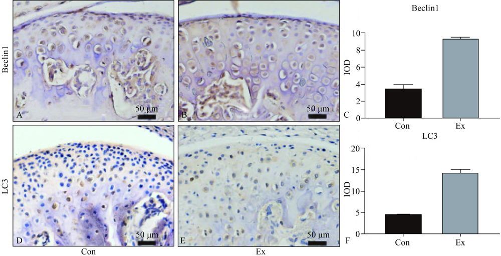
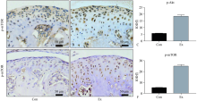
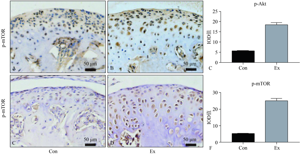
目的 探索应力诱导颞下颌关节(TMJ)退行性变早期,髁突软骨细胞增殖、凋亡和自噬行为表现及相关信号通路的变化。方法 使用小鼠强制张口模型分别对小鼠加力0 d(对照组)和10 d(实验组),加力结束后取样。髁突软骨行激光共聚焦扫描,5-乙炔基-2’脱氧尿嘧啶核苷(EdU)染色检测增殖;完整关节区切片行苏木精/伊红、甲苯胺蓝以及免疫组化染色检测增殖、凋亡、自噬及磷脂酰肌醇3-激酶/丝氨酸/苏氨酸蛋白激酶(PI3K/Akt)通路关键蛋白的表达。结果 实验组小鼠与对照组小鼠相比,髁突软骨增厚,细胞密度降低,基质分泌减少;且增殖和凋亡阳性细胞增多,自噬活性增加;伴随PI3K/Akt通路活化。结论 应力诱导TMJ退行性变早期,软骨细胞在增殖、凋亡及自噬活性上均有一定程度的激活,并伴随了PI3K/Akt通路活化。
中图分类号:
| [1] |
Wang XD, Zhang JN, Gan YH, et al. Current understanding of pathogenesis and treatment of TMJ osteoarthritis[J]. J Dent Res, 2015,94(5):666-673.
doi: 10.1177/0022034515574770 |
| [2] |
Wieckiewicz M, Boening K, Wiland P, et al. Reported concepts for the treatment modalities and pain m-anagement of temporomandibular disorders[J]. J Headache Pain, 2015,16:106.
doi: 10.1186/s10194-015-0586-5 pmid: 26644030 |
| [3] |
Ernberg M. The role of molecular pain biomarkers in temporomandibular joint internal derangement[J]. J Oral Rehabil, 2017,44(6):481-491.
doi: 10.1111/joor.12480 pmid: 28054366 |
| [4] |
Zhang SP, Teo KYW, Chuah SJ, et al. MSC exosomes alleviate temporomandibular joint osteoarthritis by attenuating inflammation and restoring matrix homeostasis[J]. Biomaterials, 2019,200:35-47.
doi: 10.1016/j.biomaterials.2019.02.006 |
| [5] |
Kurio N, Saunders C, Bechtold TE, et al. Roles of Ihh signaling in chondroprogenitor function in postnatal condylar cartilage[J]. Matrix Biol, 2018,67:15-31.
doi: 10.1016/j.matbio.2018.02.011 |
| [6] |
Koyama E, Saunders C, Salhab I, et al. Lubricin is required for the structural integrity and post-natal maintenance of TM[J]. J Dent Res, 2014,93(7):663-670.
doi: 10.1177/0022034514535807 |
| [7] |
Sperry MM, Yu YH, Kartha S, et al. Intra-articular etanercept attenuates pain and hypoxia from TMJ loading in the rat[J]. J Orthop Res, 2020,38(6):1316-1326.
doi: 10.1002/jor.v38.6 |
| [8] |
Tanaka E, Detamore MS, Mercuri LG. Degenerative disorders of the temporomandibular joint: etiology, diagnosis, and treatment[J]. J Dent Res, 2008,87(4):296-307.
pmid: 18362309 |
| [9] | 康宏. 颞下颌关节的生物力学[J]. 生物医学工程学杂志, 2000,17(3):324-327, 345. |
| Kang H. Biomechanics of temporomandibular joint[J]. J Biomed Eng, 2000,17(3):324-327, 345. | |
| [10] |
Tanaka E, Koolstra JH. Biomechanics of the temporomandibular joint[J]. J Dent Res, 2008,87(11):989-991.
doi: 10.1177/154405910808701101 |
| [11] |
Sobue T, Yeh WC, Chhibber A, et al. Murine TMJ loading causes increased proliferation and chondrocyte maturation[J]. J Dent Res, 2011,90(4):512-516.
doi: 10.1177/0022034510390810 |
| [12] |
Utreja A, Dyment NA, Yadav S, et al. Cell and matrix response of temporomandibular cartilage to mechanical loading[J]. Osteoarthritis Cartilage, 2016,24(2):335-344.
doi: 10.1016/j.joca.2015.08.010 |
| [13] |
Tanaka E, Aoyama J, Miyauchi M, et al. Vascular endothelial growth factor plays an important autocrine/paracrine role in the progression of osteoarthritis[J]. Histochem Cell Biol, 2005,123(3):275-281.
pmid: 15856277 |
| [14] | Fujisawa T, Kuboki T, Kasai T, et al. A repetitive, steady mouth opening induced an osteoarthritis-like lesion in the rabbit temporomandibular joint[J]. J D-ent Res, 2003,82(9):731-735. |
| [15] |
Ou FR, Su K, Sun JD, et al. Temporomandibular joint disorders contribute to anxiety in BalB/C mice[J]. Biochem Biophys Res Commun, 2019,516(2):339-343.
doi: 10.1016/j.bbrc.2019.06.050 |
| [16] |
Fujita M, Sato-Shigeta M, Mori H, et al. Protective effects of low-intensity pulsed ultrasound on mandibular condylar cartilage exposed to mechanical overloading[J]. Ultrasound Med Biol, 2019,45(4):944-953.
doi: 10.1016/j.ultrasmedbio.2018.12.006 |
| [17] |
Liu Q, Yang HX, Duan J, et al. Bilateral anterior elevation prosjournal boosts chondrocytes proliferation in mice mandibular condyle[J]. Oral Dis, 2019,25(6):1589-1599.
doi: 10.1111/odi.v25.6 |
| [18] |
Shen C, Cai GQ, Peng JP, et al. Autophagy protects chondrocytes from glucocorticoids-induced apoptosis via ROS/Akt/FOXO3 signaling[J]. Osteoarthritis Cartilage, 2015,23(12):2279-2287.
doi: 10.1016/j.joca.2015.06.020 |
| [19] |
Yang HX, Wen Y, Zhang M, et al. MTORC1 coordinates the autophagy and apoptosis signaling in articular chondrocytes in osteoarthritic temporomandibular joint[J]. Autophagy, 2020,16(2):271-288.
doi: 10.1080/15548627.2019.1606647 |
| [20] |
Chang J, Wang W, Zhang H, et al. The dual role of autophagy in chondrocyte responses in the pathogenesis of articular cartilage degeneration in osteoarth-ritis[J]. Int J Mol Med, 2013,32(6):1311-1318.
doi: 10.3892/ijmm.2013.1520 |
| [21] |
Zhang M, Zhang J, Lu L, et al. Enhancement of chondrocyte autophagy is an early response in the degenerative cartilage of the temporomandibular joint to biomechanical dental stimulation[J]. Apoptosis, 2013,18(4):423-434.
doi: 10.1007/s10495-013-0811-0 pmid: 23386193 |
| [22] | Chen H, Wu GY, Sun Q, et al. Hyperbaric oxygen protects mandibular condylar chondrocytes from interleukin-1β‒induced apoptosis via the PI3K/AKT signaling pathway[J]. Am J Transl Res, 2016,8(11):5108-5117. |
| [23] | Zhang QB, Lai SX, Hou XY, et al. Protective effects of PI3K/Akt signal pathway induced cell autophagy in rat knee joint cartilage injury[J]. Am J Transl Res, 2018,10(3):762-770. |
| [24] |
Cravero JD, Carlson CS, Im HJ, et al. Increased expression of the Akt/PKB inhibitor TRB3 in osteoarthritic chondrocytes inhibits insulin-like growth factor 1-mediated cell survival and proteoglycan synjournal[J]. Arthritis Rheum, 2009,60(2):492-500.
doi: 10.1002/art.v60:2 |
| [25] | Portal-Núñez S, Esbrit P, Alcaraz MJ, et al. Oxidative stress, autophagy, epigenetic changes and regulation by miRNAs as potential therapeutic targets in osteoarthritis[J]. Biochem Pharmacol, 2016,108:1-10. |
| [26] |
He Y, Zhang M, Huang AY, et al. Confocal imaging of mouse mandibular condyle cartilage[J]. Sci Rep, 2017,7:43848.
doi: 10.1038/srep43848 |
| [27] | Liu WJ, Luo HY, Wang RL, et al. Rapamycin-induced autophagy promotes the chondrogenic differentiation of synovium-derived mesenchymal stem cells in the temporomandibular joint in response to IL-1β[J]. Biomed Res Int, 2020,2020:4035306. |
| [28] |
Jing JJ, Hinton RJ, Mishina Y, et al. Critical role of Bmpr1a in mandibular condyle growth[J]. Connect Tissue Res, 2014,55(Suppl 1):73-78.
doi: 10.3109/03008207.2014.923858 |
| [29] |
Ogasawara N, Kano F, Hashimoto N, et al. Factors secreted from dental pulp stem cells show multifaceted benefits for treating experimental temporomandibular joint osteoarthritis[J]. Osteoarthritis Cartilage, 2020,28(6):831-841.
doi: 10.1016/j.joca.2020.03.010 |
| [30] | Ma DD, Kou XX, Jin J, et al. Hydrostatic compress force enhances the viability and decreases the apoptosis of condylar chondrocytes through integrin-FAK-ERK/PI3K pathway[J]. Int J Mol Sci, 2016,17(11):E1847. |
| [31] |
Grishko V, Xu M, Ho R, et al. Effects of hyaluronic acid on mitochondrial function and mitochondria-driven apoptosis following oxidative stress in human chondrocytes[J]. J Biol Chem, 2009,284(14):9132-9139.
doi: 10.1074/jbc.M804178200 |
| [32] | Yan XF, Wu HX, Wu ZY, et al. The new synthetic H2S-releasing SDSS protects MC3T3-E1 osteoblasts against H2O2-induced apoptosis by suppressing oxidative stress, inhibiting MAPKs, and activating the PI3K/akt pathway[J]. Front Pharmacol, 2017,8:7. |
| [33] | Zhang QB, Lai SX, Hou XY, et al. Protective effects of PI3K/Akt signal pathway induced cell autophagy in rat knee joint cartilage injury[J]. Am J Transl Res, 2018,10(3):762-770. |
| [34] |
Xiao ZH, Wang JK, Chen SY, et al. Autophagy promotion enhances the protective effect of morroniside on human OA chondrocyte[J]. Biosci Biotechnol Biochem, 2020,84(5):989-996.
doi: 10.1080/09168451.2020.1717925 |
| [35] |
Cao Y, Klionsky DJ. Physiological functions of Atg6/Beclin 1: a unique autophagy-related protein[J]. Cell Res, 2007,17(10):839-849.
doi: 10.1038/cr.2007.78 |
| [36] |
Arai A, Kim S, Goldshteyn V, et al. Beclin1 modulates bone homeostasis by regulating osteoclast and chondrocyte differentiation[J]. J Bone Miner Res, 2019,34(9):1753-1766.
doi: 10.1002/jbmr.v34.9 |
| [37] |
Yang ZM, Tang YX, Lu HD, et al. Long non-coding RNA reprogramming (lncRNA-ROR) regulates cell apoptosis and autophagy in chondrocytes[J]. J Cell Biochem, 2018,119(10):8432-8440.
doi: 10.1002/jcb.v119.10 |
| [38] |
Shanware NP, Bray K, Abraham RT. The PI3K, metabolic, and autophagy networks: interactive partners in cellular health and disease[J]. Annu Rev Pharmacol Toxicol, 2013,53:89-106.
doi: 10.1146/annurev-pharmtox-010611-134717 |
| [39] |
Mupparapu M, Oak S, Chang YC, et al. Conventional and functional imaging in the evaluation of temporomandibular joint rheumatoid arthritis: a systematic review[J]. Quintessence Int, 2019,50(9):742-753.
doi: 10.3290/j.qi.a43046 pmid: 31482155 |
| [40] |
Morales H, Cornelius R. Imaging approach to temporomandibular joint disorders[J]. Clin Neuroradiol, 2016,26(1):5-22.
doi: 10.1007/s00062-015-0465-0 pmid: 26374243 |
| [41] |
Bianchi J, de Oliveira Ruellas AC, Gonçalves JR, et al. Osteoarthritis of the temporomandibular joint can be diagnosed earlier using biomarkers and machine learning[J]. Sci Rep, 2020,10:8012.
doi: 10.1038/s41598-020-64942-0 |
| [1] | 古丽其合热·阿布来提,秦旭,朱光勋. 线粒体自噬在牙周炎发生发展过程中的研究进展[J]. 国际口腔医学杂志, 2024, 51(1): 68-73. |
| [2] | 孙晓倩, 张军. 机械力环境影响头颈癌生物学行为及作用机制的研究进展[J]. 国际口腔医学杂志, 2023, 50(4): 414-418. |
| [3] | 叶玉琳,江莉婷,高益鸣. 舍格伦综合征唾液腺中自噬现象的研究进展[J]. 国际口腔医学杂志, 2022, 49(5): 556-560. |
| [4] | 李归平,秦旭,朱光勋. 腺苷酸活化蛋白激酶在牙周病发生发展中的研究进展[J]. 国际口腔医学杂志, 2022, 49(3): 343-348. |
| [5] | 许琳,王如意,勾薪瑞,王晓莉,李宇. 甲状旁腺激素相关蛋白调控下颌髁突软骨的研究进展[J]. 国际口腔医学杂志, 2021, 48(5): 549-555. |
| [6] | 孟秀萍,侯建华,李怡然,孙梦瑶. 龈壁提升术材料选择及边缘设计的研究进展[J]. 国际口腔医学杂志, 2021, 48(3): 280-286. |
| [7] | 周丰,陈野,陈晨,张奕宁,耿瑞蔓,刘戟. 沉默信息调节因子1调控牙周炎发生发展的机制[J]. 国际口腔医学杂志, 2021, 48(3): 341-346. |
| [8] | 尹圆圆,马华钰,李昕怡,徐静晨,柳汀,陈嵩,何姝姝. 小鼠正畸牙移动中牙周组织自噬相关基因表达的初步研究[J]. 国际口腔医学杂志, 2020, 47(6): 627-634. |
| [9] | 李静雅,税钰森,郭永文. 循环牵张应力影响人牙周膜细胞成骨分化机制的研究进展[J]. 国际口腔医学杂志, 2020, 47(6): 652-660. |
| [10] | 朱俊瑾,周佳琦,伍颖颖. 哺乳动物雷帕霉素靶蛋白复合物1介导的自噬对骨代谢的调控[J]. 国际口腔医学杂志, 2020, 47(1): 84-89. |
| [11] | 颜丹,张锡忠,王建国. 螺纹深度对支抗微种植体和颌骨影响的三维有限元分析[J]. 国际口腔医学杂志, 2019, 46(4): 387-392. |
| [12] | 黄璐,钱捷. 三维有限元在嵌体修复中的研究进展[J]. 国际口腔医学杂志, 2018, 45(6): 728-733. |
| [13] | 张鹏, 丁一, 王琪. 炎性衰老在糖尿病牙周炎中的作用机制及研究现状[J]. 国际口腔医学杂志, 2017, 44(6): 664-668. |
| [14] | 张晓, 邓青完, 杜琼, 谢静. 主桩辅桩联合修复对前磨牙应力的有限元分析[J]. 国际口腔医学杂志, 2017, 44(5): 559-565. |
| [15] | 曹国庆, 王林霞, 杜莉平. 有限元法在桩核冠修复研究中的应用[J]. 国际口腔医学杂志, 2017, 44(2): 209-213. |
|