国际口腔医学杂志 ›› 2020, Vol. 47 ›› Issue (3): 278-285.doi: 10.7518/gjkq.2020061
马凯,李昊,赵红梅,王永亮,刘杰,柏娜( )
)
Ma Kai,Li Hao,Zhao Hongmei,Wang Yongliang,Liu Jie,Bai Na()
摘要:
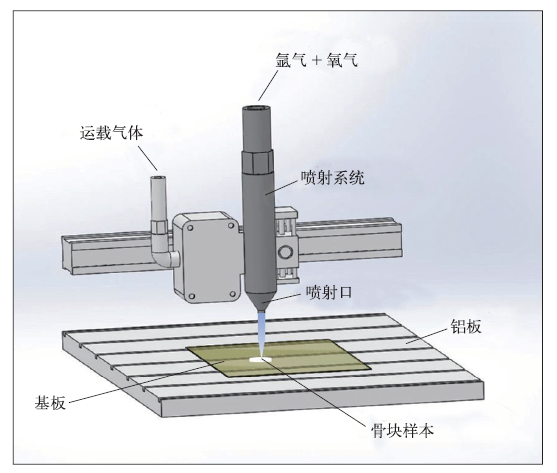
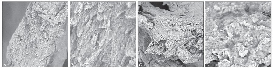
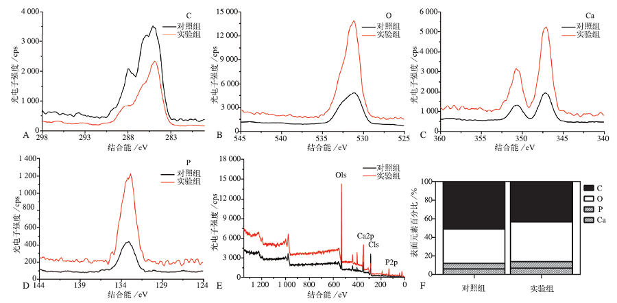
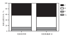
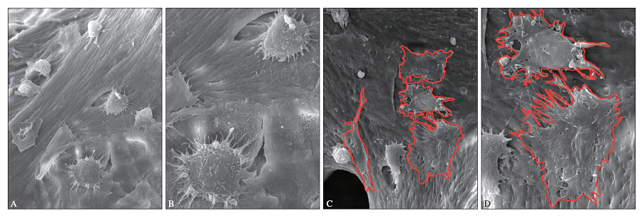
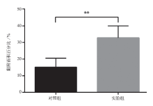
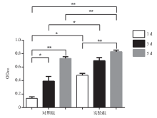
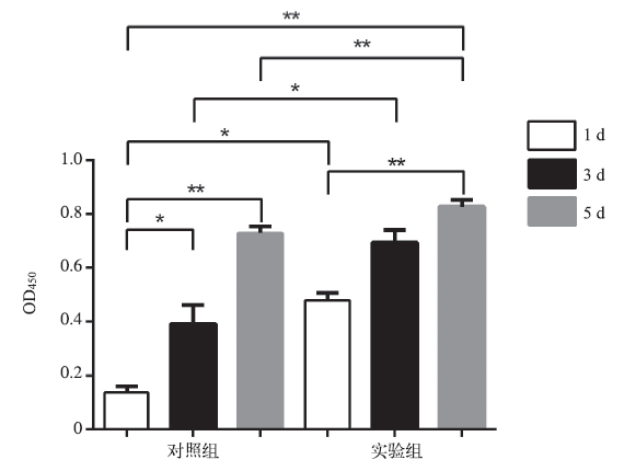
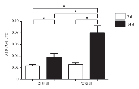
目的 研究小鼠胚胎成骨细胞MC3T3-E1在低温氩氧等离子体处理的无机牛骨上的黏附、增殖及分化特征。方法 使用低温氩氧等离子体对无机牛骨进行表面活化(实验组)后,使用扫描电子显微镜(SEM)观察无机牛骨表面形貌的变化,X射线光电子能谱分析(XPS)检测表面元素的组成。将MC3T3-E1细胞接种于低温氩氧等离子体处理的无机牛骨表面,使用SEM观察细胞的黏附形态,CCK-8法检测细胞1、3、5 d的增殖变化,碱性磷酸酶(ALP)法检测细胞7、14 d的分化状态。以不作处理的无机牛骨作为对照。结果 对照组与实验组的表面形貌无明显改变;在表面材料的元素组成上,实验组表面碳元素减少,氧、钙、磷元素均增高。实验组MC3T3-E1细胞的黏附更充分,细胞伸出伪足;培养1、3、5 d时,实验组细胞增殖数量明显高于对照组;培养14 d时,实验组ALP活性明显高于对照组(P<0.05)。结论 低温氩氧等离子体处理对无机牛骨表面小鼠胚胎成骨细胞的黏附、增殖及分化具有一定的促进作用。
中图分类号:
| [1] | Salamanca E, Pan YH, Tsai AI , et al. Enhancement of osteoblastic-like cell activity by glow discharge plasma surface modified hydroxyapatite/β-tricalcium phosphate bone substitute[J]. Materials (Basel), 2017,10(12):E1347. |
| [2] |
Tanaka M, Haniu H, Kamanaka T , et al. Physico-chemical, in vitro, and in vivo evaluation of a 3D unidirectional porous hydroxyapatite scaffold for bone regeneration[J]. Materials (Basel), 2017,10(1):E33.
doi: 10.3390/ma10010033 pmid: 28772390 |
| [3] |
Amerio P, Vianale G, Reale M , et al. The effect of deproteinized bovine bone on osteoblast growth factors and proinflammatory cytokine production[J]. Clin Oral Implants Res, 2010,21(6):650-655.
doi: 10.1111/j.1600-0501.2009.01881.x pmid: 20666792 |
| [4] |
Huh JB, Kim SE, Song SK , et al. The effect of im-mobilization of heparin and bone morphogenic pro-tein-2 to bovine bone substitute on osteoblast-like cell’s function[J]. J Adv Prosthodont, 2011,3(3):145-151.
doi: 10.4047/jap.2011.3.3.145 pmid: 22053246 |
| [5] |
Rolvien T, Barbeck M, Wenisch S , et al. Cellular mechanisms responsible for success and failure of bone substitute materials[J]. Int J Mol Sci, 2018,19(10):E2893.
doi: 10.3390/ijms19102893 pmid: 30249051 |
| [6] |
Moriguchi Y, Lee DS, Chijimatsu R , et al. Impact of non-thermal plasma surface modification on porous calcium hydroxyapatite ceramics for bone regenera-tion[J]. PLoS One, 2018,13(3):e0194303.
doi: 10.1371/journal.pone.0194303 pmid: 29538457 |
| [7] |
Yang J, Pu Y, Miao DG , et al. Fabrication of durably superhydrophobic cotton fabrics by atmospheric pressure plasma treatment with a siloxane precursor[J]. Polymers (Basel), 2018,10(4):E460.
doi: 10.3390/polym10040460 pmid: 30966495 |
| [8] |
Schmitt C, Lutz R, Doering H , et al. Bio-Oss® blocks combined with BMP-2 and VEGF for the regenera-tion of bony defects and vertical augmentation[J]. Clin Oral Implants Res, 2013,24(4):450-460.
doi: 10.1111/j.1600-0501.2011.02351.x pmid: 22092937 |
| [9] | 高媛媛, 周子谦, 柳慧芬 , 等. RGD多肽修饰的无机小牛骨粉对MC3T3-E1细胞黏附、增殖及分化的影响[J]. 口腔医学研究, 2015,31(4):354-358. |
| Gao YY, Zhou ZQ, Liu HF , et al. Adhesion, prolife-ration and differentiation of MC3T3-E1 cell on bovine bone substitute modified with RGD peptide[J]. J Oral Sci Res, 2015,31(4):354-358. | |
| [10] | Kong MG, Kroesen G, Morfill G , et al. Plasma me-dicine: an introductory review[J]. New J Phys, 2009,11(11):115012. |
| [11] |
Kim JH, Lee MA, Han GJ , et al. Plasma in dentistry: a review of basic concepts and applications in den-tistry[J]. Acta Odontol Scand, 2014,72(1):1-12.
doi: 10.3109/00016357.2013.795660 pmid: 24354926 |
| [12] |
Duske K, Koban I, Kindel E , et al. Atmospheric plasma enhances wettability and cell spreading on dental implant metals[J]. J Clin Periodontol, 2012,39(4):400-407.
doi: 10.1111/j.1600-051X.2012.01853.x pmid: 22324415 |
| [13] |
Seon GM, Seo HJ, Kwon SY , et al. Titanium surface modification by using microwave-induced argon plasma in various conditions to enhance osteoblast biocompatibility[J]. Biomater Res, 2015,19:13.
doi: 10.1186/s40824-015-0034-2 pmid: 26331083 |
| [14] |
Choi SH, Jeong WS, Cha JY , et al. Corrigendum: time-dependent effects of ultraviolet and nonthermal atmospheric pressure plasma on the biological ac-tivity of titanium[J]. Sci Rep, 2016,6:36430.
doi: 10.1038/srep36430 pmid: 27834351 |
| [15] |
Bárdos L, Baránková H . Cold atmospheric plasma: sources, processes, and applications[J]. Thin Solid Films, 2010,518(23):6705-6713.
doi: 10.1016/j.tsf.2010.07.044 |
| [16] | Lee EJ, Kwon JS, Uhm SH , et al. The effects of non-thermal atmospheric pressure plasma jet on cellular activity at SLA-treated titanium surfaces[J]. Curr Appl Phys, 2013,13:S36-S41. |
| [17] |
Hayashi R, Ueno T, Migita S , et al. Hydrocarbon deposition attenuates osteoblast activity on titanium[J]. J Dent Res, 2014,93(7):698-703.
doi: 10.1177/0022034514536578 pmid: 24868012 |
| [18] |
Pham PV . Cleaning of graphene surfaces by low-pressure air plasma[J]. R Soc Open Sci, 2018,5(5):172395.
doi: 10.1098/rsos.172395 pmid: 29892425 |
| [19] |
França R, Samani TD, Bayade G , et al. Nanoscale surface characterization of biphasic calcium pho-sphate, with comparisons to calcium hydroxyapatite and β-tricalcium phosphate bioceramics[J]. J Colloid Interface Sci, 2014,420:182-188.
doi: 10.1016/j.jcis.2013.12.055 pmid: 24559717 |
| [20] | 马楚凡, 李冬梅, 李贺军 , 等. 微弧氧化方法在钛表面注入钙磷离子及对成骨细胞早期附着的影响[J]. 第一军医大学学报, 2005,25(1):62-65. |
| Ma CF, Li DM, Li HJ , et al. Modification of titanium surface with calcium and phosphorus ions using micro-arc oxidation and its effect on osteoblast atta-chment[J]. J First Mil Med Univ, 2005,25(1):62-65. | |
| [21] | 王卫卫 . 低温等离子体对纯钛生物活化后的表面分析[D]. 青岛: 青岛大学, 2019. |
| Wang WW . Surface analysis of pure titanium bioac-tivation after low temperature plasma treatment[D]. Qingdao: Qingdao University, 2019. | |
| [22] | 谢艳婷, 李绍杰, 顾舒扬 , 等. 冷常压等离子体处理对纯钛表面性能和成骨细胞增殖迁移的影响研究[J]. 中国实用口腔科杂志, 2018,11(11):674-678, 683. |
| Xie YT, Li SJ, Gu SY , et al. Study on the effects of cold atmospheric plasma on property of pure titanium surface and proliferation and migration of osteoblasts[J]. Chin J Pract Stomatol, 2018,11(11):674-678, 683. | |
| [23] |
Shen H, Hu XX, Yang F , et al. Combining oxygen plasma treatment with anchorage of cationized gelatin for enhancing cell affinity of poly(lactide-co-glycolide)[J]. Biomaterials, 2007,28(29):4219-4230.
doi: 10.1016/j.biomaterials.2007.06.004 pmid: 17618682 |
| [24] |
Wang HY, Kwok DT, Wang W , et al. Osteoblast behavior on polytetrafluoroethylene modified by long pulse, high frequency oxygen plasma immersion ion implantation[J]. Biomaterials, 2010,31(3):413-419.
doi: 10.1016/j.biomaterials.2009.09.066 pmid: 19811820 |
| [25] |
Mwale F, Wang HT, Nelea V , et al. The effect of glow discharge plasma surface modification of polymers on the osteogenic differentiation of committed human mesenchymal stem cells[J]. Biomaterials, 2006,27(10):2258-2264.
doi: 10.1016/j.biomaterials.2005.11.006 pmid: 16313952 |
| [1] | 徐彦雪,付丽. 功能等级引导骨再生膜的研究进展[J]. 国际口腔医学杂志, 2023, 50(3): 353-358. |
| [2] | 杨立明 陈淑萍 李小菊 谢春 武斌. Bio-oss应用于种植牙唇侧骨缺损的锥形束CT研究[J]. 国际口腔医学杂志, 2015, 42(4): 420-422. |
| [3] | 杨宏丽 刘思茗综述 胡涛审校. 低温等离子体技术在口腔临床消毒中的应用[J]. 国际口腔医学杂志, 2013, 40(4): 483-485. |
| [4] | 张晓丹 胡丹青综述 平飞云审校. 牵张成骨和引导骨再生术在垂直骨增量上的比较研究[J]. 国际口腔医学杂志, 2012, 39(2): 190-193. |
| [5] | 顾晔,汪永跃. 引导骨组织再生膜应用于自身骨移植的研究现状[J]. 国际口腔医学杂志, 2008, 35(S1): -. |
|