国际口腔医学杂志 ›› 2019, Vol. 46 ›› Issue (1): 5-11.doi: 10.7518/gjkq.2019.01.002
李群,关为群( ),张杨安,黄志超
Qun Li,Weiqun Guan(),Yang’an Zhang,Zhichao. Huang
摘要:
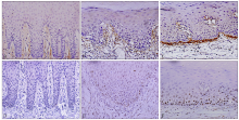
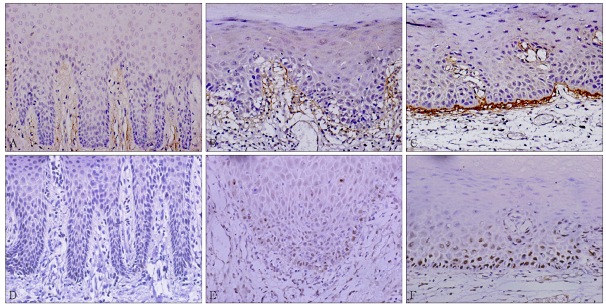
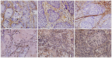
目的 探讨骨膜蛋白和p53在口腔白斑和口腔鳞状细胞癌组织中的表达、相关性及临床意义。方法 采用免疫组织化学法检测30例口腔正常黏膜、30例单纯增生白斑、38例异常增生白斑以及79例口腔鳞状细胞癌组织中骨膜蛋白和p53的表达情况,并分析表达的相关性。结果 对于骨膜蛋白阳性细胞面积指数和累积光密度值,除单纯增生与正常口腔黏膜的差异无统计学意义外,其余各组间的差异均有统计学意义(P<0.05);对于p53阳性细胞面积指数和累积光密度值,除单纯增生白斑与正常口腔黏膜的差异无统计学意义外,其余各组间的差异均有统计学意义(P<0.05)。异常增生白斑和口腔鳞状细胞癌组织中骨膜蛋白与p53阳性表达呈线性正相关(P<0.05)。骨膜蛋白和p53的阳性表达与肿瘤的分化程度、肿瘤TNM分期以及有无淋巴结转移有关(P<0.05)。结论 骨膜蛋白和p53可能共同参与口腔鳞状细胞癌的癌变过程,骨膜蛋白和p53与肿瘤侵袭有关。
中图分类号:
| [1] |
中华口腔医学会口腔黏膜病专业委员会. 口腔白斑病的定义与分级标准(试行)[J]. 中华口腔医学杂志, 2011,46(10):579-580.
doi: 10.3760/cma.j.issn.1002-0098.2011.10.002 |
|
Chinese Stomatological Association Oral Mucosal Disease Committee. Oral leukoplakia definition and grading standards[J]. Chin J Stomatol, 2011,46(10):579-580.
doi: 10.3760/cma.j.issn.1002-0098.2011.10.002 |
|
| [2] |
Neville BW, Day TA . Oral cancer and precancerous lesions[J]. CA Cancer J Clin, 2002,52(4):195-215.
doi: 10.3322/canjclin.52.4.195 pmid: 9170723 |
| [3] |
Lv H, Liu R, Fu J , et al. Epithelial cell-derived periostin functions as a tumor suppressor in gastric cancer through stabilizing p53 and E-cadherin pro-teins via the Rb/E2F1/p14ARF/Mdm2 signaling pathway[J]. Cell Cycle, 2014,13(18):2962-2974.
doi: 10.4161/15384101.2014.947203 pmid: 25486483 |
| [4] |
龙小佳, 何力, 王璐 . HPV16E6和p53在口腔癌中的表达及相关性研究[J]. 第三军医大学学报, 2016,38(21):2326-2329.
doi: 10.16016/j.1000-5404.201604111 |
|
Long XJ, He L, Wang L . Expression of HPV16E6 and p53 in oral cance[J]. J Third Mil Med Univ, 2016,38(21):2326-2329.
doi: 10.16016/j.1000-5404.201604111 |
|
| [5] |
Li B, Wang L, Chi B . Upregulation of periostin pre-vents P53-mediated apoptosis in SGC-7901 gastric cancer cells[J]. Mol Biol Rep, 2013,40(2):1677-1683.
doi: 10.1007/s11033-012-2218-3 pmid: 23076534 |
| [6] |
Michaylira CZ, Wong GS, Miller CG , et al. Periostin, a cell adhesion molecule, facilitates invasion in the tumor microenvironment and annotates a novel tumor-invasive signature in esophageal cancer[J]. Cancer Res, 2010,70(13):5281-5292.
doi: 10.1158/0008-5472.CAN-10-0704 pmid: 3274349 |
| [7] |
姜洋, 金晓明, 屠康 . 平均阳性染色面积百分比法分析免疫组化结果初探[J]. 生物医学工程学杂志, 2007,24(3):650-653.
doi: 10.3321/j.issn:1001-5515.2007.03.039 |
|
Jiang Y, Jin XM, Tu K . A primary study using the method of average positive stained area percentage to measure the immunohistochemistry result[J]. J Biomed Eng, 2007,24(3):650-653.
doi: 10.3321/j.issn:1001-5515.2007.03.039 |
|
| [8] | 汤根兄, 吴国英, 李静 . Stat3和cyclinD1在口腔黏膜癌前病变和口腔鳞癌中的表达[J]. 口腔医学研究, 2011,27(1):40-43. |
| Tang GX, Wu GY, Li J . Stat3 and cyclinD1 expre-ssion in oral mucosal precancerous lesions and oral squamous cell carcinoma[J]. J Oral Sci Res, 2011,27(1):40-43. | |
| [9] |
Eckert AW, Wickenhauser C, Salins PC , et al. Clinical relevance of the tumor microenvironment and immune escape of oral squamous cell carcinoma[J]. J Transl Med, 2016,14:85.
doi: 10.1186/s12967-016-0828-6 pmid: 4820994 |
| [10] | 胡汪来, 吴缅 . p53在肿瘤发生过程中的功能研究及进展[J]. 中国科学: 生命科学, 2017,47(1):52-58. |
| Hu WL, Wu M . Study on the function of p53 in tu-morigenesis and its progress[J]. Scienta Sinica Vitae, 2017,47(1):52-58. | |
| [11] | Hong Q, Ke C, Ji Y , et al. Expression and clinicopa-thologic significance of TUFM and p53 for the normal-adenoma-carcinoma sequence in colorectal epithelia[J]. World J Surg Oncol, 2017,15(16):238-240. |
| [12] |
Tilman G, Mattiussi M, Brasseur F , et al. Human periostin gene expression in normal tissues, tumors and melanoma: evidences for periostin production by both stromal and melanoma cells[J]. Mol Cancer, 2007,6:80.
doi: 10.1186/1476-4598-6-80 pmid: 18086302 |
| [13] |
Koh SJ, Choi Y, Kim BG , et al. Matricellular protein periostin mediates intestinal inflammation through the activation of nuclear factor κB signaling[J]. PLoS One, 2016,11(2):e0149652.
doi: 10.1371/journal.pone.0149652 pmid: 4758640 |
| [14] |
Isono T, Kim CJ, Ando Y , et al. Suppression of cell invasiveness by periostin via TAB1/TAK1[J]. Int J Oncol, 2009,35(2):425-432.
doi: 10.3892/ijo_00000355 pmid: 19578758 |
| [15] |
Kanno A, Satoh K, Masamune A , et al. Periostin, secreted from stromal cells, has biphasic effect on cell migration and correlates with the epithelial to mesenchymal transition of human pancreatic cancer cells[J]. Int J Cancer, 2008,122(12):2707-2718.
doi: 10.1002/ijc.23332 pmid: 18381746 |
| [16] |
Moniuszko T, Wincewicz A, Koda M , et al. Role of periostin in esophageal, gastric and colon cancer[J]. Oncol Lett, 2016,12(2):783-787.
doi: 10.3892/ol.2016.4692 pmid: 4950105 |
| [17] |
Landré V, Antonov A, Knight R , et al. p73 promotes glioblastoma cell invasion by directly activating POSTN (periostin) expression[J]. Oncotarget, 2016,7(11):11785-11802.
doi: 10.18632/oncotarget.7600 pmid: 4914248 |
| [18] |
Lister NC, Clemson M, Morris KV . RNA-directed epigenetic silencing of periostin inhibits cell motility[J]. R Soc Open Sci, 2015,2(6):140545.
doi: 10.1098/rsos.140545 pmid: 4632543 |
| [19] | Wang W, Sun QK, He YF , et al. Overexpression of periostin is significantly correlated to the tumor an-giogenesis and poor prognosis in patients with eso-phageal squamous cell carcinoma[J]. Int J Clin Exp Pathol, 2014,7(2):593-601. |
| [20] |
Sun L, Fang J . Epigenetic regulation of epithelial-mesenchymal transition[J]. Cell Mol Life Sci, 2016,73(23):4493-4515.
doi: 10.2174/15680096113136660103 pmid: 24168185 |
| [1] | 周金阔,张晋弘,史晓晶,刘广顺,姜磊,刘倩峰. 长链非编码RNA小核仁RNA宿主基因22调控微小RNA-27b-3p对口腔鳞状细胞癌细胞增殖、侵袭和迁移的影响[J]. 国际口腔医学杂志, 2024, 51(1): 52-59. |
| [2] | 李立恒,王蕊,王晓明,张智轶,张璇,安峰,王芹,张凡. 环状RNA hsa_circ_0085576调控微小RNA-498/B细胞特异性莫洛尼鼠白血病病毒整合位点1轴对口腔鳞状细胞癌细胞迁移和侵袭的影响[J]. 国际口腔医学杂志, 2024, 51(1): 60-67. |
| [3] | 吴佳敏,夏斌,杨禾丰,许彪. 癌相关成纤维细胞在口腔鳞状细胞癌微环境中作用的研究进展[J]. 国际口腔医学杂志, 2023, 50(6): 711-717. |
| [4] | 柳江龙, 买买提吐逊·吐尔地. 超声造影在口腔鳞状细胞癌颈部转移性淋巴结诊断中的研究进展[J]. 国际口腔医学杂志, 2023, 50(5): 514-520. |
| [5] | 盛南宁,王珏,南欣荣. 性别决定基因盒9在口腔鳞状细胞癌作用机制和治疗中的研究进展[J]. 国际口腔医学杂志, 2023, 50(3): 314-320. |
| [6] | 李潭,梁新华. 盘状蛋白结构域受体1在调控恶性肿瘤进展和治疗中的作用[J]. 国际口腔医学杂志, 2023, 50(2): 230-236. |
| [7] | 赵卓平,辛鹏飞,高阳,张彩凤,张宽收,刘青梅. 光热治疗在口腔鳞状细胞癌治疗中的研究进展[J]. 国际口腔医学杂志, 2022, 49(4): 462-470. |
| [8] | 江涵,神应强,陈谦明. 毒蕈碱受体通过Yes相关蛋白信号对口腔鳞状细胞癌生物学行为的实验研究[J]. 国际口腔医学杂志, 2022, 49(2): 138-143. |
| [9] | 蒋宇磊,夏斌,饶南荃,杨禾丰,许彪. 外泌体在口腔鳞状细胞癌恶性进展及诊疗应用的研究[J]. 国际口腔医学杂志, 2021, 48(6): 711-717. |
| [10] | 李哲儒,但红霞,陈谦明. 光动力治疗切除后复发的疣状型口腔白斑病1例[J]. 国际口腔医学杂志, 2021, 48(3): 318-321. |
| [11] | 甘建国,高攀,王晓毅. 循环肿瘤细胞与口腔鳞状细胞癌相关性的研究进展[J]. 国际口腔医学杂志, 2021, 48(2): 205-212. |
| [12] | 黄俊文,乔洁,梅子,陈茁,李杨,乔彬. 脂多糖结合蛋白在口腔鳞状细胞癌中的表达及其临床意义[J]. 国际口腔医学杂志, 2021, 48(1): 50-57. |
| [13] | 何宇晴,但红霞,陈谦明. 光动力疗法在口腔黏膜癌变防治中的应用[J]. 国际口腔医学杂志, 2020, 47(6): 669-676. |
| [14] | 郝福,宁毅,孙睿,郑晓旭. 口腔鳞状细胞癌中转化因子2β的表达及潜在的临床意义[J]. 国际口腔医学杂志, 2020, 47(2): 159-165. |
| [15] | 薛伶俐,李雅冬. 经首次根治性手术治疗口腔鳞状细胞癌患者的生存相关影响因素分析[J]. 国际口腔医学杂志, 2020, 47(2): 166-174. |
|