国际口腔医学杂志 ›› 2021, Vol. 48 ›› Issue (4): 426-432.doi: 10.7518/gjkq.2021063
伍春兰( ),唐华,陈军()
Wu Chunlan(),Tang Hua,Chen Jun()
摘要:
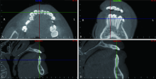
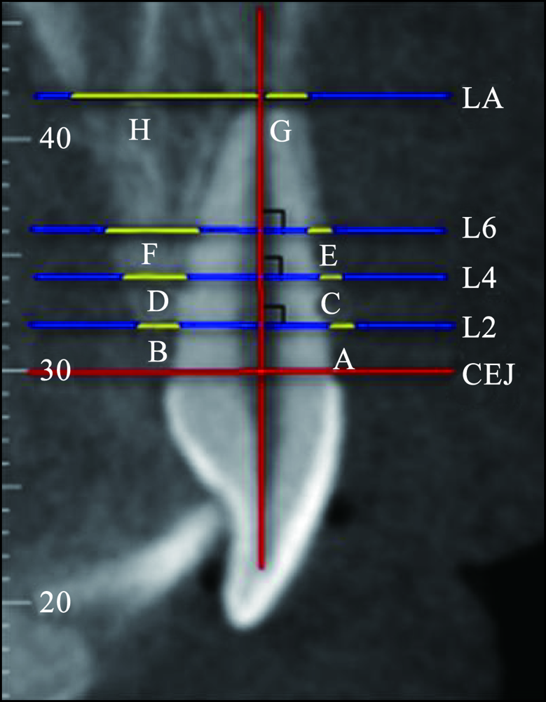
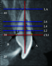
目的 运用锥形束计算机断层扫描(CBCT)对成人骨性Ⅱ类高角开牙合患者上下切牙区的牙槽骨形态进行研究分析。方法 筛选成人骨性Ⅱ类高角正畸治疗前患者58例(前牙开牙合29例、正常覆牙合29例),利用Dolphin软件测量上下切牙区牙槽骨高度和牙槽骨厚度,并进行统计分析。结果 开牙合组与对照组在切牙区牙槽骨形态的差异主要体现在舌/腭侧的牙槽骨厚度及牙槽骨高度上,开牙合患者多个切牙位点处的舌/腭侧及根尖处的牙槽骨厚度较薄、牙槽骨高度较低;Pearson检验显示覆牙合与舌/腭侧牙槽骨高度呈负相关,与牙槽骨厚度呈正相关。结论 成人骨性Ⅱ类高角开牙合患者切牙区的舌/腭侧牙槽骨厚度较薄、高度较低,矫治该类患者时应谨慎切牙过度的舌向移动,并注意转矩的控制,以避免牙周组织及牙根的损伤。
中图分类号:
| [1] | 傅民魁. 口腔正畸专科教程[M]. 北京: 人民卫生出版社, 2007: 422-430. |
| Fu MK. Textbook of orthodontics[M]. Beijing: Peo-ple’s Medical Publishing House, 2007: 422-430. | |
| [2] | Lin LH, Huang GW, Chen CS. Etiology and treatment modalities of anterior open bite malocclusion[J]. J Exp Clin Med, 2013,5(1):1-4. |
| [3] | Foosiri P, Mahatumarat K, Panmekiate S. Relationship between mandibular symphysis dimensions and mandibular anterior alveolar bone thickness as asses-sed with cone-beam computed tomography[J]. Dent Press J Orthod, 2018,23(1):54-62. |
| [4] |
Srebrzyńska-Witek A, Koszowski R, Różyło-Kalinowska I. Relationship between anterior mandibular bone thickness and the angulation of incisors and canines‒a CBCT study[J]. Clin Oral Investig, 2018,22(3):1567-1578.
doi: 10.1007/s00784-017-2255-3 pmid: 29063382 |
| [5] |
Nahás-Scocate AC, de Siqueira Brandão A, Patel MP, et al. Bone tissue amount related to upper incisors inclination[J]. Angle Orthod, 2014,84(2):279-285.
doi: 10.2319/031213-211.1 pmid: 23883305 |
| [6] |
Ma J, Huang J, Jiang JH. Morphological analysis of the alveolar bone of the anterior teeth in severe high-angle skeletal Class Ⅱ and Class Ⅲ malocclusions assessed with cone-beam computed tomography[J]. PLoS One, 2019,14(3):e0210461.
doi: 10.1371/journal.pone.0210461 |
| [7] | 王光伟, 李伟绪, 张临雪, 等. 不同错牙合类型患者上前牙区牙槽骨厚度比较[J]. 河南医学研究, 2019,28(15):2695-2698. |
| Wang GW, Li WX, Zhang LX, et al. Comparison on alveolar bone thickness of upper anterior teeth in patients with different malocclusion types[J]. Henan Med Res, 2019,28(15):2695-2698. | |
| [8] |
Timock AM, Cook V, McDonald T, et al. Accuracy and reliability of buccal bone height and thickness measurements from cone-beam computed tomography imaging[J]. Am J Orthod Dentofacial Orthop, 2011,140(5):734-744.
doi: 10.1016/j.ajodo.2011.06.021 pmid: 22051495 |
| [9] | 许天民, 刘妍, 江久汇, 等. 正畸内收上切牙对上颌牙槽骨改建的临床研究[J]. 实用口腔医学杂志, 2004,20(4):431-433. |
| Xu TM, Liu Y, Jiang JH, et al. Cephalometric study of alveolar remodeling during incisor retraction[J]. J Pract Stomatol, 2004,20(4):431-433. | |
| [10] | 季海宁, 梁源, 隋珂, 等. 成人骨性Ⅱ类错牙合不同垂直骨面型前牙区牙槽骨形态的CBCT研究[J]. 实用口腔医学杂志, 2016,32(2):268-272. |
| Ji HN, Liang Y, Sui K, et al. A cone-beam CT study on alveolar bone morphology in anterior teeth area of adult skeletal ClassⅡmalocclusion subjects with different vertical skeletal types[J]. J Pract Stomatol, 2016,32(2):268-272. | |
| [11] |
Harris EF, Butler ML. Patterns of incisor root resorption before and after orthodontic correction in cases with anterior open bites[J]. Am J Orthod Dentofacial Orthop, 1992,101(2):112-119.
doi: 10.1016/0889-5406(92)70002-R |
| [12] | 雷琦. 安氏Ⅱ类1分类和2分类上中切牙牙槽骨形态的CBCT分析[D]. 太原: 山西医科大学, 2019. |
| Lei Q. Alveolar bone morphology analysis of upper central incisor in ClassⅡdivision 1 and division 2 by cone-beam computed tomography[D]. Taiyuan: Shanxi Medical University, 2019. | |
| [13] |
Enokida M, Kaneko S, Yanagishita M, et al. Influen-ce of occlusal stimuli on the remodelling of alveolar bone in a rat hypofunction-recovery model[J]. J Oral Biosci, 2005,47(4):321-334.
doi: 10.1016/S1349-0079(05)80015-5 |
| [14] |
Liu J, Jin ZL, Li Q. Effect of occlusal hypofunction and its recovery on the three-dimensional architecture of mandibular alveolar bone in growing rats[J]. J Surg Res, 2015,193(1):229-236.
doi: 10.1016/j.jss.2014.07.015 |
| [15] |
Hayashi H, Terao A, Kunimatsu R, et al. Effects of a low level laser on periodontal tissue in hypofunctional teeth[J]. PLoS One, 2014,9(6):e100066.
doi: 10.1371/journal.pone.0100066 |
| [16] |
Motokawa M, Kaku M, Matsuda Y, et al. Effects of occlusal hypofunction and its recovery on PDL structure and expression of VEGF and bFGF in rats[J]. Clin Oral Investig, 2015,19(4):929-935.
doi: 10.1007/s00784-014-1310-6 pmid: 25209593 |
| [17] |
Mavropoulos A, Odman A, Ammann P, et al. Rehabilitation of masticatory function improves the alveolar bone architecture of the mandible in adult rats[J]. Bone, 2010,47(3):687-692.
doi: 10.1016/j.bone.2010.06.025 pmid: 20601301 |
| [18] |
Motokawa M, Terao A, Karadeniz EI, et al. Effects of long-term occlusal hypofunction and its recovery on the morphogenesis of molar roots and the perio-dontium in rats[J]. Angle Orthod, 2013,83(4):597-604.
doi: 10.2319/081812-661.1 |
| [19] |
Uehara S, Maeda A, Tomonari H, et al. Relationships between the root-crown ratio and the loss of occlusal contact and high mandibular plane angle in patients with open bite[J]. Angle Orthod, 2013,83(1):36-42.
doi: 10.2319/042412-341.1 |
| [1] | 刘盼明,李政泽,李军鹤,崔淑霞. 成人骨性Ⅱ类患者不同垂直骨面型上颌窦容积及口咽气道体积的锥形束计算机断层扫描研究[J]. 国际口腔医学杂志, 2023, 50(5): 528-537. |
| [2] | 刘晓琳,任群,高晓哲,白九平,王宇,李向军. 458例上切牙区多生牙的临床资料分析[J]. 国际口腔医学杂志, 2023, 50(1): 61-65. |
| [3] | 刘力嘉,毛婧,龙欢,蒲亚龙,王军. 二维与三维头影测量自动定点的研究进展[J]. 国际口腔医学杂志, 2022, 49(1): 100-108. |
| [4] | 周梦琪,陈学鹏,傅柏平. 正畸治疗中牙槽骨骨开窗骨开裂的预防和应对策略[J]. 国际口腔医学杂志, 2021, 48(5): 600-608. |
| [5] | 王立冬,马文,付帅,张长彬,崔庆赢,梁燕,黎明. 不同方法制作正颌手术数字化牙合板的研究及精确性分析[J]. 国际口腔医学杂志, 2021, 48(2): 156-164. |
| [6] | 张紫涵,熊鑫,王军. 三维头影测量的研究现状和应用发展[J]. 国际口腔医学杂志, 2020, 47(6): 739-744. |
| [7] | 刘晓华, 王恩博. 儿童埋伏多生牙拔除术锥形束计算机断层扫描影像学定位的应用进展[J]. 国际口腔医学杂志, 2018, 45(3): 295-300. |
| [8] | 尹一佳, 冯捷, 罗梦奇, 韩向龙. 三维立体摄影技术自然头位校正方法的研究进展[J]. 国际口腔医学杂志, 2018, 45(3): 301-306. |
| [9] | 周凤,赵玉鸣. 磨牙-切牙釉质矿化不全的研究进展[J]. 国际口腔医学杂志, 2016, 43(4): 456-461. |
| [10] | 赵朋朋,刘志顺,秦宗长. 上颌侧切牙2根2根管1例[J]. 国际口腔医学杂志, 2015, 42(5): 562-563. |
| [11] | 王波1 张桂香2 石晶3 彭惠3 张莹3. 自锁托槽矫治器矫治初期上颌异位侧切牙及牙周组织应力的三维有限元分析[J]. 国际口腔医学杂志, 2015, 42(4): 406-410. |
| [12] | 肖毅 陈思韩 熊腊平 罗霞 许艳春. 上颌中切牙透明层分布的临床分类研究[J]. 国际口腔医学杂志, 2013, 40(6): 736-738. |
| [13] | 支方静综述 莫水学审校. 切牙区黑三角与正畸治疗的研究进展[J]. 国际口腔医学杂志, 2011, 38(5): 611-613. |
| [14] | 姜世同1 王作君2 安忠军1 焦广军1 姜良坤1 罗圆圆1. 倒位埋伏阻生上中切牙的临床矫治探索[J]. 国际口腔医学杂志, 2011, 38(5): 515-517. |
| [15] | 李佳岭1 李小兵2 李佳园3. 内收下切牙对下切牙区牙槽骨改建的影响[J]. 国际口腔医学杂志, 2011, 38(4): 392-394. |
|