国际口腔医学杂志 ›› 2019, Vol. 46 ›› Issue (2): 171-176.doi: 10.7518/gjkq.2019016
黄满英1,付云2( )
)
Manying Huang1,Yun Fu2()
摘要:
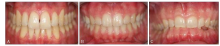
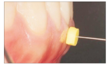
牙龈生物型泛是指牙周组织以及牙体组织的形态特征,是预测修复、种植、牙周等美学治疗术后成功率的重要参考因素之一。个体间牙龈生物型存在差异,且不同牙龈生物型对外界刺激的反应不同。研究发现:薄龈生物型更容易出现治疗后牙龈退缩,厚龈生物型更容易出现探诊出血等炎症性改变。在目前的临床工作中,正确评估牙龈生物型特别是准确测量牙龈厚度是合理选择治疗方案及预后评估的基础。然而临床治疗中尚未有统一标准的测量方法,本文就牙龈生物型测量方法作一综述,以供临床及研究工作者参考。
中图分类号:
| [1] |
Shah R, Sowmya NK, Mehta DS . Prevalence of gin-gival biotype and its relationship to clinical parame-ters[J]. Contemp Clin Dent, 2015,6(Suppl 1):S167-S171.
doi: 10.4103/0976-237X.166824 pmid: 26604569 |
| [2] |
Frost NA, Mealey BL, Jones AA , et al. Periodontal biotype: gingival thickness as it relates to probe visi-bility and buccal plate thickness[J]. J Periodontol, 2015,86(10):1141-1149.
doi: 10.1902/jop.2015.140394 pmid: 26110452 |
| [3] |
Cook DR, Mealey BL, Verrett RG , et al. Relationship between clinical periodontal biotype and labial plate thickness: an in vivo study[J]. Int J Periodontics Res-torative Dent, 2011,31(4):345-354.
doi: 10.4012/dmj.2011-028 pmid: 21837300 |
| [4] |
Younes F, Eghbali A, Raes M , et al. Relationship be- tween buccal bone and gingival thickness revisited using non-invasive registration methods[J]. Clin Oral Implants Res, 2016,27(5):523-528.
doi: 10.1111/clr.12618 pmid: 26010518 |
| [5] |
Peixoto A, Marques TM, Correia A . Gingival biotype characterization: a study in a Portuguese sample[J]. Int J Esthet Dent, 2015,10(4):534-546.
pmid: 26794050 |
| [6] |
Sin YW, Chang HY, Yun WH , et al. Association of gingival biotype with the results of scaling and root planing[J]. J Periodontal Implant Sci, 2013,43(6):283-290.
doi: 10.5051/jpis.2013.43.6.283 pmid: 3891860 |
| [7] |
Arora R, Narula SC, Sharma RK , et al. Evaluation of supracrestal gingival tissue after surgical crown leng-thening: a 6-month clinical study[J]. J Periodontol, 2013,84(7):934-940.
doi: 10.1902/jop.2012.120162 pmid: 23088528 |
| [8] |
Tao JX, Wu Y, Chen JR , et al. A follow-up study of up to 5 years of metal-ceramic crowns in maxillary central incisors for different gingival biotypes[J]. Int J Periodontics Restorative Dent, 2014,34(5):e85-e92.
doi: 10.11607/prd.2024 pmid: 25171044 |
| [9] |
Rasperini G, Acunzo R, Cannalire P , et al. Influence of periodontal biotype on root surface exposure during orthodontic treatment: a preliminary study[J]. Int J Periodontics Restorative Dent, 2015,35(5):665-675.
doi: 10.11607/prd.2239 pmid: 26357696 |
| [10] |
Kim DM, Neiva R . Periodontal soft tissue non-root coverage procedures: a systematic review from the AAP Regeneration Workshop[J]. J Periodontol, 2015,86(2 Suppl):S56-S72.
doi: 10.1902/jop.2015.130684 |
| [11] |
Chen ST, Darby IB, Reynolds EC , et al. Immediate implant placement postextraction without flap eleva-tion[J]. J Periodontol, 2009,80(1):163-172.
doi: 10.1902/jop.2009.080243 pmid: 19228102 |
| [12] |
Evans CD, Chen ST . Esthetic outcomes of immediate implant placements[J]. Clin Oral Implants Res, 2008,19(1):73-80.
doi: 10.1111/j.1600-0501.2007.01413.x pmid: 17956569 |
| [13] |
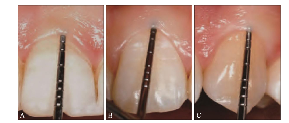
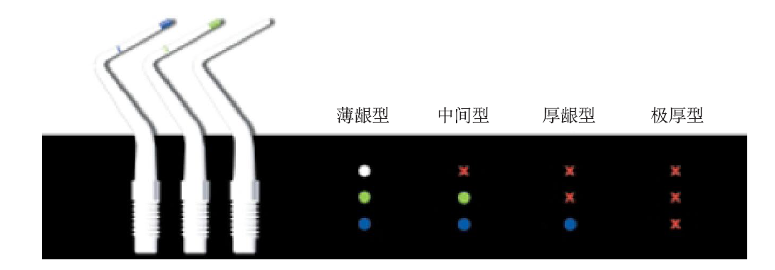
De Rouck T, Eghbali R, Collys K , et al. The gingival biotype revisited: transparency of the periodontal probe through the gingival margin as a method to discriminate thin from thick gingiva[J]. J Clin Perio-dontol, 2009,36(5):428-433.
doi: 10.1111/j.1600-051X.2009.01398.x pmid: 19419444 |
| [14] |
Eghbali A, De Rouck T, De Bruyn H , et al. The gingival biotype assessed by experienced and inexperienced clinicians[J]. J Clin Periodontol, 2009,36(11):958-963.
doi: 10.1111/j.1600-051X.2009.01479.x pmid: 19811580 |
| [15] |
Cuny-Houchmand M, Renaudin S, Leroul M , et al. Gingival biotype assessement: visual inspection re-levance and maxillary versus mandibular comparison[J]. Open Dent J, 2013,7:1-6.
doi: 10.2174/1874210601307010001 pmid: 23400554 |
| [16] |
乐迪, 张豪, 胡文杰 , 等. 牙周探诊法判断牙龈生物型的初步研究[J]. 中华口腔医学杂志, 2012,47(2):81-84.
doi: 10.3760/cma.j.issn.1002-0098.2012.02.003 |
|
Le D, Zhang H, Hu WJ , et al. Preliminary study on gingival biotype by periodontal probing[J]. Chin J Stomatol, 2012,47(2):81-84.
doi: 10.3760/cma.j.issn.1002-0098.2012.02.003 |
|
| [17] |
Kaya Y, Alkan ö, Keskin S . An evaluation of the gingival biotype and the width of keratinized gingiva in the mandibular anterior region of individuals with different dental malocclusion groups and levels of crowding[J]. Korean J Orthod, 2017,47(3):176-185.
doi: 10.4041/kjod.2017.47.3.176 pmid: 5432439 |
| [18] |
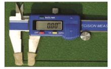
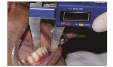
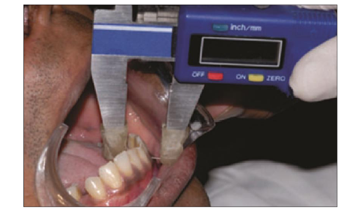
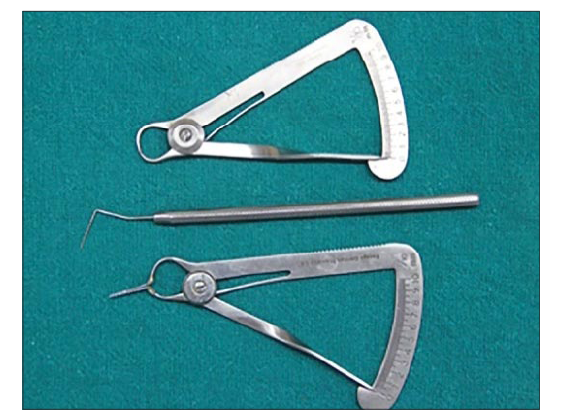
Sharma S, Thakur SL, Joshi SK , et al. Measurement of gingival thickness using digital vernier caliper and ultrasonographic method: a comparative study[J]. J Investig Clin Dent, 2014,5(2):138-143.
doi: 10.1111/jicd.12026 pmid: 23355379 |
| [19] |
Baldi C, Pini-Prato G, Pagliaro U , et al. Coronally advanced flap procedure for root coverage. Is flap thickness a relevant predictor to achieve root coverage? A 19-case series[J]. J Periodontol, 1999,70(9):1077-1084.
doi: 10.1902/jop.1999.70.9.1077 pmid: 10505811 |
| [20] |
Rathee M, Rao PL, Bhoria M . Prevalence of gingival biotypes among young dentate north indian popula-tion: a biometric approach[J]. Int J Clin Pediatr Dent, 2016,9(2):104-108.
doi: 10.5005/jp-journals-10005-1343 pmid: 27365928 |
| [21] |
Memon S, Patel JR, Sethuraman R , et al. A com-parative evaluation of the reliability of three methods of assessing gingival biotype in dentate subjects in different age groups: an in vivo study[J]. J Indian Prosthodont Soc, 2015,15(4):313-317.
doi: 10.4103/0972-4052.171830 pmid: 26929533 |
| [22] |
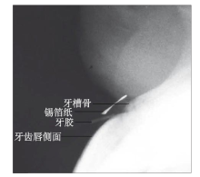
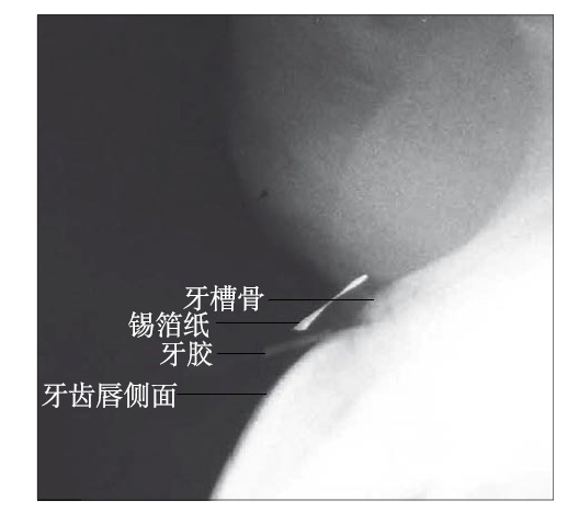
Alpiste-Illueca F . Dimensions of the dentogingival unit in maxillary anterior teeth: a new exploration technique (parallel profile radiograph)[J]. Int J Periodontics Restorative Dent, 2004,24(4):386-396.
doi: 10.1111/j.1365-2591.2004.00849.x pmid: 15446409 |
| [23] |
Galgali SR, Gontiya G . Evaluation of an innovative radiographic technique—parallel profile radiography—to determine the dimensions of dentogingival unit[J]. Indian J Dent Res, 2011,22(2):237-241.
doi: 10.4103/0970-9290.84294 pmid: 21891892 |
| [24] |
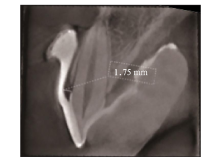
Kim YJ, Park JM, Kim S , et al. New method of asse-ssing the relationship between buccal bone thickness and gingival thickness[J]. J Periodontal Implant Sci, 2016,46(6):372-381.
doi: 10.5051/jpis.2016.46.6.372 pmid: 28050315 |
| [25] |
Esfahanizadeh N, Daneshparvar N, Askarpour F , et al. Correlation between bone and soft tissue thickness in maxillary anterior teeth[J]. J Dent (Tehran), 2016,13(5):302-308.
pmid: 5250627 |
| [26] |
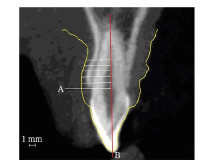
曹洁, 胡文杰, 张豪 , 等. 基于锥形束计算机体层摄影术测量牙龈厚度[J]. 北京大学学报(医学版), 2013,45(1):135-139.
doi: 10.3969/j.issn.1671-167X.2013.01.028 |
|
Cao J, Hu WJ, Zhang H , et al. Method and its app-lication of gingival thickness measurement based on cone-beam computed tomography[J]. J Peking Univ (Health Sci), 2013,45(1):135-139.
doi: 10.3969/j.issn.1671-167X.2013.01.028 |
|
| [27] |
Lee SP, Kim TI, Kim HK , et al. Discriminant ana-lysis for the thin periodontal biotype based on the data acquired from three-dimensional virtual models of Korean young adults[J]. J Periodontol, 2013,84(11):1638-1645.
doi: 10.1902/jop.2013.120594 pmid: 23305168 |
| [28] |
Malhotra R, Grover V, Bhardwaj A , et al. Analysis of the gingival biotype based on the measurement of the dentopapillary complex[J]. J Indian Soc Perio-dontol, 2014,18(1):43-47.
doi: 10.4103/0972-124X.128199 pmid: 3988642 |
| [1] | 赵喆,王富,郑秀丽,安娜,陈吉华. 功能载荷下牙移动测量方法的研究进展[J]. 国际口腔医学杂志, 2022, 49(3): 362-366. |
| [2] | 范盛梓, 谢志刚. 牙龈生物型对种植牙美学影响的研究进展[J]. 国际口腔医学杂志, 2017, 44(5): 580-582. |
| [3] | 王通,万乾炳. 牙根表面积测量方法的研究进展[J]. 国际口腔医学杂志, 2016, 43(4): 490-494. |
| [4] | 伍颖颖综述 宫苹审校. 种植体初期稳定性的研究现状与进展[J]. 国际口腔医学杂志, 2009, 36(6): 726-728. |
| [5] | 阎英综述 凌均棨审校. 牙齿磨耗的测量方法[J]. 国际口腔医学杂志, 2009, 36(4): 476-478. |
|