国际口腔医学杂志 ›› 2022, Vol. 49 ›› Issue (3): 337-342.doi: 10.7518/gjkq.2022058
黎静文( ),周力()
Li Jingwen(),Zhou Li.()
摘要:
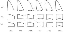
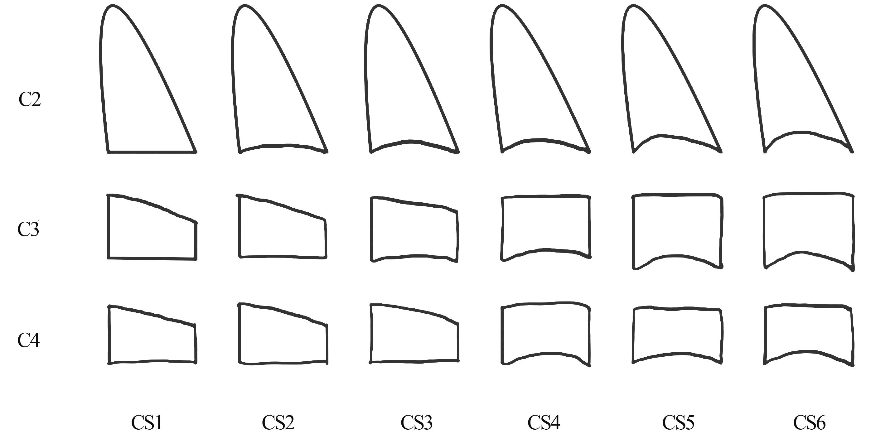
早期功能矫治及矫形治疗是改善下颌骨发育差异的重要手段,因而通过骨龄评估下颌骨生长高峰期,以此确定矫治开始时间具有重要意义。颈椎成熟(CVM)法是根据颈椎在不同生长发育阶段形状、大小有规律性的改变来预测下颌骨生长发育时期的方法。在正畸常规检查的头颅侧位片中,颈椎形态清晰可见,采用CVM法评估骨龄方便实用,相较于手腕骨法不需患者接受额外的辐射,临床应用广泛。但该方法存在主观性大、可重复性低等问题,具有一定争议。针对这些问题已有多种改进方法,包括纳入客观指标描述颈椎形态,改进试验设计及数据分析方法,使用锥形束CT影像分析颈椎形态,采用计算机辅助定点等。本文就CVM法临床应用的支持依据、相关争议以及改进方法进行综述。
中图分类号:
| 1 | Baccetti T, Franchi L, McNamara JA Jr. The cervical vertebral maturation (CVM) method for the assessment of optimal treatment timing in dentofacial orthopedics[J]. Semin Orthod, 2005, 11(3): 119-129. |
| 2 | Lamparski DG. Skeletal age assessment utilizing cervical vertebrae[D]. Pittsburgh: University of Pitts-burgh, 1972. |
| 3 | Patcas R, Wiedemeier DB, Markic G, et al. Evidence of secular trend in mandibular pubertal growth[J]. Eur J Orthod, 2017, 39(6): 680-685. |
| 4 | Mellion ZJ, Behrents RG, Johnston LE Jr. The pattern of facial skeletal growth and its relationship to various common indexes of maturation[J]. Am J Orthod Dentofacial Orthop, 2013, 143(6): 845-854. |
| 5 | Perinetti G, Contardo L, Castaldo A, et al. Diagnostic reliability of the cervical vertebral maturation method and standing height in the identification of the mandibular growth spurt[J]. Angle Orthod, 2016, 86(4): 599-609. |
| 6 | Franchi L, Nieri M, McNamara JA Jr, et al. Predicting mandibular growth based on CVM stage and gender and with chronological age as a curvilinear variable[J]. Orthod Craniofac Res, 2021, 24(3): 414-420. |
| 7 | Ball G, Woodside D, Tompson B, et al. Relationship between cervical vertebral maturation and mandibular growth[J]. Am J Orthod Dentofacial Orthop, 2011, 139(5): e455-e461. |
| 8 | Perinetti G, Braga C, Contardo L, et al. Cervical vertebral maturation: are postpubertal stages attained in all subjects[J]. Am J Orthod Dentofacial Orthop, 2020, 157(3): 305-312. |
| 9 | Gray S, Bennani H, Kieser JA, et al. Morphometric analysis of cervical vertebrae in relation to mandibular growth[J]. Am J Orthod Dentofacial Orthop, 2016, 149(1): 92-98. |
| 10 | Morris KM, Fields HW Jr, Beck FM, et al. Diagnostic testing of cervical vertebral maturation staging: An independent assessment[J]. Am J Orthod Dentofacial Orthop, 2019, 156(5): 626-632. |
| 11 | Engel TP, Renkema AM, Katsaros C, et al. The cervical vertebrae maturation (CVM) method cannot predict craniofacial growth in girls with class Ⅱ malocclusion[J]. Eur J Orthod, 2016, 38(1): 1-7. |
| 12 | Sohrabi A, Babay Ahari S, Moslemzadeh H, et al. The reliability of clinical decisions based on the cervical vertebrae maturation staging method[J]. Eur J Orthod, 2016, 38(1): 8-12. |
| 13 | Zhao XG, Lin JX, Jiang JH, et al. Validity and reliability of a method for assessment of cervical vertebral maturation[J]. Angle Orthod, 2012, 82(2): 229-234. |
| 14 | Chen LL, Xu TM, Jiang JH, et al. Quantitative cervical vertebral maturation assessment in adolescents with normal occlusion: a mixed longitudinal study[J]. Am J Orthod Dentofacial Orthop, 2008, 134(6): 720.e1-720.e7, 720-721. |
| 15 | Perinetti G, Bianchet A, Franchi L, et al. Cervical vertebral maturation: an objective and transparent code staging system applied to a 6-year longitudinal investigation[J]. Am J Orthod Dentofacial Orthop, 2017, 151(5): 898-906. |
| 16 | Gabriel DB, Southard KA, Qian F, et al. Cervical vertebrae maturation method: poor reproducibility[J]. Am J Orthod Dentofacial Orthop, 2009, 136(4): 478.e1-478.e7, 478-480. |
| 17 | Nestman TS, Marshall SD, Qian F, et al. Cervical vertebrae maturation method morphologic criteria: Poor reproducibility[J]. Am J Orthod Dentofacial Orthop, 2011, 140(2): 182-188. |
| 18 | Franchi L, Baccetti T, McNamara JA Jr. Mandibular growth as related to cervical vertebral maturation and body height[J]. Am J Orthod Dentofacial Orthop, 2000, 118(3): 335-340. |
| 19 | Perinetti G, Primozic J, Sharma B, et al. Cervical vertebral maturation method and mandibular growth peak: a longitudinal study of diagnostic reliability[J]. Eur J Orthod, 2018, 40(6): 666-672. |
| 20 | Salazar-Lazo R, Arriola-Guillén LE, Flores-Mir C. Duration of the peak of adolescent growth spurt in class i and ii malocclusion subjects using a cervical vertebrae maturation analysis[J]. Acta Odontol Latinoam, 2014, 27(2): 96-101. |
| 21 | Perinetti G, Caprioglio A, Contardo L. Visual assessment of the cervical vertebral maturation stages: a study of diagnostic accuracy and repeatability[J]. Angle Orthod, 2014, 84(6): 951-956. |
| 22 | 冯筱妍. 基于CBCT数据的自动化颈椎骨龄评估系统的初步探索[D]. 杭州: 浙江大学, 2020: 5. |
| Feng XY. A preliminary study of automated cervical vertebrae maturity assessment system based on CBCT data[D]. Hangzhou: Zhejiang University, 2020: 5. | |
| 23 | Beit P, Peltomäki T, Schätzle M, et al. Evaluating the agreement of skeletal age assessment based on hand-wrist and cervical vertebrae radiography[J]. Am J Orthod Dentofacial Orthop, 2013, 144(6): 838-847. |
| 24 | Mehta S, Dresner R, Gandhi V, et al. Effect of positional errors on the accuracy of cervical vertebrae maturation assessment using CBCT and lateral cephalograms[J]. J World Fed Orthod, 2020, 9(4): 146-154. |
| 25 | Tekın A, Cesur Aydın K. Comparative determination of skeletal maturity by hand-wrist radiograph, cephalometric radiograph and cone beam computed tomography[J]. Oral Radiol, 2020, 36(4): 327-336. |
| 26 | Byun BR, Kim YI, Yamaguchi T, et al. Quantitative assessment of cervical vertebral maturation using cone beam computed tomography in Korean girls[J]. Comput Math Methods Med, 2015, 2015: 405912. |
| 27 | Chen LL, Lan ZC, Xu XY, et al. Accuracy and repeatability of computer aided cervical vertebra landmarking in cephalogram[J].J Huazhong Univ Sci Tech (Med Sci), 2012, 32(1): 119-123. |
| 28 | 冯筱妍, 卢诗娟, 李一鸣, 等. 基于锥形线束CT数据的智能颈椎骨龄评估系统的建立[J]. 浙江大学学报(医学版), 2021, 50(2): 187-194. |
| Feng XY, Lu SJ, Li YM, et al. Establishment of an intelligent cervical vertebrae maturity assessment system based on cone beam CT data[J]. J Zhejiang Univ (Med Sci), 2021, 50(2): 187-194. | |
| 29 | Noothout JMH, De Vos BD, Wolterink JM, et al. Deep learning-based regression and classification for automatic landmark localization in medical images[J]. IEEE Trans Med Imaging, 2020, 39(12): 4011-4022. |
| 30 | Arik SÖ, Ibragimov B, Xing L. Fully automated quantitative cephalometry using convolutional neural networks[J]. J Med Imag, 2017, 4(1): 014501. |
| 31 | Dot G, Rafflenbeul F, Arbotto M, et al. Accuracy and reliability of automatic three-dimensional cephalometric landmarking[J]. Int J Oral Maxillofac Surg, 2020, 49(10): 1367-1378. |
| 32 | Eckert-Lind C, Busch AS, Petersen JH, et al. Worldwide secular trends in age at pubertal onset assessed by breast development among girls: a systematic review and meta-analysis[J]. JAMA Pediatr, 2020, 174(4): e195881. |
| 33 | Brix N, Ernst A, Lauridsen LLB, et al. Timing of puberty in boys and girls: a population-based study[J]. Paediatr Perinat Epidemiol, 2019, 33(1): 70-78. |
| [1] | 刘莉,柯华峰,武传君,田军. 上颌腭侧阻生尖牙非手术与手术助萌的比较研究[J]. 国际口腔医学杂志, 2015, 42(2): 163-165. |
| [2] | 昝琳, 林宝山, 邓潇, 陈嵩. 不同生长期安氏Ⅱ类1 分类错牙合畸形患者非拔牙矫治的疗效评估[J]. 国际口腔医学杂志, 2009, 36(3): 276-280. |
| [3] | 高辉 肖丹娜 陈扬熙 . 颈椎片判断青春快速生长发育期研究进展[J]. 国际口腔医学杂志, 2003, 30(02): 160-161. |
|