国际口腔医学杂志 ›› 2022, Vol. 49 ›› Issue (2): 153-162.doi: 10.7518/gjkq.2022038
韩婧文( ),任诗琦,刘星宇,郎鑫,储梦诗,Waseem Saleh Abdo Kaid Algumaei,郑艳()
Han Jingwen(),Ren Shiqi,Liu Xingyu,Lang Xin,Chu Mengshi,Waseem Saleh Abdo Kaid Algumaei,Zheng Yan()
摘要:
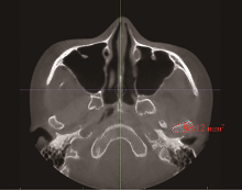
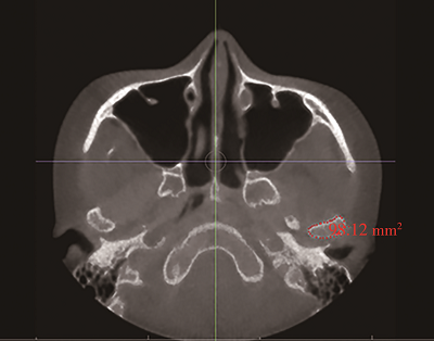
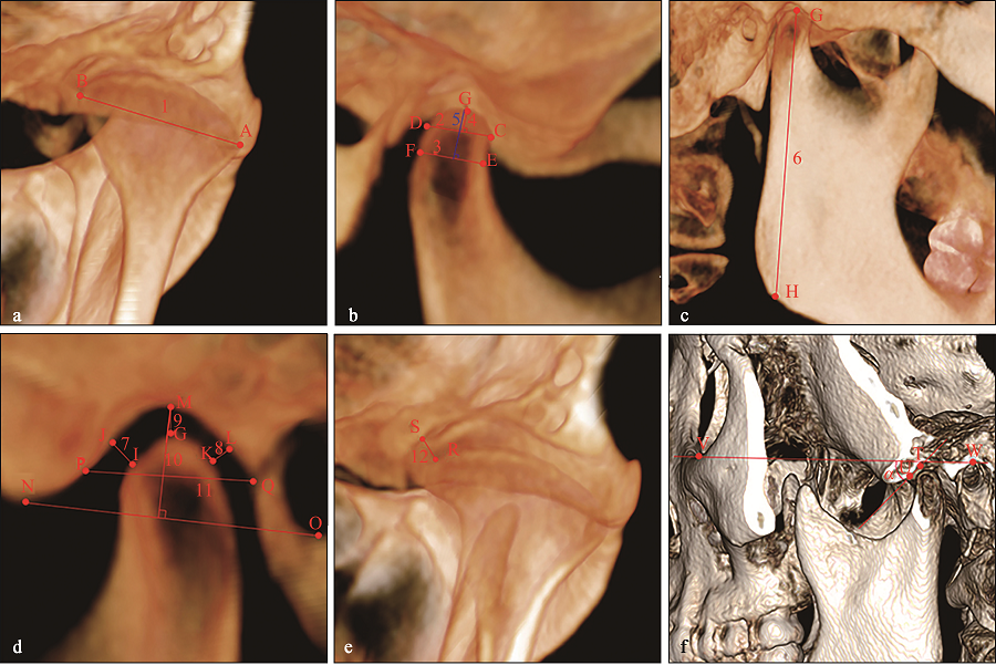
目的 通过测量分析具有不同垂直和矢状骨面型的成人髁突特征的差异,研究成人骨面型与髁突特征的相关性。方法 选取颞下颌关节正常的成人患者180例,根据∠ANB、∠MP-FH和∠MP-SN将患者分为不同的垂直及矢状骨面型,共9组,每组20例。使用Invivo6软件测量所有样本的髁突长轴径、髁突短轴径、髁突最大横截面积、髁突颈部宽度、髁突上部高度、髁突高度、下颌升支高度,关节前、后、上、内侧间隙,关节结节后斜面角度和关节窝深度、宽度。使用SPSS 23.0软件进行统计学分析,比较各组测量指标的差异。结果 低角患者髁突粗大,关节间隙大,关节窝深而窄,关节结节后斜面角度大,升支长;高角患者则相反。骨性Ⅲ类患者髁突粗大,关节间隙小,关节窝浅而宽,关节结节后斜面角度小;而骨性Ⅱ类患者则相反。结论 骨性Ⅱ类高角、骨性Ⅲ类低角患者髁突特征明显,与下颌发育有关;髁突特征与垂直骨面型、矢状骨面型之间存在关联;垂直骨面型与髁突特征的关联程度比矢状骨面型更强。在临床治疗过程中要加强对髁突及其周围结构的关注。
中图分类号:
| [1] |
Rodrigues AF, Fraga MR, Vitral RW. Computed tomography evaluation of the temporomandibular joint in class Ⅱ division 1 and class Ⅲ malocclusion patients: condyle symmetry and condyle-fossa relationship[J]. Am J Orthod Dentofacial Orthop, 2009, 136(2): 199-206.
doi: 10.1016/j.ajodo.2007.07.033 |
| [2] | 王洋, 王大为. 机械应力作用下髁突生长因子表达的研究进展[J/CD]. 中华口腔医学研究杂志(电子版), 2008, 2(4): 409-413. |
| Wang Y, Wang DW. Regulation of growth factors to the mandibular condyle under mechanical loading[J/CD]. Chin J Stomatol Res (Electron Ed), 2008, 2(4): 409-413. | |
| [3] | 朱房勇, 兰柳萍, 胡瑜, 等. 髁突颈部骨折对下颌骨生长发育的影响[J]. 中国实用口腔科杂志, 2009, 2(7): 440-442. |
| Zhu FY, Lan LP, Hu Y, et al. Effect of condylar neck fractures on growth and development of mandible[J]. Chin J Pract Stomatol, 2009, 2(7): 440-442. | |
| [4] | 李松. 下颌髁突状软骨与生长板软骨生长发育的比较研究[J]. 昆明医科大学学报, 2014, 35(2): 1-4. |
| Li S. Comparative study on the growth and development of mandibular condyle cartilage and growth plate cartilage[J]. J Kunming Med Univ, 2014, 35(2): 1-4. | |
| [5] | 陈洁, 段余峰, 涂景秋, 等. 不同垂直骨面型骨性Ⅲ类患者颞下颌关节三维形态结构的比较[J]. 中南大学学报(医学版), 2018, 43(6): 625-630. |
| Chen J, Duan YF, Tu JQ, et al. Three-dimensional morphological features of temporomandibular joint in skeletal malocclusion class Ⅲ patients with different vertical skeletal facial types[J]. J Central South Univ (Med Sci), 2018, 43(6): 625-630. | |
| [6] | Kim SE, Kim JD. A radiographic study of temporomandibular joints in skeletal class Ⅲmalocclusion[J]. Korean J Oral Maxillofac Radiol, 2003, 33(2): 85-90. |
| [7] | 田园. 青少年骨性Ⅱ类错𬌗不同垂直骨面型患者的髁突形态特点及差异性[J]. 中国药物与临床, 2009, 9(12): 1243-1244. |
| Tian Y. Condylar morphological characteristics and differences of adolescent skeletal class Ⅱ malocclusion with different vertical facial types[J]. Chin Remed Clin, 2009, 9(12): 1243-1244. | |
| [8] | 陆兴岭, 刘博, 赵丹, 等. 成人安氏Ⅰ、安氏Ⅱ和安氏Ⅲ类错𬌗畸形患者髁突位置的CBCT对比研究[J]. 中国医疗美容, 2018, 8(2): 67-70. |
| Lu XL, Liu B, Zhao D, et al. A CBCT comparative study of condylar position in adult patients with Angle classⅠ, class Ⅱ and class Ⅲ malocclusion[J]. China Med Cosmetol, 2018, 8(2): 67-70. | |
| [9] | 王欢, 丁寅. 不同垂直骨面型成年骨性Ⅲ类患者的髁突形态特点及差异[J]. 临床口腔医学杂志, 2006, 22(11): 673-675. |
| Wang H, Ding Y. Condylar morphology of the adults with different vertical facial types of skeletal Ⅲ malocclusions[J]. J Clin Stomatol, 2006, 22(11): 673-675. | |
| [10] |
Park IY, Kim JH, Park YH. Three-dimensional cone-beam computed tomography based comparison of condylar position and morphology according to the vertical skeletal pattern[J]. Korean J Orthod, 2015, 45(2): 66-73.
doi: 10.4041/kjod.2015.45.2.66 |
| [11] | 樊佳兵, 张军梅. 成年女性不同垂直骨面型下颌骨形态的测量分析[J]. 中国组织工程研究, 2021, 25(8): 1177-1183. |
| Fan JB, Zhang JM. Morphological measurement and analysis of the mandible in adult females with different vertical skeletal types[J]. Chin J Tissue Eng Res, 2021, 25(8): 1177-1183. | |
| [12] | 韩保迪, 栗震亚, 陈红. 安氏Ⅰ类错𬌗不同垂直骨面型下颌骨形态的比较研究[J]. 现代口腔医学杂志, 2008, 22(3): 239-242. |
| Han BD, Li ZY, Chen H. Morphometric study of the mandible shape of the subjects with class Ⅰ skeletal pattern in the three verticle facial types[J]. J Modern Stomatol, 2008, 22(3): 239-242. | |
| [13] | 赵营, 王建国, 魏志强. 骨性Ⅱ类错𬌗畸形患者不同垂直骨面型下颌骨三维形态特征研究[J]. 天津医科大学学报, 2015, 21(4): 342-344. |
| Zhao Y, Wang JG, Wei ZQ. Three dimensional morphological characteristics of mandible with different vertical skeletal types in patients with skeletal class Ⅱmalocclusion[J]. J Tianjin Med Univ, 2015, 21(4): 342-344. | |
| [14] | 舒艳, 刘珺, 陈杰, 等. 成人骨性Ⅲ类错𬌗不同垂直骨面型下颌骨及颏部的比较[J]. 上海口腔医学, 2011, 20(2): 191-195. |
| Shu Y, Liu J, Chen J, et al. Comparison of mandibular and chin morphology in adults with skeletal class Ⅲ malocclusion in different vertical facial types[J]. Shanghai J Stomatol, 2011, 20(2): 191-195. | |
| [15] |
Siriwat PP, Jarabak JR. Malocclusion and facial morphology is there a relationship? An epidemiolo-gic study[J]. Angle Orthod, 1985, 55(2): 127-138.
pmid: 3874569 |
| [16] |
Celikoglu M, Yavuz I, Unal T, et al. Comparison of the soft and hard tissue effects of two different protraction mechanisms in class Ⅲ patients: a randomi-zed clinical trial[J]. Clin Oral Investig, 2015, 19(8): 2115-2122.
doi: 10.1007/s00784-015-1408-5 |
| [17] |
Nakawaki T, Yamaguchi T, Tomita D, et al. Evaluation of mandibular volume classified by vertical skeletal dimensions with cone-beam computed tomography[J]. Angle Orthod, 2016, 86(6): 949-954.
doi: 10.2319/103015-732.1 |
| [18] |
Björk A. Prediction of mandibular growth rotation[J]. Am J Orthod, 1969, 55(6): 585-99.
pmid: 5253957 |
| [19] | 李放, 王建国. 不同垂直骨面型安氏Ⅰ类成年患者颞下颌关节形态特征的锥形束CT研究[J]. 国际口腔医学杂志, 2015, 42(5): 538-541. |
| Li F, Wang JG. Cone beam computed tomography analysis of temporomandibular joint morphology in adult Angle’s class Ⅰ malocclusions with different vertical skeletal features[J]. Int J Stomatol, 2015, 42(5): 538-541. | |
| [20] | 叶艳艳. 成人骨性Ⅱ类患者颅颌面特征及手术指征的判别分析[D]. 西安: 第四军医大学, 2013. |
| Ye YY. Craniomaxillofacial characteristics of adult skeletal class Ⅱ patients and discriminant analysis of surgical and non-surgical treatment[D]. Xi’an: The Fourth Millitary Medical University, 2013. | |
| [21] |
Hasebe A, Yamaguchi T, Nakawaki T, et al. Compari-son of condylar size among different anteroposterior and vertical skeletal patterns using cone-beam computed tomography[J]. Angle Orthod, 2019, 89(2): 306-311.
doi: 10.2319/032518-229.1 |
| [22] | 葛胜将, 侯凤春, 刘静, 等. 不同垂直骨面型成人骨性Ⅱ1类错𬌗女性髁突形态CBCT研究[J]. 青岛大学医学院学报, 2015, 51(4): 477-479, 482. |
| Ge SJ, Hou FC, Liu J, et al. Morphology of adult female with skeletal class Ⅱ1 malocclusions of diffe-rent vertical features: cone beam computed tomography study[J]. Acta Acad Med Qingdao Univ, 2015, 51(4): 477-479, 482. | |
| [23] |
Saccucci M, Polimeni A, Festa F, et al. Do skeletal cephalometric characteristics correlate with condylar volume, surface and shape? A 3D analysis[J]. Head Face Med, 2012, 8: 15.
doi: 10.1186/1746-160X-8-15 pmid: 22587445 |
| [24] |
Burke G, Major P, Glover K, et al. Correlations between condylar characteristics and facial morphology in class Ⅱ preadolescent patients[J]. Am J Orthod Dentofacial Orthop, 1998, 114(3): 328-336.
doi: 10.1016/S0889-5406(98)70216-1 |
| [25] |
Kubota M, Nakano H, Sanjo I, et al. Maxillofacial morphology and masseter muscle thickness in adults[J]. Eur J Orthod, 1998, 20(5): 535-542.
doi: 10.1093/ejo/20.5.535 |
| [26] | 高辉, 肖丹娜, 赵志河, 等. 成人高低角骨面型浅层咬肌不同功能位置形态的比较[J]. 实用口腔医学杂志, 2005, 21(6): 804-807. |
| Gao H, Xiao DN, Zhao ZH, et al. Comparison of superficial masseter muscle morphology between adult high-angle and low-angle facial skeletal types[J]. J Pract Stomatol, 2005, 21(6): 804-807. | |
| [27] |
Rowlerson A, Raoul G, Daniel Y, et al. Fiber-type differences in masseter muscle associated with different facial morphologies[J]. Am J Orthod Dentofacial Orthop, 2005, 127(1): 37-46.
doi: 10.1016/j.ajodo.2004.03.025 |
| [28] |
Kiliaridis S. Masticatory muscle influence on craniofacial growth[J]. Acta Odontol Scand, 1995, 53(3): 196-202.
pmid: 7572097 |
| [29] |
Chen T, Liu Z, Xue C, et al. Association of dysplastic coronoid process with long-face morphology[J]. J Dent Res, 2020, 99(3): 339-348.
doi: 10.1177/0022034519892551 pmid: 31826728 |
| [30] | 肖丹娜, 高辉. 嚼肌功能形态与垂直颅面结构的关系[J]. 国外医学·口腔医学分册, 2004, 31(2): 135-137. |
| Xiao DN, Gao H. The relationship between the functional morphology of masseter muscle and the vertical craniofacial structure[J]. Foreign Med Sci (Stomatol), 2004, 31(2): 135-137. | |
| [31] | 庾英姿, 米丛波. 咀嚼肌形态和功能与颅面形态关系研究进展[J]. 中国实用口腔杂志, 2012, 5(5): 315-318. |
| Yu YZ, Mi CB. Research progress on relationship of masticatory muscle morphology and function with craniofacial morphology[J]. Chin J Pract Stomatol, 2012, 5(5): 315-318. | |
| [32] | 李晨, 李永刚, 冯雪. 骨性Ⅱ类高角成年女性颞下颌关节骨性结构的三维研究[J]. 实用口腔医学杂志, 2016, 32(2): 239-243. |
| Li C, Li YG, Feng X. Three dimensional assessment of the temporomandibular joint in skeletal class Ⅱ malocclusion females with high vertical pattern[J]. J Pract Stomatol, 2016, 32(2): 239-243. | |
| [33] | Bench RW, Gugino CF, Hilgers JJ. Bio-progressive therapy[J]. J Clin Orthod, 1977, 11(9): 616-627. |
| [34] |
Hönicke K, Harzer W, Eckardt L. The relationships between the EMG excitation pattern of the masseter muscle and the facial skeletal morphology[J]. Fortschr Kieferorthop, 1995, 56(5): 237-244.
pmid: 7557796 |
| [35] |
Kikuchi K, Takeuchi S, Tanaka E, et al. Association between condylar position, joint morphology and craniofacial morphology in orthodontic patients without temporomandibular joint disorders[J]. J Oral Rehabil, 2003, 30(11): 1070-1075.
pmid: 14641670 |
| [36] |
Pullinger AG, Solberg WK, Hollender L, et al. Relationship of mandibular condylar position to dental occlusion factors in an asymptomatic population[J]. Am J Orthod Dentofacial Orthop, 1987, 91(3): 200-206.
doi: 10.1016/0889-5406(87)90447-1 |
| [37] |
Seren E, Akan H, Toller MO, et al. An evaluation of the condylar position of the temporomandibular joint by computerized tomography in classⅢ malocclusions: a preliminary study[J]. Am J Orthod Dentofacial Orthop, 1994, 105(5): 483-488.
doi: 10.1016/S0889-5406(94)70009-5 |
| [38] | 崔涛, 杜雨晴, 宋宇, 等. 不同矢状骨性错𬌗畸形高角型成年女性患者髁突位置的CBCT研究[J]. 口腔疾病防治, 2018, 26(3): 180-183. |
| Cui T, Du YQ, Song Y, et al. Cone-beam computed tomography study of condyle position in high-angle adult female patients with different sagittal skeletal malocclusion[J]. J Prev Treat Stomatol Dis, 2018, 26(3): 180-183. | |
| [39] |
Katsavrias EG, Halazonetis DJ. Condyle and fossa shape in class,Ⅱand class Ⅲ skeletal patterns: a morphometric tomographic study[J]. Am J Orthod Dentofacial Orthop, 2005, 128(3): 337-346.
doi: 10.1016/j.ajodo.2004.05.024 |
| [40] |
Arnett GW, Bergman RT. Facial keys to orthodontic diagnosis and treatment planning. PartⅠ[J]. Am J Orthod Dentofacial Orthop, 1993, 103(4): 299-312.
doi: 10.1016/0889-5406(93)70010-L |
| [41] |
Franchi L, Pavoni C, Faltin K, et al. Thin-plate spline analysis of mandibular shape changes induced by functional appliances in classⅡmalocclusion: a long-term evaluation[J]. J Orofac Orthop, 2016, 77(5): 325-333.
doi: 10.1007/s00056-016-0041-5 |
| [1] | 刘盼明,李政泽,李军鹤,崔淑霞. 成人骨性Ⅱ类患者不同垂直骨面型上颌窦容积及口咽气道体积的锥形束计算机断层扫描研究[J]. 国际口腔医学杂志, 2023, 50(5): 528-537. |
| [2] | 张哲,刘进,王卫红,陈志强,杨春,刘丽. 焦磷酸钙沉积症继发颞下颌关节脱位1例[J]. 国际口腔医学杂志, 2021, 48(6): 664-667. |
| [3] | 许琳,王如意,勾薪瑞,王晓莉,李宇. 甲状旁腺激素相关蛋白调控下颌髁突软骨的研究进展[J]. 国际口腔医学杂志, 2021, 48(5): 549-555. |
| [4] | 方苓力,谭玺,叶雨丝,黄兰,何瑶. 颞下颌关节退行性变早期髁突软骨细胞行为改变的实验研究[J]. 国际口腔医学杂志, 2021, 48(4): 417-425. |
| [5] | 殷晓丽,刘洋,王军. 伴发于偏颌畸形的颞下颌关节内部结构变化[J]. 国际口腔医学杂志, 2020, 47(5): 567-573. |
| [6] | 林阳阳,侯敏. 双侧下颌支矢状骨劈开术对下颌近心骨段位移变化的影响[J]. 国际口腔医学杂志, 2019, 46(6): 718-723. |
| [7] | 郭骏,费伟,李庆华. 创伤性颞下颌关节强直转归机制的动物研究[J]. 国际口腔医学杂志, 2019, 46(1): 12-19. |
| [8] | 黎静, 刘星辰, 李佳园, 李小兵. 稳定咬合板治疗慢性颞下颌关节盘不可复性移位的临床随机对照试验的系统评价[J]. 国际口腔医学杂志, 2017, 44(4): 405-410. |
| [9] | 戴智, 侯敏, 张春香. 渐进性髁突吸收致下颌后缩正颌外科的治疗进展[J]. 国际口腔医学杂志, 2017, 44(3): 359-362. |
| [10] | 罗恩. 髁突骨软骨瘤及其继发牙颌面畸形的治疗[J]. 国际口腔医学杂志, 2017, 44(2): 130-134. |
| [11] | 朱晸,史俊. 儿童下颌骨髁突骨折的治疗进展[J]. 国际口腔医学杂志, 2017, 44(2): 222-227. |
| [12] | 刘洋, 赵翰驰. 前牙重度磨损伴关节弹响患者的咬合重建[J]. 国际口腔医学杂志, 2017, 44(1): 11-18. |
| [13] | 胡欣欣,朱耀旻,郑苍尚. 特发性髁突吸收的研究进展[J]. 国际口腔医学杂志, 2016, 43(4): 412-416. |
| [14] | 李婧,樊永杰. 女性青少年不同垂直骨面型颏部形态的研究[J]. 国际口腔医学杂志, 2016, 43(4): 387-390. |
| [15] | 吕春晓 陈嵩. 颞下颌关节病的临床诊断与磁共振成像影像诊断的相关性研究[J]. 国际口腔医学杂志, 2016, 43(1): 47-. |
|