国际口腔医学杂志 ›› 2022, Vol. 49 ›› Issue (1): 27-36.doi: 10.7518/gjkq.2022026

• 论著 • 上一篇    下一篇

三维培养牙髓间充质细胞外泌体微小RNA表达谱分析

艾晓青(),窦磊,乔新,杨德琴()   

  1. 重庆医科大学附属口腔医院牙体牙髓科 口腔疾病与生物医学重庆市重点实验室 重庆市高校市级口腔生物医学工程重点实验室 重庆 401147
  • 收稿日期:2021-04-05 修回日期:2021-10-14 出版日期:2022-01-01 发布日期:2022-01-07
  • 通讯作者: 杨德琴
  • 作者简介:艾晓青,硕士,Email: 2018110850@stu.cqmu.edu.cn
  • 基金资助:
    国家自然科学基金(31970783);国家自然科学基金青年基金(81800958)

MicroRNA profile of exosomes derived from dental pulp stromal cells under three-dimensional culture condition

Ai Xiaoqing(),Dou Lei,Qiao Xin,Yang Deqin()   

  1. Dept. of Cariology and Endodontics, Stomatological Hospital of Chong-qing Medical University, Chongqing Key Laboratory of Oral Diseases and Biomedical Sciences, Chongqing Municipal Key Laboratory of Oral Biomedical Engineering of Higher Education, Chongqing 401147, China
  • Received:2021-04-05 Revised:2021-10-14 Online:2022-01-01 Published:2022-01-07
  • Contact: Deqin Yang
  • Supported by:
    This study was supported by National Natural Science Foundation of China(31970783);National Natural Science Foundation of China Youth Fund(81800958)

摘要:

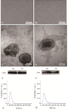
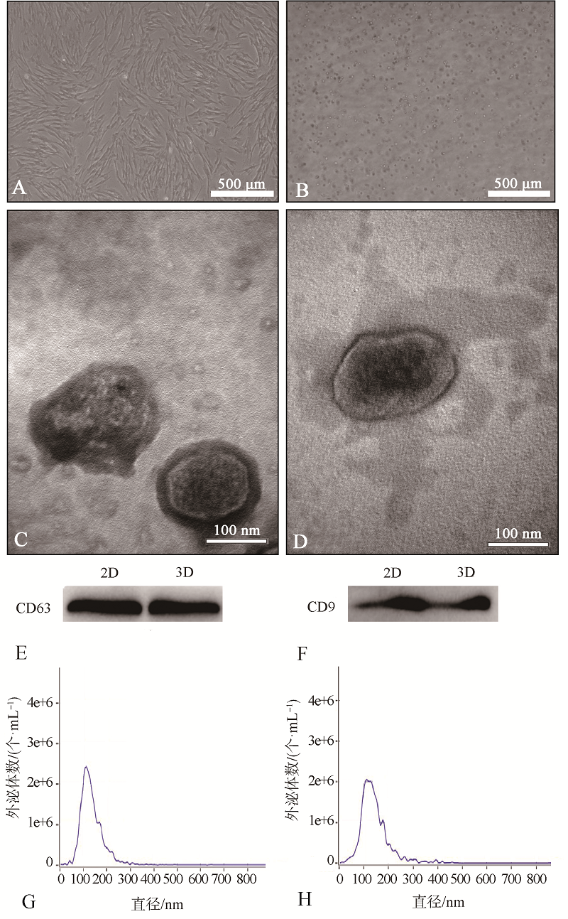
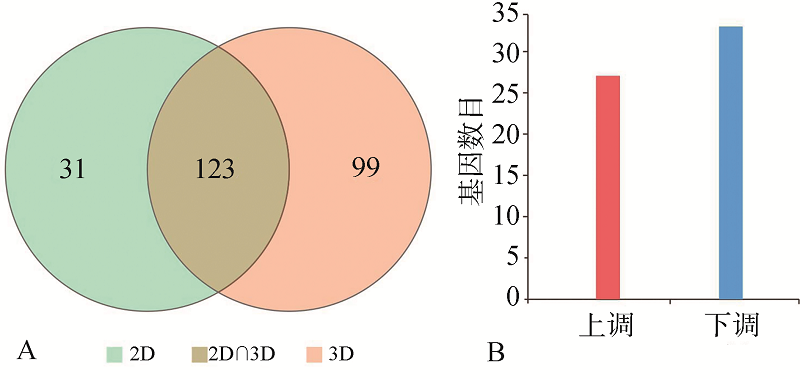
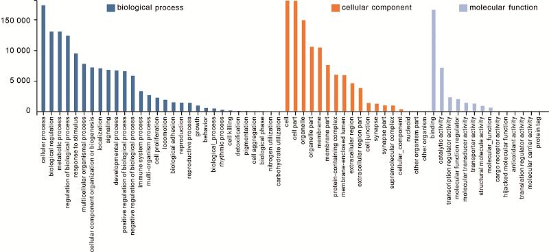
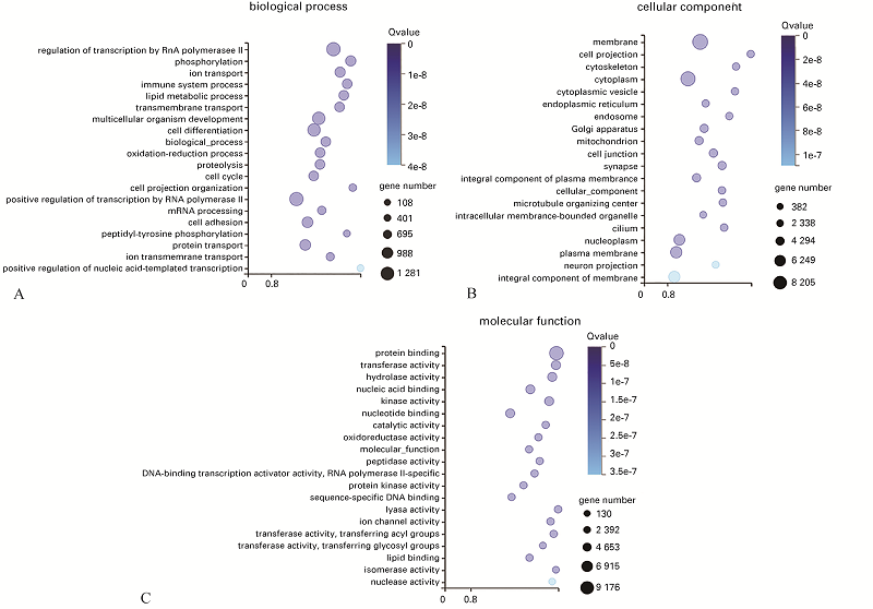
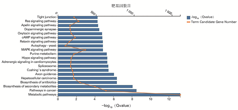
目的 分析并比较二维(2D)与三维(3D)培养条件下人牙髓间充质细胞(DPSCs)外泌体(Exo)微小RNA(miRNA)的表达谱。方法 2D与3D条件下分别培养DPSCs,提取细胞Exo,采用透射电子显微镜、蛋白质免疫印迹及纳米颗粒追踪分析等方法观察并鉴定;通过高通量测序筛选差异表达miRNA,采用Dr.Tom系统及TargetScan网站进行生物信息学分析及组织再生修复相关靶基因预测。结果 3D培养下收集的DPSC-Exo均呈现“茶托样”双层膜结构,CD63和CD9表达阳性,粒径分布符合外泌体特征,与2D培养的DPSC-Exo一致。高通量测序共计检测外泌体来源miRNA 253个,其中3D组表达222个,特有表达99个;与2D组相比,表达显著差异60个[︱log2(3D/2D)︱≥1,Qvalue≤0.001]。差异miRNA主要参与的生物学过程与分子功能分别为细胞过程和结合;京都基因与基因组数据库(KEGG)通路富集分析显示在代谢通路有显著富集。miR-302的候选靶基因有成纤维细胞生长因子(FGF)19和表皮生长因子受体等;miR-24-3p可能靶向神经元分化因子2、神经上皮细胞转化因子1、神经元分化因子1、神经元再生相关蛋白、FGF11、FGF结合蛋白3、FGF受体3、血小板衍生生长因子受体(PDGFR)β多肽、PDGFRα多肽、血管生成素、胰岛素样生长因子结合蛋白5等,以发挥组织再生修复功能。结论 相比2D培养,3D培养能调节DPSC-Exo部分miRNA表达量,上调一些与再生修复相关的miRNA,3D培养可考虑作为调节外泌体应用潜能的一种手段。

关键词: 三维培养, 牙髓间充质细胞, 外泌体测序, 微小RNA

Abstract: Objective This study aims to analyze and compare microRNA (miRNA) profiles in exosomes (Exo) derived from human dental pulp stromal cells (DPSCs) under two-dimensional (2D) and three-dimensional (3D) culture condition. Methods DPSC-Exo was extracted from 2D and 3D culture of DPSCs and identified by transmission electron microscopy, Western blot analysis, and nanoparticle tracking analysis (NTA). Differentially expressed miRNAs were screened by high-throughput sequencing for bioinformatics analysis and target gene prediction related to tissue regeneration and repair by using Dr.Tom system and TargetScan Website. Results 2D-DPSC-Exo and 3D-DPSC-Exo showed “saucer-like” double-layer membrane structure, expressed CD63 and CD9 positively and had diameter consistent with Exo characteristics. A total of 253 Exo-derived miRNAs were detected, among which 222 were in the 3D group and 99 were specifically expressed. Compared with the 2D culture, a significant difference of 60 [︱log2(3D/2D)︱≥1, Qvalue≤0.001]. Differentially expressed miRNAs were mainly involved in cellular process and binding. The Kyoto encyclopedia of genes and genomes (KEGG) pathway showed that these miRNAs were significantly enriched in metabolic pathways. Candidate target genes of miR-302 include fibroblast growth factor (FGF) 19 and epidermal growth factor receptor. Candidate target genes of miR-24-3p may play a role in tissue regeneration and repair, including neuronal differentiation (NEUROD) 2, neuroepithelial cell transforming 1, NEUROD1, neuronal regeneration-related protein, FGF11, FGF binding protein 3, FGF receptor 3, platelet-derived growth factor receptor (PDGFR) beta polypeptide, PDGFR alpha polypeptide, angiopoietin 4, and insulin-like growth factor binding protein 5. Conclusion Compared with 2D culture, 3D culture could regulate the expression of some miRNAs in DPSC-Exo. Upregulated miRNAs are mainly related to regeneration and repair, and 3D culture might be suitable to optimize the therapeutic potential of Exo.

Key words: three-dimensional culture, dental pulp stromal cells, exosomes sequencing, microRNA

图1

2D及3D培养条件下DPSCs与Exo的培养鉴定 A:2D-DPSCs 倒置显微镜;B:3D-DPSCs 倒置显微镜;C:2D-DPSC-Exo TEM;D:3D-DPSC-Exo TEM;E:CD63;F:CD9;G:2D-DPSC-Exo;H:3D-DPSC-Exo。"

图2

差异miRNA表达 A:表达量韦恩图;B:差异基因数量统计,与2D组相比,3D组上调27个,下调33个基因表达。"

表 1

部分上调差异miRNA"

基因ID 2D组表达量 3D组表达量 log2(3D/2D) Qvalue
hsa-miR-302a-3p 26 978 5.23 1.79e-221
hsa-miR-302b-3p 192 1 578 3.04 2.85e-111
hsa-miR-302c-3p 16 309 4.27 1.04e-52
hsa-miR-302d-3p 38 1 017 4.74 2.80e-201
hsa-miR-24-3p 17 702 5.37 1.23e-92
hsa-miR-27b-3p 363 989 1.44 8.07e-28
hsa-miR-34a-5p 942 2 453 1.38 1.28e-25
hsa-miR-100-5p 557 1 310 1.23 8.61e-24

图3

2D-DPSC-Exo和3D-DPSC-Exo显著差异表达miRNA聚类热图 x轴为样本的log2(表达量值+1),y轴为基因表达量,颜色越红表示表达量越高,颜色越蓝表示表达量越低。"

表2

TargetScan 网站预测hsa-miR-302a/b/c/d-3p与hsa-miR-24-3p部分靶基因"

miRNA 预测靶基因 miRNA与靶基因结合位点
预测的目标区域(顶部)和miRNA(底部)的配对情况
(下划线区域为种子区)
上下游配对
百分数/%
hsa-miR-302a/b/c/d-3p FGF19 5’...UAGAACCCUUUCCCCAGCACUUG...
3’ AGUGGUUUUGUACCUUCGUGAAU
91
EGFR 5’...GAAAAGCAAUAACAU- - -AGCACUUU
3’ AGUGGUUUUGUACCUUCGUGAAU
87
hsa-miR-24-3p NEUROD2 5’...UGCAAGGAGGCUCCACUGAGCCU...
3’ GACAAGGACGACUUGACUCGGU
74
NEUROD1 5’...AUUCAUGUAAUAAAUCUGAGCCU...
3’ GACAAGGACGACUUGACUCGGU
96
NET1 5’...AAGCCAUACUGUUUU--UGAGCCAA...
3’ GACAAGGACGACUUGACUCGGU
89
NREP 5’ ...AGAAACUAUGGGACUCUGAGCCU...
3’ GACAAGGACGACUUGACUCGGU
98
FGF11 5’...CCCUUUUCAUUGCCA--CUGAGCCA...
3’ GACAAGGACGACUUGACUCGGU
99
FGFBP3 5’...UGAGCAAUUGCCAACCUGAGCCA...
3’ GACAAGGACGACUUGACUCGGU
97
FGFR3 5’...CUCCCACACCCAAAGCUGAGCCU...
3’ GACAAGGACGACUUGACUCGGU
88
PDGFRB 5’...GGAGGCCAACUGACU--CUGAGCCA...
3’ GACAAGGACGACUUGACUCGGU
97
PDGFRA 5’...UAGUAAGUGCGAAGACUGAGCCA...
3’ GACAAGGACGACUU-GACUCGGU
95
ANGPT4 5’...GACACCCUGGGCUUCCUGAGCCA...
3’ GACAAGGACGACUU-GACUCGGU
96
IGFBP5 5’...UCCCCUGAGAAAAGACUGAGCCA...
3’GACAAGGACGACUU---GACUCGGU
95

图4

2D-DPSC-Exo与3D-DPSC-Exo显著差异表达miRNA靶基因的GO注释分类 x轴为GO功能分类,y轴为注释到GO术语(GO Term)上的基因数目。蓝色部分为biological process(生物学过程),橙色部分为cellular component(细胞组分),浅蓝色部分为molecular function(分子功能)。生物学过程包括cellular process(细胞过程)、biological regulation(生物调节)、me-tabolic process(代谢过程)、regulation of biological process(生物过程调节)、response to stimulus(对刺激的反应)、multicellular organismal process(多细胞生物过程)、cellular component organization or biogenesis(细胞成分组织或生物发生)、localization(定位)、signaling(信号)、developmental process(发育过程)、positive regulation of biological process(生物过程的正调节)、negative regulation of biological process(生物过程的负调节)、immune system process(免疫系统过程)、multi-organism process(多生物过程)、cell proliferation(细胞增殖)、locomotion(移动)、biological adhesion(生物附着)、reproduction(繁殖)、reproductive process(繁殖过程)、growth(生长)、behavior(行为)、biological_process(生物学过程)、rhythmic process(节律过程)、cell killing(细胞杀伤)、detoxification(排毒)、pigmentation(色素沉着)、cell aggregation(细胞集聚)、biological phase(生物阶段)、nitrogen utilization(氮利用)、carbohydrate utilization(糖利用)。细胞组分包括cell(细胞)、cell part(细胞部分)、organelle(细胞器)、organelle part(细胞器组分)、membrane(膜)、membrane part(膜组分)、protein-containing complex(含蛋白质复合物)、membrane-enclosed lumen(膜封闭腔)、extracellular region(胞外区域)、extracellular region part(胞外区组分)、cell junction(细胞接合)、synapse(突触)、synapse part(突触组分)、supramolecular complex(超分子复合物)、cellular_component(细胞组分)、nucleoid(类核)、other organism part(其他有机体组分)、other organism(其他有机体)。分子功能包括binding(结合)、catalytic activity(催化活性)、transcription regulator activity(转录调节活性)、molecular function regulator(分子功能调节)、molecular transducer activity(分子转导活性)、transporter activity(转运活性)、structural molecule activity(结构分子活性)、molecular_function(分子功能)、cargo receptor activity(转运受体活性)、hijacked molecular function(拦截分子功能)、antioxidant activity(抗氧化活性)、translation regulator activity(翻译调节活性)、molecular carrier activity(分子载体活性)、protein tag(蛋白标记)。"

图5

2D-DPSC-Exo与3D-DPSC-Exo显著差异表达miRNA靶基因分别在生物学过程、细胞组分和分子功能的GO富集情况(Qvalue≤0.05) x轴为富集比例,y轴为GO术语(GO Term)。图中气泡的大小表示注释到某个GO Term上的差异基因数目(gene number),颜色越蓝代表显著性值(即Qvalue值)越小,富集程度越高。A为biological process(生物学过程),B为cellular component(细胞组分),C为molecular function(分子功能)。生物学过程包括regulation of transcription by RNA polymerase Ⅱ (RNA聚合酶Ⅱ对转录的调控)、phosphorylation(磷酸化)、ion transport(离子转运)、immune system process(免疫系统过程)、lipid metabolic process(脂质代谢过程)、transmembrane transport(跨膜转运)、multicellular organism development(多细胞生物发育)、cell differentiation(细胞分化)、biological_process(生物学过程)、oxidation-reduction process(氧化还原过程)、proteolysis(蛋白酶解)、cell cycle(细胞周期)、cell projection organization(组织细胞投影)、positive regulation of transcription by RNA polymerase Ⅱ(RNA聚合酶Ⅱ对转录的正调控)、mRNA processing(mRNA加工)、cell adhesion(细胞黏附)、peptidyl-tyrosine phosphorylation(肽基酪氨酸磷酸化)、protein transport(蛋白质转运)、ion transmembrane transport(离子跨膜运输)、positive regulation of nucleic acid-templated transcription(核酸模板转录的正调控)。细胞组分包括membrane(膜)、cell projection(细胞投影)、cytoskeleton(细胞骨架)、cytoplasm(细胞质)、cytoplasmic vesicle(胞质囊泡)、endoplasmic reticulum(内质网)、endosome(核内体)、golgi apparatus(高尔基体)、mitochondrion(线粒体)、cell junction(细胞连接)、synapse(突触)、integral component of plasma membrane(质膜的组成部分)、cellular_component(细胞组分)、microtubule organizing center (微管组织中心)、intracellular membrane-bounded organelle(细胞内膜结合的细胞器)、cilium(鞭毛)、nucleoplasm(核质)、plasma membrane(质膜)、neuron projection(神经元投射)、integral component of membrane(膜的组成部分)。分子功能包括protein binding(蛋白结合)、transferase activity(转移酶活性)、hydrolase activity(水解酶活性)、nucleic acid binding(核酸结合)、kinase activity(激酶活性)、nucleotide binding(核苷酸结合)、catalytic activity(催化活性)、oxidoreductase activity(氧化还原酶活性)、molecular_function(分子功能)、peptidase activity(肽酶活性)、DNA-binding transcription activator activity, RNA polymerase Ⅱ-specific (DNA结合转录激活剂活性RNA聚合酶Ⅱ特异性)、protein kinase activity(蛋白激酶活性)、sequence-specific DNA binding(序列特异性 DNA 结合)、lyase activity(裂解酶活性)、ion channel activity(离子通道活性)、transferase activity, transferring acyl groups[转移酶活性(转移酰基)]、transferase activity, transferring glycosly groups[转移酶活性(转移糖基)]、lipid binding(脂质结合)、isomerase activity(异构酶活性)、nuclease activity(核酸酶活性)。"

图6

2D-DPSC-Exo与3D-DPSC-Exo显著差异表达miRNA靶基因 KEGG通路分析(Qvalue≤0.05) x轴代表富集比例及靶基因数目,y轴为KEGG 通路(前20)。Tight junction(紧密连接)、Ras signaling pathway(Ras信号通路)、Apelin signaling pathway(Apelin信号通路)、Dopaminergic synapse(多巴胺能突触)、Oxytocin signaling pathway(Oxytocin 信号通路)、cAMP signaling pathway(cAMP信号通路)、Relaxin signaling pathway(Relaxin信号通路)、Autophagy-yeast(自噬-酵母)、MAPK signaling pathway(MAPK信号通路)、Purine metabolism(嘌呤代谢)、Hippo signaling pathway(Hippo信号通路)、Adrenergic signaling in cardiomyocytes(心肌细胞中的肾上腺素信号)、Spliceosome(剪接体)、Cushing’s syndrome(库兴氏综合征)、Axon guidance(轴突导向)、Hepatocellular carcinoma(肝癌)、Biosynthesis of antibiotics(抗生素的生物合成)、Biosynthesis of secondary metabolites(次生代谢物的生物合成)、Pathways in cancer(癌症相关通路)、Metabolic pathways(代谢通路)。"

[1] Altanerova U, Benejova K, Altanerova V, et al. Dental pulp mesenchymal stem/stromal cells labeled with iron sucrose release exosomes and cells applied intra-nasally migrate to intracerebral glioblastoma[J]. Neoplasma, 2016,63(6):925-933.
[2] Colombo M, Raposo G, Théry C. Biogenesis, secretion, and intercellular interactions of exosomes and other extracellular vesicles[J]. Annu Rev Cell Dev Biol, 2014,30:255-289.
[3] Xian XH, Gong QM, Li C, et al. Exosomes with highly angiogenic potential for possible use in pulp regeneration[J]. J Endod, 2018,44(5):751-758.
[4] Huang CC, Narayanan R, Alapati S, et al. Exosomes as biomimetic tools for stem cell differentiation: applications in dental pulp tissue regeneration[J]. Biomaterials, 2016,111:103-115.
[5] Qazi TH, Mooney DJ, Duda GN, et al. Niche-mi-micking interactions in peptide-functionalized 3D hydrogels amplify mesenchymal stromal cell paracrine effects[J]. Biomaterials, 2020,230:119639.
[6] di Liegro CM, Schiera G, di Liegro I. Extracellular vesicle-associated RNA as a carrier of epigenetic information[J]. Genes, 2017,8(10):240.
[7] Bu NU, Lee HS, Lee BN, et al. In vitro characterization of dental pulp stem cells cultured in two microsphere-forming culture plates[J]. J Clin Med, 2020,9(1):242.
[8] Dou L, Yan QF, Liang PP, et al. iTRAQ-based proteomic analysis exploring the influence of hypoxia on the proteome of dental pulp stem cells under 3D culture[J]. Proteomics, 2018,18(3/4):1700215.
[9] Swanson WB, Gong T, Zhang Z, et al. Controlled release of odontogenic exosomes from a biodegra-dable vehicle mediates dentinogenesis as a novel biomimetic pulp capping therapy[J]. J Control Release, 2020,324:679-694.
[10] Li Y, Yang YY, Ren JL, et al. Exosomes secreted by stem cells from human exfoliated deciduous teeth contribute to functional recovery after traumatic brain injury by shifting microglia M1/M2 polarization in rats[J]. Stem Cell Res Ther, 2017,8:198.
[11] Gao Z, Zhu X, Dou Y. The miR-302/367 cluster: a comprehensive update on its evolution and functions[J]. Open Biol, 2015,5(12):150138.
[12] Zhang ZH, Hong YF, Xiang D, et al. MicroRNA-302/367 cluster governs hESC self-renewal by dually regulating cell cycle and apoptosis pathways[J]. Stem Cell Rep, 2015,4(4):645-657.
[13] Herbst RS. Review of epidermal growth factor receptor biology[J]. Int J Radiat Oncol, 2004,59(2):S21-S26.
[14] Li J, Yu J, Zhang H, et al. Exosomes-derived MiR-302b suppresses lung cancer cell proliferation and migration via TGFβRII inhibition[J]. Cell Physiol Biochem, 2016,38(5):1715-1726.
[15] 陈伟, 欧和生. miR-24对内皮细胞功能的调节及其在心血管疾病发生发展中的作用[J]. 生理学报, 2016,68(2):201-206.
Chen W, Ou HS. Regulation of miR-24 on vascular endothelial cell function and its role in the development of cardiovascular disease[J]. Acta Physiolog Sinica, 2016,68(2):201-206.
[16] Shen SJ, Song Y, Ren XY, et al. MicroRNA-27b-3p promotes tumor progression and metastasis by inhi-biting peroxisome proliferator-activated receptor ga-mma in triple-negative breast cancer[J]. Front Oncol, 2020,10:1371.
[17] Jiang Q, Xing W, Cheng J, et al. Long non-coding RNA TP73-AS1 promotes the development of lung cancer by targeting the miR-27b-3p/LAPTM4B axis[J]. Onco Targets Ther, 2020,13:7019-7031.
[18] Bouchard A, Witalis M, Chang J, et al. Hippo signal transduction mechanisms in T cell immunity[J]. Immune Netw, 2020,20(5):e36.
[19] Luo P, Jiang C, Ji P, et al. Exosomes of stem cells from human exfoliated deciduous teeth as an anti-inflammatory agent in temporomandibular joint chondrocytes via miR-100-5p/mTOR[J]. Stem Cell Res Ther, 2019,10(1):216.
[20] Tian F, Wang J, Zhang Z, et al. LncRNA SNHG7/miR-34a-5p/SYVN1 axis plays a vital role in proli-feration, apoptosis and autophagy in osteoarthritis[J]. Biol Res, 2020,53(1):9.
[21] Duan Y, Li X, Zhang S, et al. Therapeutic potential of HERS spheroids in tooth regeneration[J]. The-ranostics, 2020,10(16):7409-7421.
[22] Pan J, Deng J, Luo Y, et al. Thermosensitive hydrogel delivery of human periodontal stem cells overexpressing platelet-derived growth factor-BB enhan-ces alveolar bone defect repair[J]. Stem Cells Dev, 2019,28(24):1620-1631.
[23] Yin W, Liu YL, Bian Z. MG53 inhibits the progression of tongue cancer cells through regulating PI3K-AKT signaling pathway: evidence from 3D cell culture and animal model[J]. Small, 2019,15(8):1805492.
[1] 周金阔,张晋弘,史晓晶,刘广顺,姜磊,刘倩峰. 长链非编码RNA小核仁RNA宿主基因22调控微小RNA-27b-3p对口腔鳞状细胞癌细胞增殖、侵袭和迁移的影响[J]. 国际口腔医学杂志, 2024, 51(1): 52-59.
[2] 李立恒,王蕊,王晓明,张智轶,张璇,安峰,王芹,张凡. 环状RNA hsa_circ_0085576调控微小RNA-498/B细胞特异性莫洛尼鼠白血病病毒整合位点1轴对口腔鳞状细胞癌细胞迁移和侵袭的影响[J]. 国际口腔医学杂志, 2024, 51(1): 60-67.
[3] 洪娅娅,陈学鹏,姒蜜思. 非编码RNA调控牙囊干细胞成骨分化的研究进展[J]. 国际口腔医学杂志, 2022, 49(3): 263-271.
[4] 钱素婷,丁玲敏,纪雅宁,林军. 微小RNA在牙周炎龈沟液中的表达差异及对牙周炎的调控机制[J]. 国际口腔医学杂志, 2022, 49(3): 349-355.
[5] 孙坚炜,雷利红,谭静怡,陈莉丽. 微小RNA 155对骨免疫的调控及其在牙周炎中作用的研究进展[J]. 国际口腔医学杂志, 2020, 47(5): 607-615.
[6] 周婕妤,刘琳,吴亚菲,赵蕾. 微小RNA介导的牙周炎与动脉粥样硬化相关机制的研究进展[J]. 国际口腔医学杂志, 2020, 47(1): 76-83.
[7] 刘志凯,王淳艺,李春洁. 胚胎小鼠颌下腺分支形态发生及其影响因素[J]. 国际口腔医学杂志, 2019, 46(1): 43-47.
[8] 冯顶丽,卓丽丹,芦笛,郭红延. 微小RNA调节间充质干细胞软骨分化机制的研究进展[J]. 国际口腔医学杂志, 2018, 45(6): 640-645.
[9] 方川,李雅冬. 微小RNA在口腔鳞状细胞癌中的研究进展[J]. 国际口腔医学杂志, 2018, 45(6): 646-651.
[10] 郝奕霖, 房付春, 吴补领. 微小RNA在人牙周膜来源细胞成骨分化中的作用[J]. 国际口腔医学杂志, 2018, 45(1): 46-49.
[11] 刘润恒,刘于冬,陈卓凡. 微小RNA在骨分化过程中的作用机制[J]. 国际口腔医学杂志, 2017, 44(1): 108-113.
[12] 耿奉雪,潘亚萍. 微小RNA-203的生物学功能及其在口腔疾病中的作用[J]. 国际口腔医学杂志, 2016, 43(6): 685-689.
[13] 李龙,黄洪章. 微小RNA-205在肿瘤化学治疗耐药中的作用和机制[J]. 国际口腔医学杂志, 2016, 43(6): 734-738.
[14] 李泓钰,黄洪章. 蜗牛蛋白调控腭部发生发育的机制[J]. 国际口腔医学杂志, 2016, 43(4): 468-472.
[15] 刘佳佳 吴圆琴 曾昕 周瑜. 微小RNA在口腔扁平苔藓中的作用[J]. 国际口腔医学杂志, 2015, 42(1): 48-53.
Viewed
Full text


Abstract

Cited

  Shared   
  Discussed   
[1] 张新春. 桩冠修复与无髓牙的保护[J]. 国际口腔医学杂志, 1999, 26(06): .
[2] 王昆润. 长期单侧鼻呼吸对头颅发育有不利影响[J]. 国际口腔医学杂志, 1999, 26(05): .
[3] 彭国光. 颈淋巴清扫术中颈交感神经干的解剖变异[J]. 国际口腔医学杂志, 1999, 26(05): .
[4] 杨凯. 淋巴化疗的药物运载系统及其应用现状[J]. 国际口腔医学杂志, 1999, 26(05): .
[5] 康非吾. 种植义齿下部结构生物力学研究进展[J]. 国际口腔医学杂志, 1999, 26(05): .
[6] 柴枫. 可摘局部义齿用Co-Cr合金的激光焊接[J]. 国际口腔医学杂志, 1999, 26(04): .
[7] 孟姝,吴亚菲,杨禾. 伴放线放线杆菌产生的细胞致死膨胀毒素及其与牙周病的关系[J]. 国际口腔医学杂志, 2005, 32(06): 458 -460 .
[8] 费晓露,丁一,徐屹. 牙周可疑致病菌对口腔黏膜上皮的粘附和侵入[J]. 国际口腔医学杂志, 2005, 32(06): 452 -454 .
[9] 赵兴福,黄晓晶. 变形链球菌蛋白组学研究进展[J]. 国际口腔医学杂志, 2008, 35(S1): .
[10] 庞莉苹,姚江武. 抛光和上釉对陶瓷表面粗糙度、挠曲强度及磨损性能的影响[J]. 国际口腔医学杂志, 2008, 35(S1): .