国际口腔医学杂志 ›› 2022, Vol. 49 ›› Issue (4): 420-425.doi: 10.7518/gjkq.2022020
吴文智( ),冯达兴,陈垂壮,周丽鹃
Wu Wenzhi(),Feng Da-xing,Chen Chuizhuang,Zhou Lijuan.
摘要:
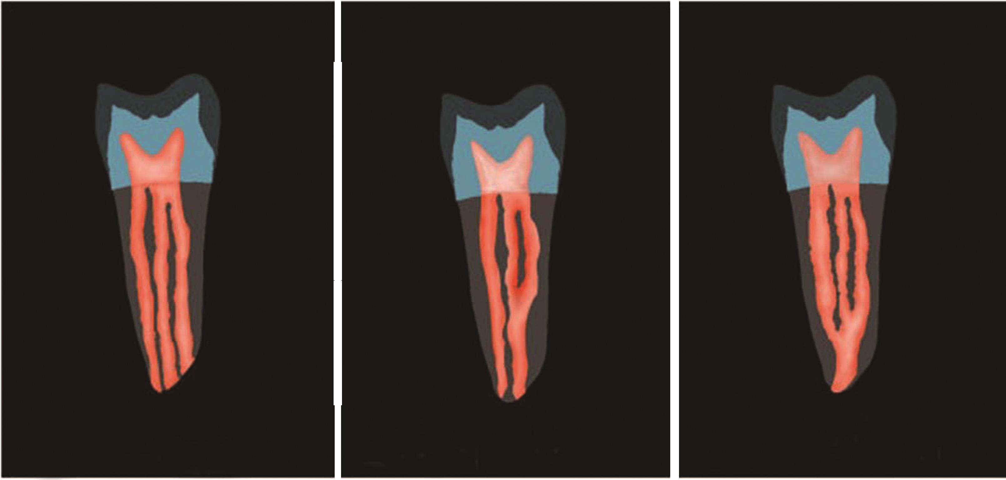
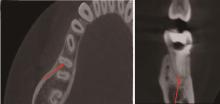
目的 评估海口地区人群下颌第一恒磨牙近中中央根管(MMCs)发生率及其影响因素。方法 回顾分析2018年1月—2020年12月1 032例患者的1 964颗下颌第一恒磨牙锥形束CT的影像资料,观察其近中根管的形态特点,统计MMCs的发生率,收集记录患者的年龄、性别、民族、居住环境、牙位、近颊(MB)-近舌(ML)根管口间距、根管峡区(RCIs)等信息,并对影响因素进行logistic回归分析。结果 海口地区人群下颌第一恒磨牙MMCs发生率为22.3%,以附加型(3-2)为主(63.8%),RCIs发生率为50.5%,经logistic回归分析显示年龄≤40岁(OR=2.667,95%CI:1.185~6.004),居住城市(OR=1.456,95%CI:1.036~2.048),合并RCIs (OR=3.808,95%CI:1.353~10.718),MB-ML根管口间距<3 mm(OR=3.374,95%CI:1.168~9.741),均是第一恒磨牙发生MMCs的独立危险因素。结论 海口地区第一恒磨牙MMCs发生率较高。年龄≤40岁、居住城市、合并RCIs、MB-ML根管口间距<3 mm均是第一恒磨牙发生MMCs的独立危险因素。
中图分类号:
| 1 | 李米雪子, 张琛. 椅旁计算机辅助设计/计算机辅助制作髓腔固位冠修复根管治疗后磨牙的临床考量[J]. 国际口腔医学杂志, 2021, 48(3): 274-279. |
| Li MXZ, Zhang C. Clinical consideration on the application of computer-aided design/computer-aided manufacturing endocrown in molar restoration after root canal therapy[J]. Int J Stomatol,2021, 48(3): 274-279. | |
| 2 | Hu XL, Huang ZJ, Huang ZW, et al. Presence of isthmi in mandibular mesial roots and associated factors: an in vivo analysis[J]. Surg Radiol Anat, 2019, 41(7): 815-822. |
| 3 | 张博森, 刘敏, 吴家媛. 贵州地区人下颌第一恒磨牙近中中央根管充填率调查及遗漏原因分析[J]. 临床口腔医学杂志, 2018, 34(7): 412-415. |
| Zhang BS, Liu M, Wu JY. Treatment survey of the middle mesial root canals in permanent mandibular first molars of Chinese individuals in Guizhou[J]. J Clin Stomatol, 2018, 34(7): 412-415. | |
| 4 | 陆亚倩, 杨晨露, 刘亚文, 等. 下颌第一磨牙根管形态的CBCT研究[J]. 口腔医学, 2020, 40(5): 443-447. |
| Lu YQ, Yang CL, Liu YW, et al. Root and canal configuration of mandibular first molars:an in vivo cone-beam computed tomographic study[J]. Stomatology, 2020, 40(5): 443-447. | |
| 5 | Vertucci FJ. Root canal anatomy of the human permanent teeth[J]. Oral Surg Oral Med Oral Pathol, 1984, 58(5): 589-599. |
| 6 | 刘玮佳, 孙琼. 下颌第一磨牙近中及远中中央根管的研究现状与展望[J]. 安徽医学, 2021, 42(4): 460-463. |
| Liu WJ, Sun Q. Research status and prospect of mesial and distal central root canals of mandibular first molars[J]. Anhui Med J, 2021, 42(4): 460-463. | |
| 7 | 刘忠俊, 张治勇, 邝锐芳, 等. CBCT检测下颌第一磨牙近中中根管和峡区的发生率[J]. 口腔疾病防治, 2018, 26(11): 717-721. |
| Liu ZJ, Zhang ZY, Kuang RF, et al. CBCT detection of the incidence of middle mesial canal and isthmus in the mandibular first molar[J]. J Dent Prevent Treat, 2018, 26(11): 717-721. | |
| 8 | 刘佼佼, 王晨, 王汝卉, 等. 下颌第一恒磨牙近中中央根管发生的相关影响因素及分型的锥形束CT研究[J]. 中华老年口腔医学杂志, 2020, 18(5): 257-260, 296. |
| Liu JJ, Wang C, Wang RH, et al. Presence and rela-ted factors of middle mesial canals in mandibular permanent first molars: an in vivo cone-beam computed tomographic study[J]. Chin J Geriatr Dent, 2020, 18(5): 257-260, 296. | |
| 9 | 倪彩霞, 胡雪凌, 余程. 下颌第一磨牙近中中央根管的发生率及其相关影响因素[J]. 临床口腔医学杂志, 2020, 36(1): 36-38. |
| Ni CX, Hu XL, Yu C. Incidence of middle mesial canals in mandibular first molars and the related factors[J]. J Clin Stomatol, 2020, 36(1): 36-38. | |
| 10 | 王晨, 李彦青, 王汝卉, 等. 应用CBCT对下颌第一恒磨牙近中中央根管发生率及其相关因素的研究[J]. 实用口腔医学杂志, 2020, 36(5): 809-813. |
| Wang C, Li YQ, Wang RH, et al. Incidence and related factors of middle mesial canals in mandibular permanent first molars examined by CBCT[J]. J Pract Stomatol, 2020, 36(5): 809-813. | |
| 11 | Qiao X, Zhu HL, Yan YJ, et al. Prevalence of middle mesial canal and Radix entomolaris of mandibular first permanent molars in a western Chinese po-pulation: an in vivo cone-beam computed tomogra-phic study[J]. BMC Oral Health, 2020, 20(1): 224. |
| 12 | TAN Boon Kit, 邬微微, 孟亚军, 等. 中国人下颌第一恒磨牙近中根根尖根管及断面解剖形态的显微CT研究[J]. 临床口腔医学杂志, 2020, 36(3): 143-147. |
| TAN BK, Wu WW, Meng YJ, et al. Apical root canal and cross-section anatomy morphology of me-sial roots of mandibular first molar teeth by means of micro-computed tomography in Chinese population[J]. J Clin Stomatol, 2020, 36(3): 143-147. | |
| 13 | Srivastava S, Alrogaibah NA, Aljarbou G. Cone-beam computed tomographic analysis of middle mesial canals and isthmus in mesial roots of mandibular first molars-prevalence and related factors[J]. J Conserv Dent, 2018, 21(5): 526-530. |
| 14 | Keleş A, Keskin C, Karataşlıoğlu E, et al. Middle mesial canal preparation enhances the risk of fracture in mesial root of mandibular molars[J]. J Endod, 2020, 46(9): 1323-1329. |
| 15 | Honap MN, Devadiga D, Hegde MN. To assess the occurrence of middle mesial canal using cone-beam computed tomography and dental operating microscope: an in vitro study[J]. J Conserv Dent, 2020, 23(1): 51-56. |
| 16 | Tahmasbi M, Jalali P, Nair MK, et al. Prevalence of middle mesial Canals and isthmi in the mesial root of mandibular molars: an in vivo cone-beam computed tomographic study[J]. J Endod, 2017, 43(7): 1080-1083. |
| 17 | Versiani MA, Ordinola-Zapata R, Keleş A, et al. Middle mesial canals in mandibular first molars: a micro-CT study in different populations[J]. Arch Oral Biol, 2016, 61: 130-137. |
| [1] | 杨雨楠,刘鹏,王虎,游梦. 上颌窦黏膜增厚的锥形束CT影像分析[J]. 国际口腔医学杂志, 2023, 50(3): 302-307. |
| [2] | 叶泽林,刘璐,龙虎,游梦. 弯曲前牙的影像评价及治疗的研究进展[J]. 国际口腔医学杂志, 2022, 49(2): 173-181. |
| [3] | 田浩楠,林敏,谢丛蔓,任嫒姝. 上颌腭侧阻生尖牙与寰椎后桥相关性的锥形束CT研究[J]. 国际口腔医学杂志, 2021, 48(5): 536-540. |
| [4] | 施丹妮,杨鑫,吴建勇. 锥形束CT三维头影测量参考坐标系的研究进展[J]. 国际口腔医学杂志, 2021, 48(4): 398-404. |
| [5] | 丁张帆,郭陟永,苗诚,李春洁,宣鸣,王晓毅,张壮. 基于锥形束CT的三维可视化技术在颌骨囊性病变手术中的应用[J]. 国际口腔医学杂志, 2021, 48(2): 180-186. |
| [6] | 王奔,许喆桢,韦曦. 数字化微创技术在牙髓根尖周病学中的应用与进展[J]. 国际口腔医学杂志, 2021, 48(1): 110-118. |
| [7] | 唐蓓,赵文俊,王虎,郑广宁,游梦. 根管超填导致下牙槽神经损伤2例[J]. 国际口腔医学杂志, 2020, 47(3): 293-296. |
| [8] | 章婷婷,胡常红,彭燕,周文翘,张慧聪,刘蝶. 300例不同年龄段有牙颌人群上唇软组织侧貌的锥形束CT三维测量分析[J]. 国际口腔医学杂志, 2020, 47(2): 182-188. |
| [9] | 王春林,刘从华,宋思吟,周丽淑,林丽佳. 运用锥形束CT诊断上下颌横向发育不调的研究进展[J]. 国际口腔医学杂志, 2020, 47(1): 121-124. |
| [10] | 黎祺, 黄少宏. 岭南地区广府民系人群下颌第二恒磨牙牙根和根管形态的锥形束CT研究[J]. 国际口腔医学杂志, 2019, 46(6): 640-649. |
| [11] | 曹焜,李家锋,孙玉华,鲍强,卢秋宁,唐巍. 下颌下窝的锥形束CT影像分析[J]. 国际口腔医学杂志, 2019, 46(2): 209-212. |
| [12] | 孟怡彤,张晓东. 成人个别正常颌上气道不同软件三维测量的比较研究[J]. 国际口腔医学杂志, 2018, 45(6): 690-694. |
| [13] | 徐迅, 黄建生, 甘泽坤, 罗震. 上颌第一磨牙区腭侧骨板的锥形束CT测量结果及其临床意义[J]. 国际口腔医学杂志, 2017, 44(6): 686-690. |
| [14] | 袁艺航, 张成晓雪, 王扬, 何双双, 宋雪娟, 王虎. 成都正常人群上颌前牙区鼻腭管相关解剖结构的锥形束CT研究[J]. 国际口腔医学杂志, 2017, 44(5): 566-572. |
| [15] | 吴杉杉, 张茹, 侯本祥. 钙化根管的诊断与治疗[J]. 国际口腔医学杂志, 2017, 44(3): 279-283. |
|