国际口腔医学杂志 ›› 2022, Vol. 49 ›› Issue (1): 55-59.doi: 10.7518/gjkq.2022004
章善( ),沈树平,张舫,杨卫东()
Zhang Shan(),Shen Shuping,Zhang Fang,Yang Weidong()
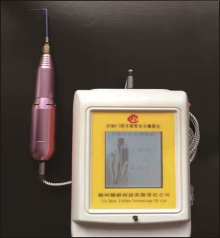
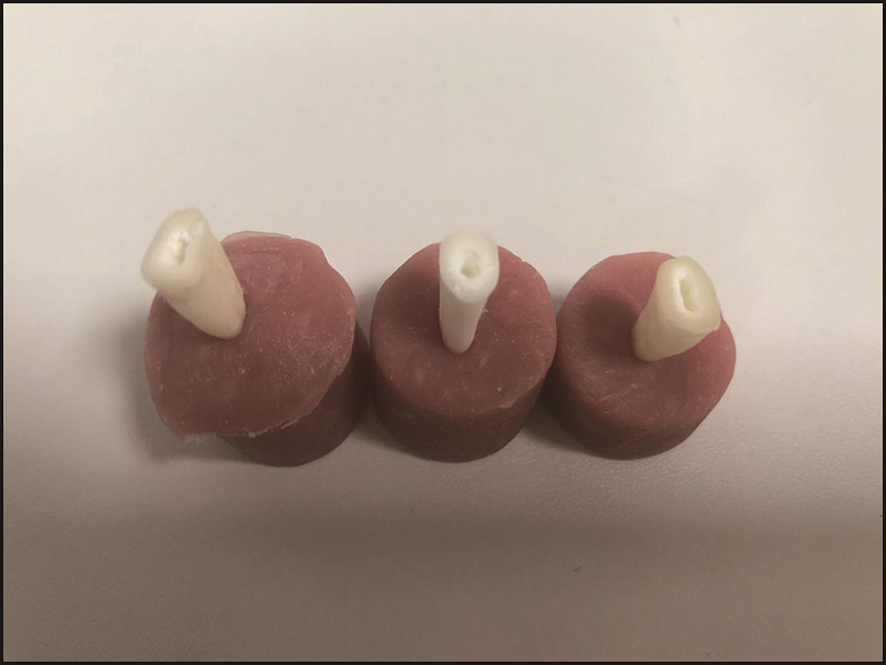
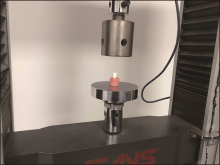
摘要: 目的 探讨PIPS-Er: YAG激光技术(光子激活光声流)治疗后根管壁牙本质失水状况及对牙根抗压强度的影响。方法 选取2018年7—9月南京市口腔医院口腔颌面外科门诊拔除的120颗健康正畸减数牙(单根牙)进行离体牙实验,按随机数字表法将其均分为A组(PIPS-Er: YAG激光冲洗组)、B组(注射器冲洗组)、C组(超声荡洗组)和D组(空白对照组,未治疗),比较治疗后各组根管壁牙本质失水状况和治疗前后牙根抗压强度的差异。结果 1)A~D组内各标本根管上、中、下段根管壁牙本质含水量依次递增,但差异无统计学意义(P>0.05);根管壁上段:A、C组低于B、D组(P<0.05),A组略低于C组(P>0.05),B组低于D组(P<0.05);根管壁中段:A组低于B~D组(P<0.05),A组略低于C组(P>0.05),B组低于D组(P<0.05);根管壁下段:A组低于B~D组(P<0.05)、A组略低于C组(P>0.05),B组低于D组(P<0.05)。2)A、B、C组与D组间的抗压强度差异无统计学意义(P>0.05)。结论 使用Er:YAG激光PIPS技术能降低根管壁牙本质的含水量,且不会降低牙根的抗压强度,有良好的临床应用前景。
| [1] | DiVito E, Peters OA, Olivi G. Effectiveness of the Erbium: YAG laser and new design radial and strip-ped tips in removing the smear layer after root canal instrumentation[J]. Lasers Med Sci, 2012,27(2):273-280. |
| [2] | Arslan H, Capar ID, Saygili G, et al. Effect of photon-initiated photoacoustic streaming on removal of apically placed dentinal debris[J]. Int Endod J, 2014,47(11):1072-1077. |
| [3] | Nasher R, Franzen R, Gutknecht N. The effectiveness of the Erbium: Yttrium aluminum garnet PIPS technique in comparison to different chemical solutions in removing the endodontic smear layer-an in vitro profilometric study[J]. Lasers Med Sci, 2016,31(9):1871-1882. |
| [4] | Arslan H, Akcay M, Ertas H, et al. Effect of PIPS technique at different power settings on irrigating solution extrusion[J]. Lasers Med Sci, 2015,30(6):1641-1645. |
| [5] | Golob BS, Olivi G, Vrabec M, et al. Efficacy of photon-induced photoacoustic streaming in the reduction of Enterococcus faecalis within the root canal: different settings and different sodium hypochlorite concentrations[J]. J Endod, 2017,43(10):1730-1735. |
| [6] | Turkel E, Onay EO, Ungor M. Comparison of three final irrigation activation techniques: effects on canal cleanness, smear layer removal, and dentinal tubule penetration of two root canal sealers[J]. Photomed Laser Surg, 2017,35(12):672-681. |
| [7] | 段睿, 王婷, 赵颖煊, 等. Er: YAG激光联合次氯酸钠对粪肠球菌杀菌效果的研究[J]. 口腔医学研究, 2017,33(4):449-452. |
| Duan R, Wang T, Zhao YX, et al. Antimicrobial ef-fects of Enterococcus faecalis in root canal using Er: YAG laser combined with different concentra-tions of sodium hypochlorite irrigants[J]. J Oral Sci Res, 2017,33(4):449-452. | |
| [8] | Balić M, Lucić R, Mehadžić K, et al. The efficacy of photon-initiated photoacoustic streaming and so-nic-activated irrigation combined with QMiX solution or sodium hypochlorite against intracanal E. fae-calis biofilm[J]. Lasers Med Sci, 2016,31(2):335-342. |
| [9] | Meire MA, Havelaerts S, De Moor RJ. Influence of lasing parameters on the cleaning efficacy of laser-activated irrigation with pulsed erbium lasers[J]. Lasers Med Sci, 2016,31(4):653-658. |
| [10] | 刘敏, 彭彬. 两种功率PIPS-Er: YAG激光对根管内玷污层去除效果的比较研究[J]. 口腔医学研究, 2018,34(10):1067-1071. |
| Liu M, Peng B. Comparison of photon-initiated pho-toacoustic streaming with two kinds of power set-tings on removal of smear layer[J]. J Oral Sci Res, 2018,34(10):1067-1071. | |
| [11] | Silvestrin T, Torabinejad M, Handysides R, et al. Effect of apex size on the leakage of gutta-percha and sealer-filled root canals[J]. Quintessence Int, 2016,47(5):373-378. |
| [12] | 陈永菊, 李新, 潘亚萍, 等. 根管湿润度对根管封闭性的影响[J]. 实用口腔医学杂志, 2019,35(1):81-86. |
| Chen YJ, Li X, Pan YP, et al. The influence of root canal moisture on the canal sealing[J]. J Pract Sto-matol, 2019,35(1):81-86. | |
| [13] | Bachmann L, Diebolder R, Hibst R, et al. Changes in chemical composition and collagen structure of dentine tissue after erbium laser irradiation[J]. Spectrochim Acta A Mol Biomol Spectrosc, 2005,61(11/12):2634-2639. |
| [14] | Virdee SS, Seymour DW, Farnell D, et al. Efficacy of irrigant activation techniques in removing intracanal smear layer and debris from mature permanent teeth: a systematic review and meta-analysis[J]. Int Endod J, 2018,51(6):605-621. |
| [15] | 田甜甜, 余擎. 不同根管壁处理方法对根管封闭剂性能影响的研究进展[J]. 牙体牙髓牙周病学杂志, 2016,26(9):564-568. |
| Tian TT, Yu Q. The effect of different root canal wall treatments on the properties of root canal sea-lers[J]. Chin J Conserv Dent, 2016,26(9):564-568. |
| [1] | 孙旭,邓振南,文才,赵颖. Er: YAG激光照射种植体表面微形貌变化的扫描电子显微镜观察[J]. 国际口腔医学杂志, 2023, 50(6): 669-673. |
| [2] | 陈凤英,王贻宁,黄翠. 桩冠粘接的研究进展[J]. 国际口腔医学杂志, 2004, 31(01): 67-68. |
|