国际口腔医学杂志 ›› 2021, Vol. 48 ›› Issue (2): 238-242.doi: 10.7518/gjkq.2021010
唐蓓( ),王扬,王虎,游梦()
Tang Bei(),Wang Yang,Wang Hu,You Meng()
摘要:
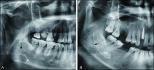
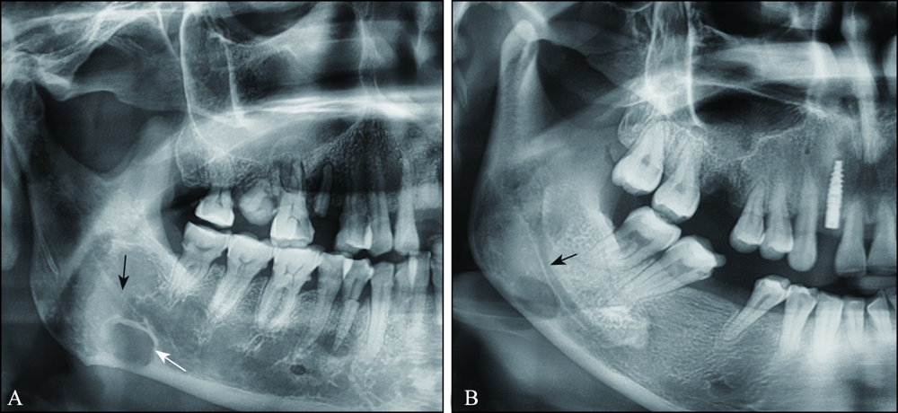
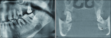
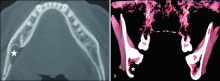
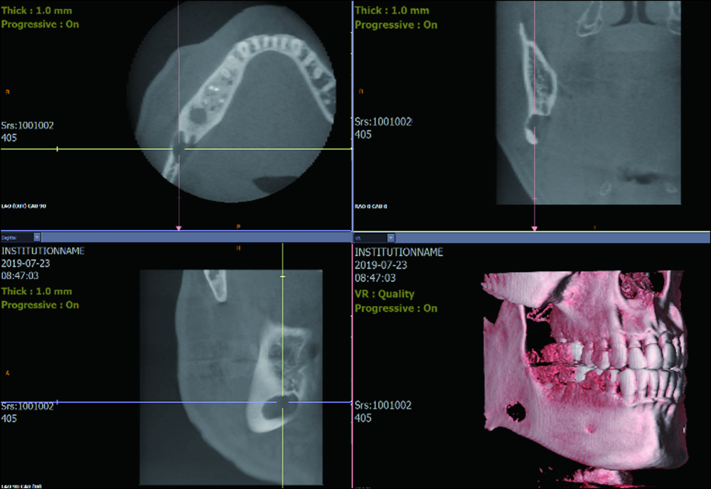
Stafne骨腔(SBC)是一种罕见的下颌骨骨质的凹陷缺损,常发生在下颌骨后份,在临床上极易被误诊为颌骨囊肿或肿瘤而进行不必要的手术治疗。本文通过回顾国内外研究,对SBC的病因、临床表现、影像学特点进行综述,总结其典型的影像学表现,介绍其影像表现的多样性,为临床准确诊断提供指导。
中图分类号:
| [1] |
Stafne EC. Bone cavities situated near the angle of the mandible[J]. J Am Dent Assoc, 1942,29(17):1969-1972.
doi: 10.14219/jada.archive.1942.0315 |
| [2] |
Branstetter BF, Weissman JL, Kaplan SB. Imaging of a Stafne bone cavity: what MR adds and why a new name is needed[J]. AJNR Am J Neuroradiol, 1999,20(4):587-589.
pmid: 10319966 |
| [3] |
Quesada-Gómez C, Valmaseda-Castellón E, Berini-Aytés L, et al. Stafne bone cavity: a retrospective study of 11 cases[J]. Med Oral Patol Oral Cir Bucal, 2006,11(3):E277-E280.
pmid: 16648768 |
| [4] |
Schneider T, Filo K, Locher MC, et al. Stafne bone cavities: systematic algorithm for diagnosis derived from retrospective data over a 5-year period[J]. Br J Oral Maxillofac Surg, 2014,52(4):369-374.
doi: 10.1016/j.bjoms.2014.01.017 pmid: 24560588 |
| [5] |
Sisman Y, Miloglu O, Sekerci AE, et al. Radiogra-phic evaluation on prevalence of Stafne bone defect: a study from two centres in Turkey[J]. Dentomaxillofac Radiol, 2012,41(2):152-158.
doi: 10.1259/dmfr/10586700 pmid: 22074869 |
| [6] |
Reuter I. An unusual case of Stafne bone cavity with extra-osseous course of the mandibular neurovascular bundle[J]. Dentomaxillofac Radiol, 1998,27(3):189-191.
doi: 10.1038/sj/dmfr/4600349 pmid: 9693534 |
| [7] |
Lello GE, Makek M. Stafne,s mandibular lingual co-rtical defect. Discussion of aetiology[J]. J Maxillofac Surg, 1985,13(4):172-176.
doi: 10.1016/s0301-0503(85)80042-4 pmid: 3860595 |
| [8] |
Shimizu M, Osa N, Okamura K, et al. CT analysis of the Stafne's bone defects of the mandible[J]. Dentomaxillofac Radiol, 2006,35(2):95-102.
doi: 10.1259/dmfr/71115878 pmid: 16549436 |
| [9] |
Philipsen HP, Takata T, Reichart PA, et al. Lingual and buccal mandibular bone depressions: a review based on 583 cases from a world-wide literature survey, including 69 new cases from Japan[J]. Dentomaxillofacial Radiol, 2002,31(5):281-290.
doi: 10.1038/sj.dmfr.4600718 |
| [10] | Minowa K, Inoue N, Sawamura T, et al. Evaluation of static bone cavities with CT and MRI[J]. Dentomaxillofac Radiol, 2003,32(1):2-7. |
| [11] | Minowa K, Inoue N, Izumiyama Y, et al. Static bone cavity of the mandible: computed tomography fin-dings with histopathologic correlation[J]. Acta Ra-diol, 2006,47(7):705-709. |
| [12] | Bastos DC, Bufalino A, Ferraz E, et al. Stafne,s bone cavity in the anterior mandible: a case report emphasizing their etiopathogenetic mechanisms[J]. Oral Maxillofac Pathol, 2017,124(2):e74. |
| [13] | Hisatomi M, Munhoz L, Asaumi J, et al. Stafne bone defects radiographic features in panoramic radiographs: assessment of 91 cases[J]. Med Oral Patol Oral Cir Bucal, 2019,24(1):e12-e19. |
| [14] | Lee KH, Thiruchelvam JK, McDermott P. An unu-sual presentation of stafne bone cyst[J]. J Maxillofac Oral Surg, 2015,14(3):841-844. |
| [15] | Hisatomi M, Munhoz L, Asaumi J, et al. Parotid mandibular bone defect: a case report emphasizing imaging features in plain radiographs and magnetic resonance imaging[J]. Imaging Sci Dent, 2017,47(4):269-273. |
| [16] |
Etöz M, Etöz OA, Sahman H, et al. An unusual case of multilocular Stafne bone cavity[J]. Dentomaxillofac Radiol, 2012,41(1):75-78.
pmid: 22184629 |
| [17] |
Kim H, Seok JY, Lee S, et al. Bilateral stafne bone cavity in the anterior mandible with heterotopic salivary gland tissue: a case report[J]. Korean J Pathol, 2014,48(3):248-249.
pmid: 25013425 |
| [18] | Ozaki H, Ishikawa S, Kitabatake K, et al. A case of simultaneous unilateral anterior and posterior stafne bone defects[J]. Case Rep Dent, 2015,2015:1-5. |
| [19] | Aguiar LBV, Neves FS, Bastos LC, et al. Multiple stafne bone defects: a rare entity[J]. ISRN Dent, 2011,2011:1-3. |
| [20] | Ariji E, Fujiwara N, Tabata O, et al. Stafne's bone cavity. Classification based on outline and content determined by computed tomography[J]. Oral Surg Oral Med Oral Pathol, 1993,76(3):375-380. |
| [21] | de Araújo Neto FB, Carneiro SGB, Fugita DYA, et al. Stafne's bone defect in atypical location: between the subcondylar region and neck of the mandible ramus[J]. Open J Med Imaging, 2017,7(3):124-129. |
| [1] | 赵文俊,刘媛媛,郝晓琪,王凯利,任家银,郭文豪,郑广宁. 46例患者第三磨牙区多生牙的影像学分析[J]. 国际口腔医学杂志, 2019, 46(1): 20-25. |
| [2] | 赵文俊, 王凯利, 刘莉, 郭文豪, 郑广宁. 2例下颌髁突动脉瘤样骨囊肿[J]. 国际口腔医学杂志, 2018, 45(3): 307-312. |
| [3] | 李唐 李春洁 门乙 杨文宾 韩波 李龙江. Meta分析评价全景片在口腔癌下颌骨侵犯中的诊断效能[J]. 国际口腔医学杂志, 2016, 43(1): 26-. |
| [4] | 任洪芋 陈昕. 不同分辨率锥形束CT和全景片在根尖吸收诊断中的对比研究[J]. 国际口腔医学杂志, 2014, 41(5): 514-517. |
| [5] | 刘太琪 周力 王艳民 . 全景片和锥形束CT评价牙根近远中倾斜度的现状和进展[J]. 国际口腔医学杂志, 2013, 40(1): 102-104. |
| [6] | 任家银 王虎 李果 赵书平 吴万红 袁珊珊. 成都地区12 岁青少年第三磨牙发育的X 线分析[J]. 国际口腔医学杂志, 2012, 39(3): 297-300. |
|