国际口腔医学杂志 ›› 2020, Vol. 47 ›› Issue (5): 538-546.doi: 10.7518/gjkq.2020086
付世锦( ),曾刊,李鑫,杨静,汪成林,叶玲()
Fu Shijin(),Zeng Kan,Li Xin,Yang Jing,Wang Chenglin,Ye Ling()
摘要:
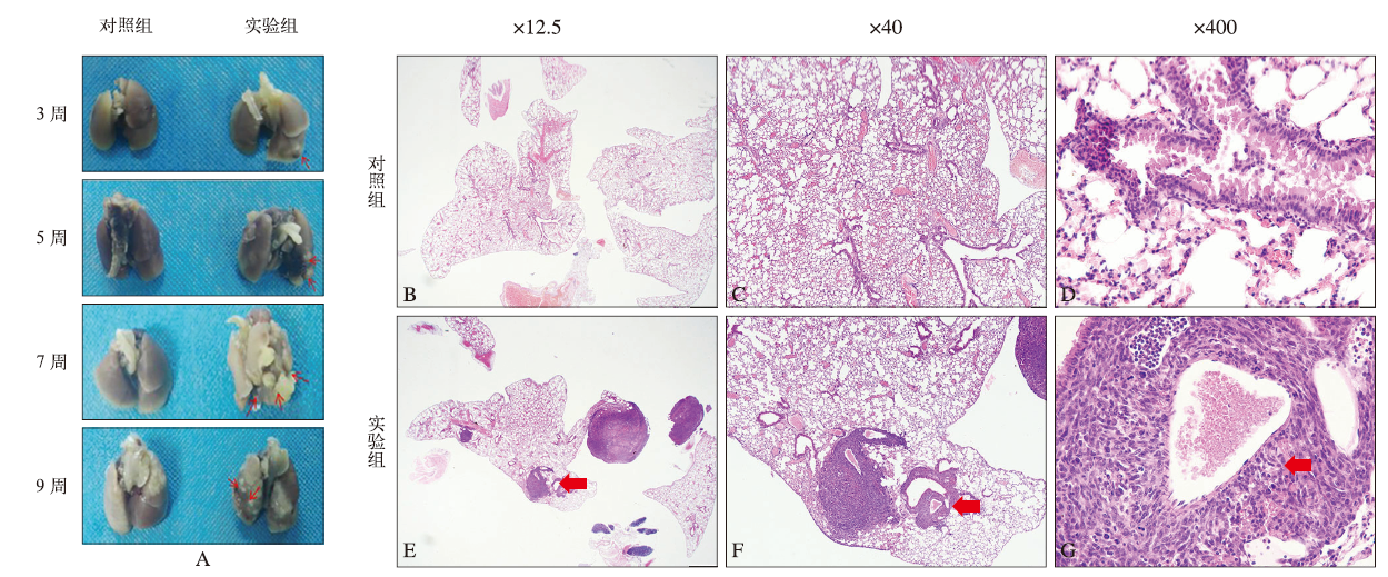
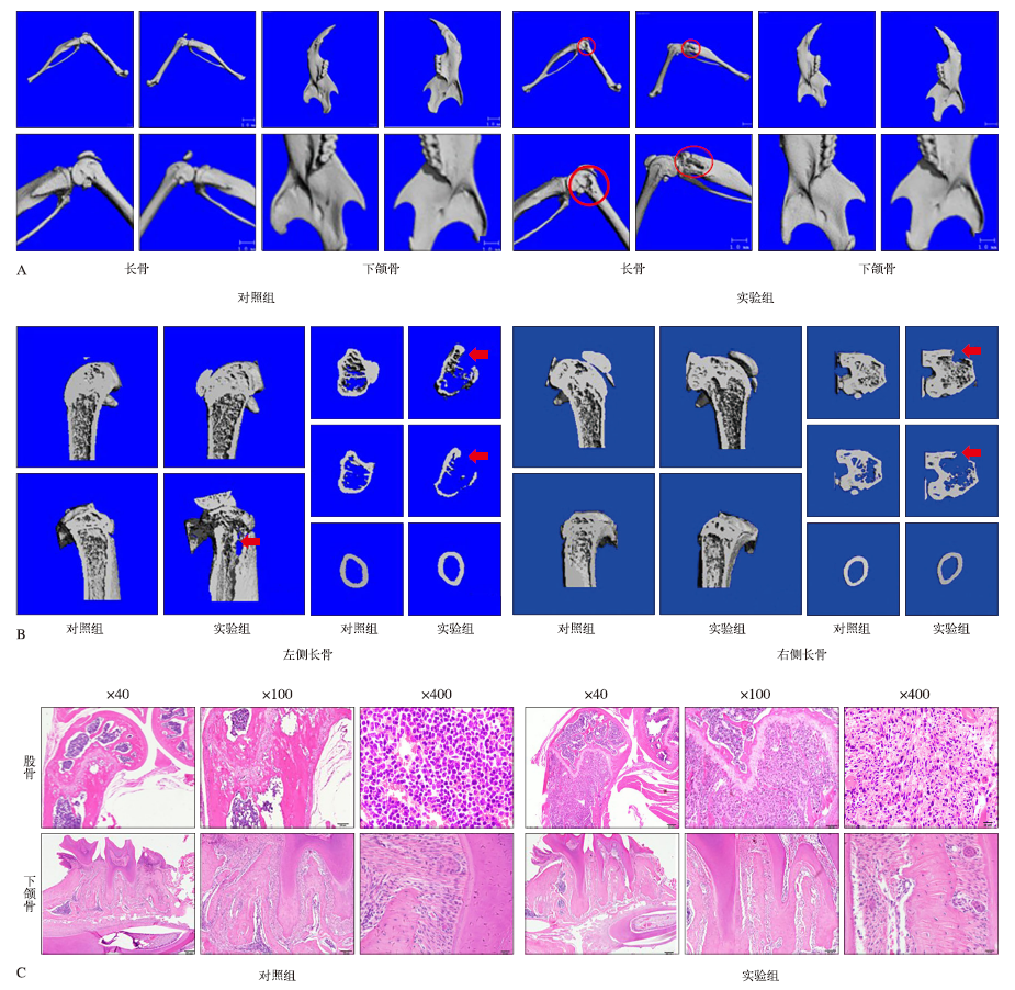
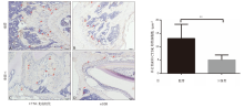
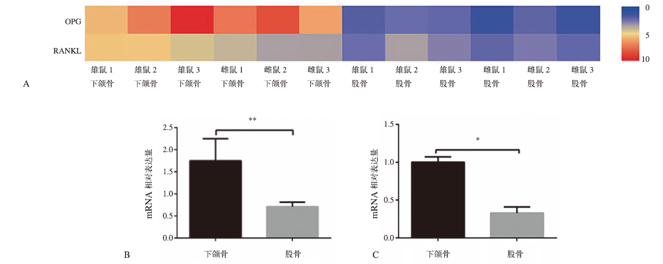
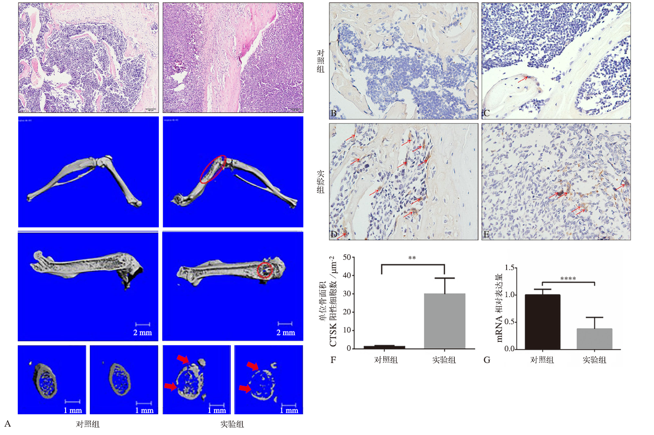
目的 探究下颌骨和股骨中骨保护素(OPG)与核因子κB受体活化因子配体(RANKL)的比值是否参与调控肺癌细胞骨转移部位差异。方法 采用小鼠Lewis肺癌细胞构建全身性肺癌细胞骨转移小鼠模型及股骨局部肺癌细胞骨转移模型,苏木素-伊红染色与显微CT(MicroCT)检测肿瘤细胞定植与骨组织破坏情况,组织蛋白酶K免疫组织化学染色检测骨组织中破骨细胞活化情况,转录组测序(RNA-seq)及实时定量聚合酶链反应(RT-qPCR)检测骨组织中OPG mRNA/RANKL mRNA比值。 结果 全身性肺癌细胞骨转移小鼠模型证实:相较于股骨,下颌骨不易发生肺癌细胞骨转移。分析生理情况下小鼠下颌骨与股骨的差异,发现在肺癌细胞骨转移低发的下颌骨中破骨细胞数目明显少于转移高发的股骨,且OPG mRNA/RANKL mRNA比值明显高于股骨(P<0.01)。进一步分析发生肿瘤细胞转移的骨组织中破骨细胞活化情况及OPG mRNA/RANKL mRNA比值发现,肺癌细胞骨转移引起的骨破坏主要发生在组织蛋白酶K阳性破骨细胞集中分布的区域,发生肿瘤细胞转移的骨组织中破骨细胞数目明显多于非骨转移组(P<0.01),且OPG mRNA/RANKL mRNA比值明显低于非骨转移组(P<0.000 1)。结论 在小鼠肺癌细胞骨转移低发的下颌骨与高发的股骨中,破骨细胞活化情况及OPG mRNA/RANKL mRNA比值存在明显差异,提示骨组织可能通过OPG/RANKL影响破骨细胞活化进而参与调控肺癌细胞骨转移部位差异。
中图分类号:
| [1] |
Meyer I, Shklar G. Malignant tumors metastatic to mouth and jaws[J]. Oral Surg Oral Med Oral Pathol, 1965,20:350-362.
pmid: 14342927 |
| [2] |
Sánchez Aniceto G, García Peñín A, de la Mata Pages R, et al. Tumors metastatic to the mandible: analysis of nine cases and review of the literature[J]. J Oral Maxillofac Surg, 1990,48(3):246-251.
doi: 10.1016/0278-2391(90)90388-i pmid: 2406399 |
| [3] |
Hernandez RK, Wade SW, Reich A, et al. Incidence of bone metastases in patients with solid tumors: analysis of oncology electronic medical records in the United States[J]. BMC Cancer, 2018,18(1):44.
doi: 10.1186/s12885-017-3922-0 pmid: 29306325 |
| [4] |
Bray F, Ferlay J, Soerjomataram I, et al. Global cancer statistics 2018: GLOBOCAN estimates of incidence and mortality worldwide for 36 cancers in 185 countries[J]. CA Cancer J Clin, 2018,68(6):394-424.
doi: 10.3322/caac.21492 pmid: 30207593 |
| [5] |
Tsuya A, Kurata T, Tamura K, et al. Skeletal metas-tases in non-small cell lung cancer: a retrospective study[J]. Lung Cancer, 2007,57(2):229-232.
doi: 10.1016/j.lungcan.2007.03.013 pmid: 17451841 |
| [6] | 北京医学奖励基金会肺癌青年专家委员会, 中国胸外科肺癌联盟. 肺癌骨转移诊疗专家共识(2019版)[J]. 中国肺癌杂志, 2019,22(4):187-207. |
| Youth Specialists Committee of Lung Cancer, Beijing Medical AwardFoundaton, Chinese Lung Cancer Union. Expert consensus on the diagnosis and treat-ment of bone metastasis in lung cancer (2019 version)[J]. Chin J Lung Cancer, 2019,22(4):187-207. | |
| [7] |
Clain A. Secondary malignant disease of bone[J]. Br J Cancer, 1965,19(1):15-29.
doi: 10.1038/bjc.1965.3 |
| [8] |
Ueki Y, Tiziani V, Santanna C, et al. Mutations in the gene encoding c-Abl-binding protein SH3BP2 cause cherubism[J]. Nat Genet, 2001,28(2):125-126.
doi: 10.1038/88832 pmid: 11381256 |
| [9] |
Simonds WF, James-Newton LA, Agarwal SK, et al. Familial isolated hyperparathyroidism: clinical and genetic characteristics of 36 kindreds[J]. Medicine (Baltimore), 2002,81(1):1-26.
doi: 10.1097/00005792-200201000-00001 |
| [10] | Ruggiero SL, Mehrotra B, Rosenberg TJ, et al. Osteo-necrosis of the jaws associated with the use of bis-phosphonates: a review of 63 cases[J]. J Oral Maxil-lofac Surg, 2004,62(5):527-534. |
| [11] |
Yoneda T, Hiraga T. Crosstalk between cancer cells and bone microenvironment in bone metastasis[J]. Biochem Biophys Res Commun, 2005,328(3):679-687.
doi: 10.1016/j.bbrc.2004.11.070 pmid: 15694401 |
| [12] |
Fidler IJ. The pathogenesis of cancer metastasis: the ‘seed and soil’ hypojournal revisited[J]. Nat Rev Cancer, 2003,3(6):453-458.
doi: 10.1038/nrc1098 pmid: 12778135 |
| [13] |
Paget S. The distribution of secondary growths in cancer of the breast. 1889[J]. Cancer Metastasis Rev, 1989,8(2):98-101.
pmid: 2673568 |
| [14] |
Zhuang XQ, Zhang H, Li XY, et al. Differential ef-fects on lung and bone metastasis of breast cancer by Wnt signalling inhibitor DKK1[J]. Nat Cell Biol, 2017,19(10):1274-1285.
doi: 10.1038/ncb3613 pmid: 28892080 |
| [15] |
Wang H, Yu CJ, Gao X, et al. The osteogenic niche promotes early-stage bone colonization of disse-minated breast cancer cells[J]. Cancer Cell, 2015,27(2):193-210.
doi: 10.1016/j.ccell.2014.11.017 pmid: 25600338 |
| [16] |
Martin TJ, Sims NA. RANKL/OPG; Critical role in bone physiology[J]. Rev Endocr Metab Disord, 2015,16(2):131-139.
doi: 10.1007/s11154-014-9308-6 pmid: 25557611 |
| [17] |
Dougall WC. Molecular pathways: osteoclast-depen-dent and osteoclast-independent roles of the RANKL/ RANK/OPG pathway in tumorigenesis and metastasis[J]. Clin Cancer Res, 2012,18(2):326-335.
pmid: 22031096 |
| [18] |
Wu JB, Yin LJ, Shi CH, et al. MAOA-dependent activation of shh-IL6-RANKL signaling network promotes prostate cancer metastasis by engaging tumor-stromal cell interactions[J]. Cancer Cell, 2017,31(3):368-382.
pmid: 28292438 |
| [19] | Dougall WC, Chaisson M. The RANK/RANKL/OPG triad in cancer-induced bone diseases[J]. Cancer Me-tastasis Rev, 2006,25(4):541-549. |
| [20] |
Grimaud E, Soubigou L, Couillaud S, et al. Receptor activator of nuclear factor kappaB ligand (RANKL)/osteoprotegerin (OPG) ratio is increased in severe osteolysis[J]. Am J Pathol, 2003,163(5):2021-2031.
pmid: 14578201 |
| [21] | 羊惠君. 实地解剖学[M]. 北京: 人民卫生出版社, 2011. |
| Yang HJ. Practical anatomy teaching[M]. Beijing: People’s Medical Publishing House, 2011. | |
| [22] | 邹仲之. 组织学与胚胎学[M]. 北京: 人民卫生出版社, 2013. |
| Zou ZZ. Histology and embryology[M]. Beijing: People’s Medical Publishing House, 2013. | |
| [23] |
Irizarry AR, Yan GR, Zeng QQ, et al. Defective enamel and bone development in sodium-dependent citrate transporter (NaCT) Slc13a5 deficient mice[J]. PLoS One, 2017,12(4):e0175465.
pmid: 28406943 |
| [24] |
Jiang XB, Iseki S, Maxson RE, et al. Tissue origins and interactions in the mammalian skull vault[J]. Dev Biol, 2002,241(1):106-116.
pmid: 11784098 |
| [25] |
Hirshberg A, Berger R, Allon I, et al. Metastatic tumors to the jaws and mouth[J]. Head Neck Pathol, 2014,8(4):463-474.
doi: 10.1007/s12105-014-0591-z pmid: 25409855 |
| [1] | 余岳霖,孔卫东. 甲状旁腺激素受体1基因相关与原发性牙齿萌出障碍的研究进展[J]. 国际口腔医学杂志, 2023, 50(5): 573-580. |
| [2] | 尹一佳,杨瑾廷,申建琪,黄凌依,井岩,官秋玥,韩向龙. 钙黏蛋白5驱动内皮细胞特异性过表达Dickkopf 1影响骨形成[J]. 国际口腔医学杂志, 2022, 49(6): 641-647. |
| [3] | 黎静文,周力. 颈椎成熟法评估下颌骨骨龄的研究进展[J]. 国际口腔医学杂志, 2022, 49(3): 337-342. |
| [4] | 安宁,李姣,梅志丹. 骨保护素/核因子-κB受体活化因子/核因子κB-受体活化因子配体信号分子调控牙萌出的研究进展[J]. 国际口腔医学杂志, 2022, 49(1): 116-120. |
| [5] | 吕辉,王华,孙雯. 辅助性T细胞17与牙周炎骨免疫[J]. 国际口腔医学杂志, 2020, 47(6): 661-668. |
| [6] | 孙坚炜,雷利红,谭静怡,陈莉丽. 微小RNA 155对骨免疫的调控及其在牙周炎中作用的研究进展[J]. 国际口腔医学杂志, 2020, 47(5): 607-615. |
| [7] | 杨佩佩,杨羽晨,张强. 尼古丁对牙槽骨破骨细胞的作用及其机制的研究进展[J]. 国际口腔医学杂志, 2020, 47(5): 616-620. |
| [8] | 朱俊瑾,周佳琦,伍颖颖. 哺乳动物雷帕霉素靶蛋白复合物1介导的自噬对骨代谢的调控[J]. 国际口腔医学杂志, 2020, 47(1): 84-89. |
| [9] | 卢可心,张迪亚,吴燕岷. 蛋白酶激活受体在牙周组织细胞中相关作用的研究进展[J]. 国际口腔医学杂志, 2019, 46(6): 657-662. |
| [10] | 林阳阳,侯敏. 双侧下颌支矢状骨劈开术对下颌近心骨段位移变化的影响[J]. 国际口腔医学杂志, 2019, 46(6): 718-723. |
| [11] | 张誉泓,戚孟春,董伟,孙红. CaMKi>Ⅱδ基因沉默对破骨细胞分化功能及c-fosi>/c-juni>/CREBi>基因的影响[J]. 国际口腔医学杂志, 2019, 46(4): 420-425. |
| [12] | 王小萌,王晓,史册,孙宏晨,黄洋. 骨形态发生蛋白信号通路及其交叉对话对下颌骨发育的影响[J]. 国际口腔医学杂志, 2019, 46(3): 258-262. |
| [13] | 胡巍,王译凡,袁一方,李影,郭斌. 节律基因调控成骨和破骨活动机制的研究进展[J]. 国际口腔医学杂志, 2019, 46(3): 302-307. |
| [14] | 高鑫,曾融生. 骨保护素在口腔领域的研究进展[J]. 国际口腔医学杂志, 2019, 46(3): 316-319. |
| [15] | 刘曼, 孟耀, 何勇, 魏宁, 张强. 口腔全景片评价更年期妇女骨质疏松的研究[J]. 国际口腔医学杂志, 2017, 44(2): 165-169. |
|